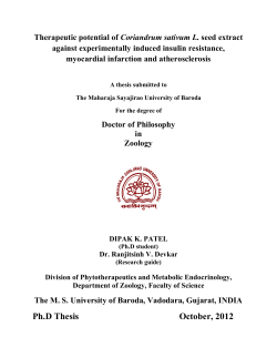
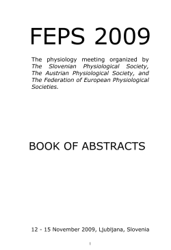
Volume 6, Issue 4 тАв Winter 2011 - 2012
Publications Agreement Number 40025049 | 1911-1606
Practical Approach to Isolated
Right-Sided Heart Failure
Bizios and Howlett
Aldosterone Antagonist Therapy
in Heart Failure
www.csimonline.com
Barry et al.
тАЬOPINION
IS GOOD.
BUT EVIDENCE
*
IS BETTER.тАЭ
BRILINTA (ticagrelor), co-administered with
acetylsalicylic acid (ASA), is indicated for the
secondary prevention of atherothrombotic events
in patients with Acute Coronary Syndromes (ACS)
(unstable angina [UA], nonтАУST elevation myocardial
infarction [NSTEMI] or ST elevation myocardial
infarction [STEMI]) who are to be managed medically,
and those who are to be managed with percutaneous
coronary intervention (PCI) (with or without stent)
and/or coronary artery bypass graft (CABG).
Based on a relationship observed in PLATO between
maintenance ASA dose and relative efficacy of
BRILINTA compared to clopidogrel, BRILINTA is
recommended to be co-administered with low
maintenance dose ASA (75-150 mg daily).
BRILINTA is contraindicated in patients who: are
hypersensitive to this medication or to any ingredient
in the formulation, have active pathological bleeding
such as peptic ulcer or intracranial hemorrhage, have
a history of intracranial hemorrhage, have moderate
to severe hepatic impairment or are also taking
strong CYP3A4 inhibitors.
BRILINTA should be used with caution in patients
with a propensity to bleed (e.g., due to recent trauma,
recent surgery, active or recent gastrointestinal
bleeding, or moderate hepatic impairment) and in
patients requiring oral anticoagulants (e.g., warfarin)
and/or fibrinolytics agents (within 24 hours of
BRILINTA dosing). Caution should also be used in
patients with concomitant administration of medicinal
products that may increase the risk of bleeding (e.g.,
non-steroidal anti-inflammatory drugs [NSAIDs]).
Co-administration of BRILINTA and high maintenance
dose ASA (>150 mg daily) is not recommended.
In the PLATO study, bleeding events associated with
BRILINTA vs. clopidogrel included total major (11.6%
vs. 11.2%) and combined total major + minor (16.1%
vs. 14.6%). When minor bleeding was included,
combined PLATO-defined major and minor bleeding
events were significantly higher on BRILINTA than on
clopidogrel (p=0.0084). There were few fatal bleeding
events in the study, 20 (0.2%) for BRILINTA and 23
(0.3%) for clopidogrel. The most common adverse
events associated with BRILINTA vs. clopidogrel were
dyspnea (12.0% vs. 6.5%), headache (6.5% vs. 5.8%)
and nosebleed (6.0% vs. 3.4%).
See the Product Monograph for full contraindications,
warnings, precautions, dosing and administration.
Reference: 1. BRILINTA┬о Product Monograph.
AstraZeneca Canada Inc. May 26, 2011.
*Fictitious quote. May not be representative
of all healthcare professionals.
BR032E
08/12
BRILINTA┬о and the AstraZeneca logo are registered trademarks
of the AstraZeneca group of companies. ┬й AstraZeneca 2011
158
CONTENTS
Volume 6, Issue 4
2011
Volume 6, Issue 4 тАв 2011
Publications Agreement Number 40025049
EDITOR-IN-CHIEF
Hector Baillie
EDITORIAL BOARD
Ranjani Aiyar, Simona Bar, Pat Bergin,
Peter Brindley, Kaberi Dasgupta, Don Echenberg,
Don Farquhar, Bert Govig, Luc Lanthier,
Alex Leung, Suzanne Morin, Jock Murray,
Kathryn Myers, Glen Pearson, Louise Pilote,
Colin Powell, Margot Roach, Linda Snell,
Matthieu Touchette, George Veenhuyzen
C S I M S TA F F
Ms. Domenica Utano
Ms. Katie Beal
MANAGING EDITOR
Susan Harrison
ART DIRECTOR
Andrea Brierley
Message from the President тАв
Message du pr├йsident
129
Pharm-GIM
138 Aldosterone Antagonist Therapy in
Heart Failure: The Trilogy of Trials!
Maria Bacchus MD
Arden Barry PharmD, Glen J. Pearson PharmD,
Sheri L. Koshman PharmD
CSIM News
129 Thank You to Our Sponsors
137 Congratulations
141 Edmonton Physician Receives Honour
from Mayo Clinic
Donald Farquhar MD
131 Practical Approach to the Diagnosis
and Management of Isolated
Right-Sided Heart Failure
Scott Bryant
Marie Dumont
ADVERTISING
John Birkby
(905) 628-4309
[email protected]
ACCOUNTING
Susan McClung
GROUP PUBLISHER
John D. Birkby
_______________________________________
Subscription Rates:
Per year: $15.00; per issue: $3.75
__________________________________
Return undeliverable Canadian Addresses to:
135 Le lymphome intravasculaire: une
pathologie sournoise et agressive
├а inclure dans le diagnostic diff├йrentiel
dтАЩune fi├иvre dтАЩ├йtiologie ind├йtermin├йe
Maxime Berthelot-Richer MD, Chantal Vall├йe MD,
Rabia Temmar MD, Hans Knecht MD, Rami Kotb MD,
Martine Chamberland MD
144 WegenerтАЩs Granulomatosis Causing
Gastrointestinal Hemorrhage and
Perforation of the Small Intestine:
A Case Report and Review of the
Literature
Sankalp V. Bhavsar MD
151 Dr. Julian Derwent Loudon and
CanadaтАЩs First Electrocardiograph
Peter A. Kopplin MD
Guidelines Review
156 2011 Canadian Hypertension
Education Program Update
Resident GIM
157 Internal Medicine: Life in the
Fast Lane
William Connors MD
Monika Pawlowska MD, Eric M. Yoshida MD,
David Collins MD, Edward Jones MD
149 Q Fever: A Rare Cause of
Community-Acquired Pneumonia?
Stephen Pizzale MD, Cheryl Main MD,
Atreyi Mukherji MD
115 King St W., Suite 220, Dundas, ON L9H 1V1
Canadian Journal of General Internal Medicine is published
four times a year by Andrew John Publishing Inc., with
offices located at 115 King Street West, Suite 220, Dundas,
ON L9H 1V1.
153 Atypical Femur Fracture in a Liver
Transplant Recipient on Long-Term
Bisphosphonate Therapy
We welcome editorial submissions but cannot assume
responsibility or commitment for unsolicited material.Any editorial material, including photographs that are accepted from
an unsolicited contributor, will become the property of
Andrew John Publishing Inc.
Daniel Banaszek BSc, Suzanne Morin MD
The publisher and the Canadian Society of Internal Medicine
shall not be liable for any of the views expressed by the
authors published in Canadian Journal of General Internal
Medicine, nor shall these opinions necessarily reflect those of
the publisher.
147 Giant Cell Arteritis
History of Medicine
Case Reviews тАв R├йexamen des cas
C I R C U L AT I O N C O O R D I N AT O R
Brenda Robinson
[email protected]
Clinical Review
Anna Bizios MD, Jonathan Howlett MD
Scott Bryant, Michael Peebles
T R A N S L AT O R
143 A FatherтАЩs Gift
Perspectives in GIM
COPY EDITORS
PROOFREADER
Humanities
We acknowledge the financial support of the Government of Canada
through the Canada Periodical Fund of the Department of Canadian Heritage.
тАвтАвтАвтАвтАв
Every effort has been made to ensure that the information
provided herein is accurate and in accord with standards
accepted at the time of printing. However, readers are
advised to check the most current product information provided by the manufacturer of each drug to verify the recommended dose, the method and duration of administration,
and contraindications. It is the responsibility of the licensed
prescriber to determine the dosages and best treatment for
each patient. Neither the publisher nor the editor assumes
any liability for any injury and/or damage to persons or
property arising from this publication.
128
ABOUT THE COVER
This issueтАЩs cover photo was taken by J.J. Guy Longtin, a career fire chief who enjoys photography as a way to
relieve stress. This photo was captured at the annual powwow of the Pikw├аkanag├аns of Golden Lake, Ontario.
For Instructions to Authors, please visit
www.andrewjohnpublishing.com/CGJIM/instructionstoauthors.html
Volume 6, Issue 4, Winter 2011-2012
Canadian Journal of General Internal Medicine
CSIM Members of Council
Me ssag e from t he President тАв Messa ge de la pr ├йsid ente
Dr. Maria Bacchus
President
Calgary, AB
Dr. Finlay McAlister
Past-President
Edmonton, AB
Maria Bacchus MD
Dr. Stephen Hwang
About the Author
Maria Bacchus is an associate professor in the Department of Medicine at the
University of Calgary, in Calgary, Alberta. Correspondence may be directed to
[email protected].
Secretary-Treasurer, Ontario Region Representative
Toronto, ON
Dr. Nadine Lahoud
Quebec Region Representative
LaSalle, QC
Au sujet de lтАЩauteur
Maria Bacchus est professeure agr├йg├йe de m├йdecine ├а la facult├й de m├йdecine de
lтАЩUniversit├й de Calgary en Alberta. Veuillez adresser la correspondance ├а
[email protected]
Dr. Patrick Bergin
Eastern Region Representative
Charlottetown, PEI
Dr. David Simpson
Eastern Region Representative
Halifax, NS
Dr. Donald Echenberg
Vice-President, Education Committee
Quebec Region Representative
Sherbrooke, QC
Dr. Bert Govig
Vice-President, Health Promotion Committee
Quebec Region Representative
Amos, QC
Dr. Tom Maniatis
Quebec Region Representative
Montreal, QC
Dr. Benjamin Chen
Ontario Region Representative
Napanee, ON
Dr. William Coke
Ontario Region Representative
Toronto, ON
Dr. Ameen Patel
Ontario Region Representative
Hamilton, ON
Dr. Neil Gibson
Western Region Representative
St. Albert, AB
Dr. Amy Hendricks
Western Region Representative
Yellowknife, NT
Dr. Anne Marie PausJenssen
Dear Colleagues,
Cher coll├иgues,
I am honoured to be your advocate for general
internal medicine (GIM) as the 15th president of
the Canadian Society of Internal Medicine. I am
excited to hear your input and suggestions as we
forge our identity as a subspecialty.
I would like to thank our past-president, Finlay
McAlister, for his exemplary leadership as he
steered us through groundbreaking changes
including the recognition of general internal
medicine as a subspecialty.
Special thanks also to David Simpson and the
members of the Annual Meeting Committee for
the successful organization and implementation of
our recent meeting in Halifax, Nova Scotia. Under
the guidance of Drs. Nadine Lahoud and Kathleen
Raby, we are enthusiastically planning our next
Annual Meeting, to be held October 17тАУ20, 2012,
in Quebec City, Quebec.
This is an exhilarating time for GIM, and I invite
you to use this opportunity to shape the future of
our subspecialty. Your suggestions are valuable. We
encourage you to make your voice heard. Please
email me or your council members at
[email protected].
JтАЩai lтАЩimmense privil├иge de promouvoir les int├йr├кts
de la m├йdecine interne g├йn├йrale en votre nom ├а
titre de 15e pr├йsidente de la Soci├йt├й canadienne de
m├йdecine interne (SCMI). Je suis impatiente de
conna├оtre votre opinion, vos id├йes et vos
suggestions alors que la discipline forge son
identit├й de sursp├йcialit├й.
Je tiens ├а remercier le pr├йsident sortant,
Finlay McAlister, de son leadership exemplaire
durant une p├йriode de changement sans pr├йc├йdent
qui a d├йbouch├й notamment sur la reconnaissance
de la sursp├йcialit├й de la m├йdecine interne g├йn├йrale.
Mes remerciements vont ├йgalement ├а David
Simpson et aux membres du comit├й du congr├иs
annuel qui ont organis├й et tenu de main de ma├оtre
le dernier congr├иs ├а Halifax en Nouvelle-├Йcosse.
Sous la direction des Dres Nadine Lahoud et
Kathleen Raby, nous planifions d├йj├а avec beaucoup
dтАЩenthousiasme le prochain congr├иs qui aura lieu
dans la ville de Qu├йbec du 17 au 20 octobre 2012.
Nous abordons sans doute lтАЩ├вge dтАЩor de la
m├йdecine interne g├йn├йrale, et je vous invite ├а
participer ├а cette entreprise fascinante qui consiste
├а fa├зonner lтАЩavenir de la sursp├йcialit├й. Nous
accordons ├йnorm├йment dтАЩimportance ├а votre
opinion et ├а vos suggestions. NтАЩh├йsitez surtout pas
├а les exprimer. Vous pouvez communiquer avec les
membres du conseil dтАЩadministration ou avec moi
par courriel ├а [email protected].
Western Region Representative
Saskatoon, SK
Dr. Ranjani Aiyar
Resident Representative
Calgary, AB
Other CSIM Positions
Dr. Gary Victor
Vice-President, Membership Affairs
Kelowna, BC
CSIM News
Dr. Bill Ghali
Vice-President, Research and Awards Committee
Calgary, AB
Dr. Kathleen Raby
Co-Chair 2012 Annual Meeting Committee
Quebec City, QC
Dr. Jim Nishikawa
Representative on the RCPSC Specialty Committee in
Internal Medicine
Ottawa, ON
To contact a member of CSIM Council,
please e-mail [email protected].
The CSIM gratefully acknowledges support of our scientific program by educational grants from:
PLATINUM SPONSORSHIP
Abbott
AstraZeneca
Bayer Healthcare
Eli Lilly Canada
NovoNordisk
Merck
Canadian Journal of General Internal Medicine
GOLD SPONSORSHIP
Boehringer-Ingelheim
Sanofi Aventis
Thank you to AstraZeneca for their support of the
Osler Awards and the Osler Lecture.
Volume 6, Issue 4, Winter 2011-2012
129
e
b
r
e
h
t
IтАЩ d ra
!
e
r
e
h
t
h
r ig
Dr. Isabelle C
ochrane and
husband
Nelson, BC
ENRICH YO
YOUR
OUR CAREER.
CAREER. ENHANC
ENHANCEE YOUR QUALITY OF LIF
LIFE.
E.
ch
H ea lt h M at
I ha d he ard of
ke pt their
B C an d ha d
e
fi le. Whe n th
in fo rmat ion on
eir
en t th ro ug h th
time ca me I w
e
fo r a full- tim
si te, lo ok ing
the
om po si tion in
emerge nc y ro
e
ha tтАЩs w he re w
mou nt ains. T
fo r
- a pa ra dise
fo un d N elson
sk iing.
ba ck co un tr y
Join the hundred
hundredss of physicians who have moved to British
B
Columbia to enjoy a quality of life
that is envied aro
around
ound the world.
world Find out how our phy
physician
ysician services team can assist you in
matching your lif
lifestyle
festyle interests with exciting career opportunities. Register
Register online today!
todayy!
healthmatchbc.org
healt
thmatchbc.org
thmatchbc.
org
FIND A JOB IN BC
Health
H
ealth Match
Match BC
BC is
is a free
free health
health professional
professional recruitment
recruitment service
service funded
funded by
by the
the Government
Government of
of British
British Columbia,
Columbia, Canada
Canada
TTEL:
EL: 6604.736.5920
0 4.736.5920
s
TTOLL-FREE:
OL L-FREE: 11.877.867.3061
.877.867.3061
s
EEMAIL:
M AIL: [email protected]
welcome @ healthmatchbc.org
C oc hrane
D r. Is ab elle
Pe rs p e c t i v e s i n G I M
Practical Approach to the Diagnosis and Management of Isolated Right-Sided
Heart Failure
Anna Bizios MD, Jonathan Howlett MD
About the Authors
Anna Bizios is a cardiology fellow in the Department of Cardiac Sciences, University of Calgary, in Calgary,
Alberta. Jonathan Howlett is a clinical professor of medicine at the Libin Cardiovascular Institute and
University of Calgary. He is a member of the Canadian Cardiovascular Society Heart Failure Guidelines
Primary Panel. Correspondence may be directed to [email protected].
Clinical Vignette
A 65-year-old obese, diabetic female was admitted to hospital with
symptoms of exercise intolerance, shortness of breath on exertion, weight
gain, and edema. She had suffered a myocardial infarction (MI) 2 years
previously and was on appropriate medical therapy. She was known to have
stage 3 diabetic kidney disease.
On admission, she had the following vitals: heart rate 80 beats per minute,
blood pressure 155/95 mm Hg, and oxygen saturation 96% on room air. She
was obese (body mass index 35 kg/m2), had 6 cm jugular venous distension,
and had moderate peripheral edema. Her heart sounds were normal, but a
fourth heart sound was present. She also had a right ventricular heave.
Auscultation of the chest was normal, and because of obesity, hepatomegaly
was not evident clinically. An electrocardiogram (ECG) revealed sinus
rhythm, right atrial enlargement, right ventricular hypertrophy, and old
inferior Q waves. She was treated with intravenous furosemide 40 mg/d, and
she lost 5 kg over the next 4 days. She then developed dizziness when
standing, worsening lethargy, and ongoing shortness of breath. She had a
postural drop in systolic blood pressure (110 mm Hg lying and 90 mm Hg
standing). Her serum creatinine rose from 160 to 245 mmol/L. Her chest
radiograph showed cardiomegaly only. Her urinary protein excretion was
600 mg protein per day. Her complete blood count was unremarkable, and
her electrolytes remained within the normal range.
An echocardiogram showed a dilated right ventricle (RV) with septal
flattening and a right ventricular systolic pressure (RVSP) of 45 mm Hg (but
acoustic windows were poor because of obesity). There was no valvular
dysfunction, but inferior wall hypokinesis was evident. A right heart
catheterization showed a pulmonary artery (PA) pressure of 78/36 mm Hg
and a mean pulmonary capillary wedge pressure (PCWP) of 10 mm Hg. A
Persantine MIBI scan showed an old inferior MI, no ischemia, and normal
LV (left ventricle) function. A subsequent investigation confirmed the
presence of severe obstructive sleep apnea. Her condition improved
following the introduction of continuous positive airway pressure (CPAP)
therapy.
Discussion
Heart failure is an important clinical syndrome affecting the Canadian
population: an estimated 500,000 individuals are living with the disease,
and another 50,000 patients are newly diagnosed each year.1 A significant
proportion of these individuals suffer from right-sided heart failure (RHF),
which is an independent predictor of mortality in patients with heart
failure and may occur as part of the biventricular heart failure syndrome
or in the absence of left-sided heart failure (LHF).2 Isolated RHF (iRHF)
frequently occurs as a consequence of a unique set of causes and should
be treated differently. Thus, recognition of iRHF is essential. A practical
approach to the diagnosis and management of RHF is outlined below.
Canadian Journal of General Internal Medicine
Congestive
gestive heart failure
diagnosed
Clinical features
suggest iRHF
s#AUTIOUSDIURESIS
s)NVESTIGATEFORPOTENTIAL
CAUSE
o RHFF,, and treat once deterrmined
of
s,OWTHRESHOLDFOR
hemodynamic assessmeent
(right heart catheterization)
on)
Clinical
inical
IMPROOVEMENT
Coontinue
Clinicall features
suggest LHF
s)NVEST
TIGATEFORCAUSESOF,(&
s RIALOFTHERAPY
s4
,ACKOFIM
MPROVEMENT
renal inssufficiency
early inn diuresis,
or
POORLYT
TOLERATED
THEERAPY
)MPROVEM
MENT
EXPECTEED
outcome
me
No
IMPROVEMEENT
Reeassess
PATTIENTAND
initiaal diagnosis
Continue
ue
Consider
der isolated RHF
WITHL OWTHRESHOLDFOR
right heeart catheterization
Figure 1. Generalized approach to congestive heart failure. iRHF = isolated
right-sided heart failure; LHF = left-sided heart failure; RHF = right-sided heart
failure.
Step 1: Making the Diagnosis of Heart Failure
iRHF generally refers to the clinical presentation of signs and symptoms of
heart failure without the specific findings of LHF. This means the patientтАЩs
clinical condition is not due to elevated left-sided filling pressures, which can
be difficult to demonstrate without invasive assessments. The first step in the
identification of iRHF is the diagnosis of heart failure in general (Figure 1
and Table 1). During the initial clinical assessment, clues may be present to
suggest RHF in the absence of left-sided involvement. For example, a history
of long-standing chronic obstructive pulmonary disease or other severe lung
disease may be elicited. Symptoms such as fatigue, abdominal distension, or
anorexia in the absence of dyspnea, orthopnea, and paroxysmal nocturnal
dyspnea should raise suspicions for RHF. On physical examination,
predominant right-sided findings (e.g., pulsatile liver, ascites, elevated jugular
venous pulsation with KussmaulтАЩs sign) may be present without pulmonary
crackles. A chest radiograph may fail to show pulmonary congestion or LV
Volume 6, Issue 4, Winter 2011-2012
131
P r a c t i c a l A p p r o a c h t o I s o l a t e d R i g h t - S i d e d H e a r t Fa i l u r e
Table 1. Signs and Symptoms Suggestive of Right-Sided
Heart Failure
evidence of elevated right-sided filling pressures. A clinician may be alerted
to iRHF only after unsuccessful initial therapy for presumed LHF.
Symptoms
Step 2: Identifying the Etiology of RHF
Shortness of breath
Fatigue
Anorexia or early satiety
Right upper quadrant discomfort
Peripheral edema
Signs
Elevated jugular venous pulsation
Positive hepatojugular reflux
KussmaulтАЩs sign*
Peripheral edema or ascites
Hepatomegaly
Right-sided S3
Tricuspid regurgitation murmur
Right ventricular lift
*KussmaulтАЩs sign is observed when the jugular venous pulsation rises on inspiration.
Source: Adapted from the Canadian Cardiovascular Society Heart Failure Guidelines Pocket
Card, 2011.
Table 2. BNP or NT-proBNP Reference Levels*
BNP
<100 pg/mL,
decompensated HF unlikely
>500 pg/mL,
decompensated HF likely
NT-proBNP
<300 pg/mL, decompensated HF unlikely
>900 pg/mL, decompensated HF likely
(ages 50тАУ75 y)
>1,800 pg/mL, decompensated HF likely
(age >75 y)
BNP = B-type natriuretic peptide; HF = heart failure; NT-proBNP = N-terminal pro B-type
natriuretic peptide.
*Note that renal failure may cause the BNP and NT-proBNP to rise in the absence of heart
failure.
Source: Adapted from the Canadian Cardiovascular Society Heart Failure Guidelines Pocket
Card, 2011.
dilatation, but signs of RHF may be evident, such as RV enlargement,
dilatation of the azygous vein, and an enlarged PA.
One cannot rule out compensated LHF on the basis of a history and physical
examination alone. In fact, up to 25% of patients with congestive heart failure
have a normal chest radiograph upon initial assessment.3 Transthoracic
echocardiography (TTE) should be used routinely to assess cardiac chamber
size and function in patients with heart failure. In the setting of iRHF, TTE
will confirm two major points: (1) a lack of evidence of increased left
ventricular filling pressures, systolic dysfunction, or valvular disease and
(2) evidence of abnormal right ventricular size, function, right-sided valvular
heart disease, or filling pressures. An elevated B-type natriuretic peptide
(BNP) or NT-proBNP is frequently seen (Table 2) but is usually lower than
values observed in LHF.4 Once the diagnosis is made, attention must be
turned to determining the underlying etiology of iRHF.
An important clinical clue to the presence of iRHF is the lack of symptomatic
improvement in patients despite diuresis. Additionally, patients may
demonstrate systemic hypotension or worsening renal function despite
132
Volume 6, Issue 4, Winter 2011-2012
Identifying the cause of RHF should be approached from two perspectives.
Firstly, the clinician should obtain an assessment of the cardiac structures
and hemodynamics. Secondly, potential non-cardiac etiologies should be
investigated with ancillary testing.
In addition to traditional assessment, the TTE should include an attempt to
find evidence of increased LV filling pressures or diastolic dysfunction,
especially if the LV ejection fraction is >40%. If the TTE reveals an enlarged
PA, a dilated RV, and no significant tricuspid or pulmonary regurgitation
(particularly in the presence of a right bundle branch block on
electrocardiography), an atrial septal defect (ASD) should be excluded via
transesophageal echocardiography (TEE). A high-quality TTE does not
exclude the presence of an ASD.
Further imaging with cardiac computed tomographic angiography allows
for the assessment of cardiac function, valvular or pericardial involvement,
and any coronary artery disease that may be contributing to iRHF.
Alternatively, cardiac magnetic resonance imaging can be tailored to specific
disease states such as RV infarction, congenital heart disease, pulmonary
arterial hypertension (PAH), and arrhythmogenic right ventricular
cardiomyopathy (ARVC).5 Interestingly, while cardiac ischemia commonly
presents with diastolic LHF, it is rarely seen as a cause of iRHF,6 so cardiac
ischemic work-up is less of a priority in the absence of demonstrable left
ventricular dysfunction.
Despite advances in non-invasive cardiac imaging, several studies have shown
that echocardiographically derived estimates of RVSP are incorrect in more
than 30% of cases.7 Right heart catheterization continues to play a critical
role in the assessment of heart failure тАУ in particular the measurement of PA
pressure and PCWP. It is often required to accurately quantify the extent of
hemodynamic disruption and to confirm pulmonary arterial hypertension
(PAH), which is defined by a mean PA pressure greater than 25 mm Hg at
rest. It is the gold standard for diagnosing cor pulmonale, which refers
specifically to RHF caused by PAH. If clinical assessment and non-invasive
imaging have failed to yield a definitive diagnosis for iRHF, prompt referral
for right heart catheterization is recommended.
Additional non-cardiac testing should be selected based on clinical suspicion.
Severe lung disease such as obstructive sleep apnea (OSA), chronic
thromboembolic disease, and restrictive lung disease can cause sufficiently
high PA pressures to cause RHF; if only mild or moderate pulmonary disease
is present, a different etiology must be sought. Ancillary tests may include,
but are not limited to pulmonary function testing, chest CT, ventilationperfusion scans, polysomnography, and arterial blood gases. Systemic
disorders known to cause RHF, such as sickle cell disease, thalassemia major,
and connective tissue disorders, should be considered and investigated if
supported by the clinical picture. Uncommonly, poor RV function without
an obvious underlying cause may be due to ARVC, which may be familial.
Step 3: Treating RHF
The management of RHF depends on whether it is isolated or secondary to
LHF. If present, treatment of this condition should be initiated according to
the published guidelines. Conversely, the management of isolated RHF is
largely empiric, with few randomized studies available for guidance. Diuretics
are widely employed to treat fluid retention but must be used judiciously to
maintain adequate LV filling pressures. ACE inhibitors, beta-blockers, and
Canadian Journal of General Internal Medicine
Bizios and Howlett
Table 3. Clinical Pearls Relating to RHF
The chest radiograph may be negative for pulmonary edema in up to 25% of patients presenting with congestive heart failure.3
Doppler echocardiography frequently yields inaccurate estimates of PA pressure and cardiac output in the evaluation of pulmonary hypertension.7
BNP levels rise in isolated RHF, though generally not as high as in LHF. Values typically rise to 300тАУ400 pg/mL, in comparison to LHF values of
>700тАУ1,100 pg/mL for those presenting with acute symptoms.4
Although classically involving the RV alone, some involvement of the LV has been found in >80% of subjects in a cohort study of familial ARVC.9
Only severe pulmonary disease can cause iRHF тАУ in a patient with mild or moderate lung disease, a different etiology for iRHF must be sought.
In an elderly female with iRHF and tricuspid regurgitation, OSA should be considered and investigated if supported by the clinical picture.
RV dilatation with right bundle branch block on ECG should raise concern for an ASD until proven otherwise. A TEE should be performed to rule out
an ASD.
Ventilation-perfusion lung scans should be preferred over helical computed tomographic pulmonary angiography for the diagnosis of chronic thromboembolic
disease in patients with pulmonary hypertension (sensitivity of 96% versus 51%, respectively).10
Constrictive pericarditis can cause signs and symptoms similar to RHF: peripheral edema, ascites, and liver congestion are common presentations. The TTE
may be reported as normal unless specific respiratory variations in tricuspid and mitral inflows are requested.
If hypotension and pre-renal azotemia develop during early diuresis of presumed LHF, RHF should be strongly considered.
ARVC = arrhythmogenic right ventricular cardiomyopathy; BNP = B-type natriuretic peptide; ECG = electrocardiogram; iRHF = isolated right-side heart failure; LHF = left-side heart failure;
LV = left ventricle; OSA = obstructive sleep apnea; PA = pulmonary artery; RHF = right-side heart failure; RV = right ventricle; TTE = transthoracic echocardiography.
digoxin have no proven benefit. An effort should be made to maintain
normal sinus rhythm whenever possible as many patients with RHF do not
tolerate loss of atrioventricular synchrony.
Individualized treatment regimens must also target the underlying cause of
RHF. For example, therapeutic options for primary PAH may include
prostanoids, endothelin receptor blockers, phosphodiesterase-5 (PDE-5)
inhibitors, and anticoagulation.8 Because access to these treatments is limited,
referral of such patients to a pulmonary hypertension clinic is suggested.
Thromboembolic disease is treated with systemic anticoagulation. An acute
pulmonary embolus that is hemodynamicallly significant may also be
amenable to thrombolysis. Pulmonary endarterectomy may play a role in
the treatment of chronic thromboembolic pulmonary hypertension. OSA
may be managed with weight loss and CPAP. Congenital or valvular disorders
may require surgical intervention. Patients with ARVC may be at high risk
for sudden death and require referral to a centre with experience in treatment
of this disorder.
Recently, there has been investigation into two specific therapies targeting
the RV: PDE-5 inhibitors and metabolic modulators.8 In brief, it has been
noted that PDE-5 inhibitors increase contractility in the hypertrophied RV
of rats, separate from the effect of increased PA dilation and decreased PA
remodelling. Indeed, clinical trials of PDE-5 inhibitors in patients with LV
systolic dysfunction and pulmonary hypertension refractory to standard
therapy suggest that improvements in PA pressure, exercise tolerance, and
HF symptoms may result. Metabolic modulation by the molecule
dichloroacetate has also been found to increase RV inotropy and decrease
PA remodelling. While these therapies are being further researched, it is
hopeful that RV-specific treatments will provide a breakthrough in the
management of RHF. Clinical pearls relating to RHF are presented in Table 3.
Conclusion
The diagnostic approach to RHF begins with the identification of the failing
RV in the setting of a normally functioning left heart. Thereafter, the choice
of advanced cardiac imaging and ancillary non-cardiac testing must be
tailored to the individual patient. The role of right heart catheterization,
however, should not be underestimated in obtaining an accurate assessment
of cardiac and pulmonary hemodynamics.
While the management of RHF is largely empiric, new therapeutic modalities
Canadian Journal of General Internal Medicine
are being developed. Currently, cautious diuresis, the maintenance of sinus
rhythm whenever possible and the management of the underlying cause
remain the mainstay of therapy for RHF.
References
1.
Ross H, Howlett J, Arnold JMO, et al. Treating the right patient at
the right time: access to heart failure care. Can J Cardiol
2006;22:749тАУ54.
2. Ghio S, Gavazzi A, Campana C, et al. Independent and additive
prognostic value of right ventricular systolic function and
pulmonary artery pressure in patients with chronic heart failure. J
Am Coll Cardiol 2001;37:183тАУ8.
3. Fonseca C, Mota T, Morais H, et al. The value of the
electrocardiogram and chest x-ray for confirming or refuting a
suspected diagnosis of heart failure in the community. Eur J Heart
Fail 2004;6:807тАУ12.
4. Hobbs RE. Using BNP to diagnose, manage, and treat heart failure.
Cleve Clin J Med 2003;70:333тАУ6.
5. Goetschalckx K, Rademakers F, Bogaert J. Right ventricular
function by MRI. Curr Opin Cardiol 2010;25:451тАУ5.
6. Haji SA, Movahed A. Right ventricular infarction тАУ diagnosis and
treatment. Clin Cardiol 2000;23:473тАУ82.
7. Fisher MR, Forfia PR, Chamera E, et al. accuracy of Doppler
echocardiography in the hemodynamic assessment of pulmonary
hypertension. Am J Respir Crit Care Med 2009;179:615тАУ21.
8. Haddad F, Ashley E, Michelakis ED. New insights for the diagnosis
and management of right ventricular failure, from molecular
imaging to targeted right ventricular therapy. Curr Opin Cardiol
2010;25:131тАУ40.
9. Sen-Chowdhry S, Syrris P, Ward D, et al. Clinical and genetic
characterization of families with arrhythmogenic right ventricular
dysplasia/cardiomyopathy provides novel insights into patterns of
disease expression. Circulation 2007;115:1710.
10. Tunarin N, Gibbs SJ, Win Z, et al. Ventilation-perfusion scintigraphy
is more sensitive than multidetector CTPA in detecting chronic
thromboembolic pulmonary disease as a treatable cause of
pulmonary hypertension. J Nucl Med 2007;48:680.
Volume 6, Issue 4, Winter 2011-2012
133
1-877-522-9722
[email protected]
Cranbrook
Kamloops
Nelson
Vernon
Williams Lake
LifeтАЩs better here for...
Internists
Credibility. Compassion. Caring.
1][SVOMWKVEXMJ]MRK1]TEXMIRXWEVI┬╛SYVMWLMRK7SMW
my family.
/MHWPSZIMX┬пFMOMRKWOMMRK┬╜WLMRK8LISYXHSSV
STTSVXYRMXMIWEVIIRHPIWW-┬┤ZIXVEHIHVYWLLSYVJSVXMQI
[MXLQ]JEQMP]
-X┬┤WETIVJIGXFEPERGIFIX[IIRGEVMRKERHGEVIJVII
Interior Health is seeking General Internists and an
Internal Medicine/Respirologist for several communities
throughout BCтАЩs sunny interior. May be eligible for
additional incentives. Must be eligible for licensure in
BC.
Communities within Interior Health offer exceptional
outdoor recreational opportunities such as skiing,
WRS[QSFMPMRKLMOMRKQSYRXEMRFMOMRKKSP┬╜RKERH
┬╜WLMRK
LifeтАЩs better here. Find out why.
R├йexamen des cas
Le lymphome intravasculaire: une pathologie sournoise et agressive ├а inclure dans le
diagnostic diff├йrentiel dтАЩune fi├иvre dтАЩ├йtiologie ind├йtermin├йe
Maxime Berthelot-Richer MD, Chantal Vall├йe MD, Rabia Temmar MD, Hans Knecht MD, Rami Kotb MD,
Martine Chamberland MD
├А propos des auteurs
Maxime Berthelot-Richer est r├йsident en cardiologie ├а lтАЩUniversit├й Laval. Il a fait son tronc commun de m├йdecine interne ├а la
Facult├й de m├йdecine et des sciences de la sant├й de lтАЩUniversit├й de Sherbrooke. Martine Chamberland est interniste au Centre
Hospitalier Universitaire de Sherbrooke (CHUS). Chantal Vall├йe est interniste ├а lтАЩH├┤pital Charles LeMoyne. Rabia Temmar est
pathologiste au CHUS. Hans Knetch est h├йmatologue au CHUS. Rami Kotb est h├йmatologue au CHUS. Martine Chamberland,
Chantal Vall├йe, Rabia Temmar, Hans Knetch, et Rami Kotb sont aussi professeurs ├а la Facult├й de m├йdecine et des sciences de la
sant├й, Universit├й de Sherbrooke. Correspondance peut ├кtre envoy├й ├а [email protected].
L
a fi├иvre dтАЩ├йtiologie ind├йtermin├йe (FEI) est un probl├иme clinique
fr├йquemment rencontr├й par lтАЩinterniste. Le lymphome intravasculaire
(LIV) est une forme rare et agressive de lymphome non-hodgkinien extranodal1. La maladie est limit├йe ├а la microcirculation, sans masse tumorale
d├йtectable. La fi├иvre prolong├йe avec ou sans autres sympt├┤mes B en est
souvent la manifestation initiale2. Un diagnostic rapide est important car il
existe un traitement curatif potentiel3тАУ5. Malheureusement, la pr├йsentation
non sp├йcifique, lтАЩabsence de masse tumorale d├йtectable, la raret├й de cette
pathologie et sa m├йconnaissance de la part des cliniciens font en sorte que
la majorit├й des diagnostics sont faits tardivement, voire en post-mortem3,6.
Nous rapportons dans cet article deux cas de lymphomes intravasculaires
ayant eu comme manifestation principale une fi├иvre dтАЩ├йtiologie
ind├йtermin├йe. Ces cas illustrent ├а la fois les atteintes fr├йquentes de cette
entit├й, les pi├иges et les avenues diagnostiques de m├кme que lтАЩ├йvolution fatale
lorsque non reconnue pr├йcocement.
Premier cas
Une femme de 79 ans, sans ant├йc├йdent pertinent, se pr├йsente ├а lтАЩurgence
pour un tableau de fi├иvre vesp├йrale ├йvoluant depuis 1 semaine accompagn├йe
dтАЩune diminution de son ├йtat g├йn├йral. LтАЩhistoire et lтАЩexamen physique sont
par ailleurs non r├йv├йlateurs. Le bilan d├йmontre un syndrome inflammatoire
important avec une vitesse de s├йdimentation (VS) ├а 70 mm/h, une prot├йine
C r├йactive ├а 180 mg/L, de m├кme quтАЩune an├йmie normochrome
normocytaire ar├йg├йn├йrative ├а 88 gr/dl qui progressera et n├йcessitera d├иs lors
la transfusion dтАЩun culot globulaire sur une base hebdomadaire. On note
├йgalement une augmentation des lactates d├йshydrog├йnase (LDH) ├а 450 UI/L
(N <250). Le bilan microbiologique exhaustif est n├йgatif. Le bilan
immunologique est ├йgalement enti├иrement n├йgatif, de m├кme quтАЩune biopsie
de lтАЩart├иre temporale. La biopsie de moelle osseuse (BMO) met en ├йvidence
une cellularit├й augment├йe et une dysplasie m├йdullaire. La tomographie
axiale de lтАЩabdomen d├йmontre une l├йg├иre h├йpatom├йgalie ├а 18 cm et la rate
est mesur├йe ├а 12 cm. Une tomodensitom├йtrie par ├йmission de positrons
(TEP) d├йmontre un hyperm├йtabolisme l├йger et non sp├йcifique de la moelle
osseuse et de la rate.
Un diagnostic par d├йfaut de syndrome inflammatoire possiblement
secondaire ├а une vasculite syst├йmique est pos├й et un traitement avec de la
prednisone ├а 1 mg/kg est institu├й. La fi├иvre sтАЩamende et du m├йthotrexate 15
mg sc par semaine est introduit pour consolider le traitement et permettre
la d├йcroissance de la corticoth├йrapie. Cependant, apr├иs une am├йlioration
transitoire de son ├йtat g├йn├йral, la patiente se red├йt├йriore au cours des 2 mois
suivants. Son poids passe de 96 ├а 76 kilogrammes. Elle d├йveloppe des
troubles cognitifs, une polyneuropathie axonale sensitivo-motrice des 4
membres, un anasarque et une hyponatr├йmie suggestive dтАЩun SIADH. La
Canadian Journal of General Internal Medicine
ferritine, initialement ├а 300 ┬╡g/L, est maintenant ├а 5300 ┬╡g/L. Le dosage des
triglyc├йrides est ├а 2,19 mmol/L et le fibrinog├иne est ├а 8,6 g/L. Le dosage de
la b├кta-2-microglobuline ├а 724 nmol/L.
Devant ces nouveaux ├йl├йments, le diagnostic de lymphome intravasculaire
est ├йvoqu├й. Le TEP/CT scan de contr├┤le est enti├иrement normal. La BMO
de contr├┤le ne d├йmontre pas dтАЩinfiltration lymphomateuse ni
dтАЩh├йmophagocytose. LтАЩexamen cutan├й est sans particularit├й hormis un
├йryth├иme plantaire bilat├йral non sp├йcifique. Des biopsies cutan├йes incluant
lтАЩhypoderme sont effectu├йes sur peau saine au niveau de la cuisse et de
lтАЩabdomen. LтАЩanalyse pathologique d├йmontre une infiltration des petits
capillaires par des immunoblastes de ph├йnotype B et confirme le diagnostic
de lymphome intravasculaire (Figure 1). La patiente ├йtant grabataire, elle
opte pour des soins de confort et d├йc├иde dans les jours suivants.
Deuxi├иme cas
Un homme de 65 ans est transf├йr├й dтАЩun h├┤pital p├йriph├йrique pour une fi├иvre
├йvoluant depuis un mois accompagn├йe de fatigue et sudation nocturne
profuse. Il se plaint aussi de troubles neurologiques, rapportant notamment
des paresth├йsies et une faiblesse progressive des membres inf├йrieurs ainsi
que des troubles dтАЩ├йquilibre et une difficult├й ├а la marche. Finalement, il
rapporte un tableau douloureux dorsolombaire. ├А lтАЩexamen, on objective
une atteinte de la pallesth├йsie et de la proprioception aux membres
inf├йrieurs. Le reste de lтАЩexamen est sans particularit├й. Le bilan d├йmontre une
pancytop├йnie (GB: 1,2 ├Ч 109/L; neutrophiles: 0,400 ├Ч 109/L; Hb: 97 g/L; Plt:
75 ├Ч 109/L). La prot├йine C r├йactive est ├йlev├йe ├а 100 mg/L et les LDH sont ├а
800 UI/L. Le bilan microbiologique extensif ne met en ├йvidence aucune
infection et le bilan immunologique est n├йgatif. La biopsie de moelle, hormis
une l├йg├иre hyperplasie de la lign├йe ├йrythro├пde, est sans particularit├й.
Un bilan radiologique d├йmontre une spl├йnom├йgalie ├а 18 cm. Le TEP scan
r├йv├иle un m├йtabolisme augment├й de la surr├йnale gauche et une discr├иte
captation aux apex pulmonaires. Une cytoponction de la surr├йnale gauche
est effectu├йe et oriente vers le diagnostic dтАЩun ph├йochromocytome mais
cette hypoth├иse est ├йlimin├йe par une scintigraphie au MIBG et une recherche
de cat├йcholamines et m├йtan├йphrines urinaires qui sтАЩav├иrent n├йgatives.
La r├йsonnance magn├йtique (IRM) c├йr├йbrale d├йmontre une augmentation
de volume de la tige hypophysaire. Une IRM dorsolombaire d├йmontre de
petites anomalies de signal ├а divers niveaux. Un ├йlectromyogramme (EMG)
met en ├йvidence une polyneuropathie axonale ├а pr├йdominance sensitive.
Une neurosarco├пdose est ├йvoqu├йe comme diagnostic de travail. Le patient
est mis sous prednisone 1,5 mg/kg et on note imm├йdiatement une
am├йlioration de son ├йtat g├йn├йral de m├кme quтАЩune r├йsolution de la fi├иvre. Il
re├зoit son cong├й.
Il revient un mois plus tard avec r├йcidive de temp├йrature et d├йt├йrioration
Volume 6, Issue 4, Winter 2011-2012
135
Le lymphome intravasculaire
Figure 1. Lymphome B ├а grandes cellules intravasculaire. A,
Capillaires du derme en peau saine distendus par des
cellules tumorales occupant lтАЩespace intravasculaire (HE
├Ч400). B, Les cellules tumorales sont positives pour le
marqueur CD20 (├Ч200). C, Les cellules sont ├йgalement
positives pour le marqueur MUM1 (├Ч400).
de son ├йtat caract├йris├йe par une perte de poids, un oed├иme des membres
inf├йrieurs important et une incontinence urinaire et f├йcale nouvelle. Durant
lтАЩhospitalisation, une acidose lactique s├йv├иre inexpliqu├йe (jusquтАЩ├а 15
mmol/L) est pr├йsente en lтАЩabsence dтАЩhypotension ou dтАЩhypoperfusion
clinique. Les paresth├йsies, la faiblesse des membres inf├йrieurs et la
pancytop├йnie progressent. Un tableau confusionnel fluctuant est not├й. Une
├йruption papuleuse violac├йe appara├оt au niveau du tronc. Une biopsie
cutan├йe de ces l├йsions d├йmontre une infiltration des capillaires par des
immunoblastes B, compatible avec le diagnostic de lymphome
intravasculaire. Le patient d├йc├иde cette m├кme journ├йe dтАЩinsuffisance
multiorganique.
Discussion
Le lymphome intravasculaire est un sous-type rare de lymphome extranodal non-hodgkinien diffus ├а grandes cellules. La plupart des cas sont de
ph├йnotype B. Cette entit├й se distingue par sa prolif├йration cellulaire presque
exclusivement limit├йe ├а la lumi├иre des capillaires. Depuis sa description en
19597, un peu plus de 300 cas ont ├йt├й rapport├йs dans la litt├йrature.
Deux sous-types cliniques existent: la forme asiatique, caract├йris├йe par une
association forte avec le syndrome h├йmophagocytaire et un envahissement
de la moelle osseuse, et la forme occidentale, qui pr├йsente moins
fr├йquemment ces particularit├йs mais davantage dтАЩatteinte neurologique.
Au sujet de la forme occidentale, lтАЩ├вge moyen au diagnostic est de 70 ans2.
La maladie se pr├йsente de fa├зon agressive et est g├йn├йralement diss├йmin├йe au
moment du diagnostic. La majorit├й des patients pr├йsentent des sympt├┤mes
B, notamment de la fi├иvre2. LтАЩatteinte cutan├йe est fr├йquente, quoique tr├иs
h├йt├йrog├иne. La plus classique est une ├йruption papuleuse violac├йe. Pr├иs du
tiers des patients pr├йsentent dans leur ├йvolution des atteintes neurologiques
multifocales rapidement progressives, centrales ou p├йriph├йriques2. Des
infiltrations de la microcirculation pulmonaire, h├йpatique et r├йnale sont
aussi possibles avec ou sans d├йfaillance multiorganique. Des atteintes
endocriniennes vari├йes, y compris le SIADH et lтАЩinfiltration de la tige
hypophysaire, ont ├йgalement ├йt├й rapport├йes8.
Au bilan, une augmentation des LDH ou de la b├кta-2-microglobuline est
observ├йe dans plus de 80% des cas2. On retrouve g├йn├йralement une an├йmie
136
Volume 6, Issue 4, Winter 2011-2012
normocytaire ar├йg├йn├йrative et un syndrome inflammatoire.
Le diagnostic de cette pathologie peut sтАЩav├йrer ardu. DтАЩune part, il nтАЩy a
g├йn├йralement pas de masse tumorale d├йtectable puisque les cellules
n├йoplasiques prolif├иrent quasi exclusivement dans la lumi├иre des capillaires.
Contrairement ├а la majorit├й des autres lymphomes, la biospie de moelle, le
frottis p├йriph├йrique et le TEP/CT scan sont souvent n├йgatifs. Avec ses
manifestations vari├йes, cette pathologie peut mimer diverses autres
affections9. Elle est parfois confondue avec une vasculite syst├йmique ou du
syst├иme nerveux central, puisquтАЩelle entra├оne des foyers isch├йmiques
multifocaux.
En lтАЩabsence de l├йsions cutan├йes visibles, des biopsies en peau saine peuvent
permettre de poser pr├йcocement le diagnostic10,11. Les sp├йcimens biopsiques
doivent inclure lтАЩhypoderme car cette zone semble pr├йf├йrentiellement
affect├йe11. Les sites ├а privil├йgier sont les cuisses et lтАЩabdomen. Le nombre
exact de biopsies n├йcessaire au diagnostic nтАЩest pas connu mais il est
recommand├й dтАЩen faire plusieurs12. Alternativement, une biopsie dтАЩun
organe atteint, notamment la biopsie c├йr├йbrale, peut mener au diagnostic.
Le traitement de cette pathologie inclut une chimioth├йrapie ├а base
dтАЩanthracycline, plus souvent un r├йgime CHOP1. Malheureusement, le
pronostic reste sombre compte tenu du caract├иre dтАЩembl├йe diss├йmin├й de la
maladie et de la fr├йquence des r├йcidives. LтАЩutilisation du rituximab a
r├йcemment apport├й de bons r├йsultats avec quelques gu├йrisons dans des
rapports de cas r├йfractaires au CHOP et ouvre la voie vers de meilleurs
succ├иs th├йrapeutiques4,5.
Conclusion
Le lymphome intravasculaire est une entit├й ├а suspecter chez les patients
avec une FEI qui demeure sans diagnostic apr├иs un bilan habituel. Les
atteintes cutan├йes ou neurologiques sont ├а rechercher. Un syndrome
inflammatoire, lтАЩaugmentation des LDH, de la b├кta-2-microglobuline et
une an├йmie doivent orienter le clinicien vers ce diagnostic. En lтАЩabsence
de l├йsions cutan├йes ├а lтАЩexamen, des biopsies multiples en peau saine au
niveau des cuisses et de lтАЩabdomen doivent ├кtre obtenues afin de poser
rapidement le diagnostic et de proposer au patient un traitement
potentiellement curatif.
Canadian Journal of General Internal Medicine
Berthelot-Richer et al.
Bibliographie
1.
2.
3.
4.
5.
Gatter KC, Warnke RA. Intravascular large B-cell lymphoma. In: Jaffe
ES, Harris NL, Stein H, Vardiman JW, editors. World Health
Organization Classification of Tumours: Pathology and Genetics of
Tumours of Haematopoietic and Lymphoid Tissues. Lyon: IARC
Press, 2001:177тАУ8.
Ferreri A, Campo E, Seymour J, et al. Intravascular lymphoma: clinical
presentation, natural history, management and prognostic factors in
a series of 38 cases, with special emphasis on the cutaneous variant. B
J Haematol 2004;127:173тАУ83.
Ponzoni M, Ferreri AJ, Campo E, et al. Definition, diagnostic, and
management of intravascular large B-cell lymphoma: proposals and
perspectives from an international consensus meeting. J Clin Oncol
2007;25(21):3168тАУ73.
Bashenova L, Higginbottom P, Mason J. Intravascular lymphoma: a
role for single-agent rituximab. Leuk Lymphoma 2006;47(2):337тАУ41.
Shimada K, Matsue K, Yamamoto K, et al. Retrospective analysis of
intravascular large B-cell lymphoma treated with rituximabcontaining chemotherapy as reported by the IVL group in Japan. J
Clin Oncol 2008;26:3189тАУ95.
6.
7.
8.
9.
10.
11.
12.
Bozzoli V, Chiara M, DтАЩAlo F, et al. Intravascular large B cell
lymphoma: when lymphoma is suspected but routine diagnostic
work-up is negative. Leuk Lymphoma 2009;50(11):1900тАУ3.
Pfleger L, Tappeiner J. On the recognition of systematized
endotheliomatosis of the cutaneous blood vessels
(reticuloendotheliosis). Hautarzt 1959;10:359тАУ63.
Kraus M, Jones D, Bartlett N. Intravascular lymphoma associated with
endocrine dysfunction: a report of four cases and a review of the
literature. Am J Med 1999;107:169тАУ76.
Anastasi J. Another great medical mimic: intravascular lymphoma.
Leuk Lymphoma 2009;50(11):1742тАУ3.
Asada N, Odawara J, Kimura S, et al. Use of random skin biopsy for
diagnosis of intravascular large B-cell lymphomas. Mayo Clinc Proc
2007;82:1525тАУ7.
Kiyohara T, Kumakiri M, Kobayashi H, et al. A case of intravascular
large B-cell lymphoma mimicking erythema nodosum: the
importance of multiple skin biopsies. J Cutan Pathol 2000;27:413тАУ8.
Gill S, Melosky B, Haley L, ChanYan C. Use of random skin biopsy to
diagnose intravascular lymphoma presenting as fever of unknown
origin. Am J Med 2003;114:56тАУ8.
CSIM News
The Canadian Society of Internal Medicine congratulates...
Dr. William Ghali, Calgary, Alberta, for winning the 2011 Dr. David Sackett
Senior Investigator Award.
Dr. Ayelet Kuper, Toronto, Ontario, for winning the 2011 New Investigator
Award.
Dr. Martha Spencer, Vancouver, British Columbia, for being the recipient of
the 2011 Hui Lee Health Promotion Scholarship.
Dr. Jane Lemaire, Calgary, Alberta, as the 2011 CSIM/Royal College Osler
Lecturer.
The recipients of the CSIM Osler Awards:
Dr. Hector Baillie, Nanaimo, British Columbia
Dr. Daniel Panisko, Toronto, Ontario
Dr. Barrie Phillips, Terrace, British Columbia
Dr. Belluru S. Satyanarayana, Bathurst, New Brunswick
Dr. Mark Whalen, Campbellton, New Brunswick
Awards for Excellence in Research
Congratulations to the winners of the 2011 CSIM/CAPM Research
Awards.
Oral Research Presentations
First Place:
Second Place:
Third Place:
Dr. Alexander Leung, Brigham & WomenтАЩs Hospital,
Harvard Medical School
тАЬImpact of Vendor Computerized Physician Order
Entry in Community HospitalsтАЭ
Dr. Anita Au, University of Alberta
тАЬPreoperative Use of Thienopyridines and Outcomes
After Surgery: A Systematic ReviewтАЭ
Dr. Guillaume Babin, Laval University
тАЬAdaptive Servo-Ventilation in Patients with Heart
Failure and Sleep Apnea : A Systematic Review and
Meta-AnalysisтАЭ
Poster Research Presentations
Ted Giles Clinical Vignettes
Congratulations to the winners of the 2011 Ted Giles Clinical Vignettes
Prize Competition:
First Place:
Dr. Benjamin Sehmer, University of Saskatchewan
тАЬSoda Induced Paralysis: A Case of Graves' Disease
Presenting as Thyrotoxic Periodic Paralysis
Second Place:
Dr. Karen Okrainec, McGill University
тАЬPOEMS syndrome: finding rhyme and reason in a
complex, debilitating and uncommon diagnosisтАЭ
Third Place (tie): Dr. Julie Gilmour, Queen's University
тАЬA Diagnostic Challenge: Cushing's in PregnancyтАЭ
Third Place (tie): Dr. Faizan Amin, McMaster University
тАЬA Young Man with Lymphadenopathy, Atrioventricular Block, and a Potentially Reversible Cause
of Heart FailureтАЭ
Canadian Journal of General Internal Medicine
First Place:
Second Place:
Third Place:
Dr. David Campbell, University of Calgary
тАЬThe Effect of Primary Care Networks on Diabetes
in AlbertaтАЩs Underserved PopulationsтАЭ
Dr. Malcolm Wells, University of Western Ontario
тАЬVasoactive Medications for the Treatment of Acute
Variceal Bleeds: A Systematic Review And MetaAnalysisтАЭ
Dr. Alison Walzak, University of Calgary
тАЬDevelopment of a Comprehensive Set of Assessment
Tools for Evaluation of Procedural Skills in Internal
MedicineтАЭ
Volume 6, Issue 4, Winter 2011-2012
137
Pharm-GIM
Aldosterone Antagonist Therapy in Heart Failure: The Trilogy of Trials!
Arden Barry PharmD, Glen J. Pearson PharmD, Sheri L. Koshman PharmD
About the Authors
Arden Barry (far left) is a postdoctoral clinical pharmacotherapy practice fellow in cardiology, Glen J.
Pearson an associate professor of medicine, and Sheri Koshman an assistant professor of medicine, all at
the Mazankowski Alberta Heart Institute, University of Alberta, in Edmonton, Alberta. Correspondence
may be directed to [email protected].
Background
Hollywood seems to have run out of original ideas. Nowadays, the only
thing better than a blockbuster movie is an even bigger blockbuster sequel.
There was a time when sequels were rare; now, it seems like all successful
movie franchises are composed of at least a trilogy. If one is good, then
three or four are bound to better. In health care research, clinical trials
investigating heart failure (HF) treatment are not devoid of a similar
outcome. When the results of the first trials demonstrating the benefits
of angiotensin-converting enzyme (ACE) inhibitors and ╬▓-blockers in HF
were published, the number of subsequent trials involving different agents
multiplied rapidly. Surprisingly, aldosterone antagonists did not follow
suit after the publication of the results of the Randomized Aldactone
Evaluation Study (RALES), which demonstrated a significant benefit in
mortality with spironolactone.1 Between 1999 and 2010, only two studies
were published. How could a treatment with such promise be denied the
spotlight for so long? Recently, a third installment in the saga of
aldosterone antagonist therapy in HF was published: the Eplerenone in
Mild Patients Hospitalization and Survival Study in Heart Failure
(EMPHASIS-HF).
The treatment of HF has evolved dramatically over the past 50 years. In
the 1960s, standard treatment consisted of salt restriction, digitalis,
thiazide diuretics, and strict bed rest.2,3 Around that time, it was postulated
that a novel pharmacologic agent, spironolactone, could potentially
benefit patients with refractory HF by blocking aldosterone-mediated
retention of sodium and water. Initial case reports demonstrated
conflicting evidence with regard to efficacy when spironolactone was
dosed at 400тАУ600 mg per day in combination with thiazide diuretics.2,3
Undeterred, researchers demonstrated additional physiologic benefits of
spironolactone beyond the renin-aldosterone-angiotensin system, such as
inhibiting cardiac fibroblast proliferation, thereby prohibiting and
reversing cardiac remodelling.4 However, the clinical use of spironolactone
in HF was limited until 1999.
RALES was the first large-scale randomized controlled trial to be
conducted with spironolactone among patients with HF.1 The trial
enrolled 1,663 patients with systolic HF (left ventricular ejection fraction
[LVEF] тЙд35% [mean 26%]) and New York Heart Association (NYHA)
class IIIтАУIV symptoms. Concurrent pharmacologic therapy included ACE
inhibitors and loop diuretics. The rate of ╬▓-blocker use was low (10%),
as the trial was conducted in the era prior to this medicationтАЩs being
recommended for standard use in HF. The trial compared spironolactone
25тАУ50 mg (mean dose 26 mg) daily to placebo for a period of 24 months.
The results demonstrated benefit in the primary outcome of death from
any cause (34.5% vs. 45.9%; hazard ratio [HR] 0.70 [95% CI 0.60тАУ0.82])
138
Volume 6, Issue 4, Winter 2011-2012
as well as the secondary end points of death and hospitalization from
cardiac causes. Although there was a high rate of reported adverse events
in both groups, the only significant one was gynecomastia in men (10%
vs. 1%, p < .001) in the spironolactone group. Serious hyperkalemia (тЙе6.0
mmol/L) was rare and not different between groups; however, patients
with significant renal impairment (serum creatinine >221 ╬╝mol/L) or
hyperkalemia (serum potassium >5.0 mmol/L) were excluded from the
study. This singular trial led to the recommended use of spironolactone
in patients with NYHA class IIIтАУIV HF, which remained essentially
unchanged and unchallenged for the following 11 years. However, the
inevitable sequel saw a new protagonist, eplerenone, arrive on the scene.
Eplerenone
Eplerenone is a more selective aldosterone antagonist that was introduced
to the Canadian market in 2009. On the basis of the results of the
Eplerenone Post-Acute Myocardial Infarction Heart Failure Efficacy and
Survival Study (EPHESUS; Table 1), it is currently indicated as adjunctive
therapy for patients with systolic dysfunction (LVEF тЙд40%) after acute
myocardial infarction (MI).5
EPHESUS
The first large randomized controlled trial involving eplerenone was
EPHESUS, which compared eplerenone 25тАУ50 mg daily to placebo in
6,632 patients within 3 to 14 days of an acute MI (LVEF тЙд40% [mean
33%]) and clinical evidence of HF.6 Patients with diabetes were included
if they met the LVEF criteria without evidence of HF due to their increased
risk of cardiovascular (CV) events. Patients were treated concomitantly
with appropriate medical therapy for MI and were observed for 16
months. The two primary outcomes тАУ death from any cause (absolute risk
reduction [ARR], 2.3%; relative risk [RR] 0.85 [95% CI 0.75тАУ0.96]) and
the composite of CV death and first CV hospitalization (ARR 3.3%; RR
0.87 [95% CI 0.79тАУ0.95]) тАУ were both significantly improved with
eplerenone. Hyperkalemia (тЙе6.0 mmol/L) occurred more commonly with
eplerenone (5.5% vs. 3.9%, p = .002) but no difference was seen in the
rate of gynecomastia. This was the first trial to highlight the role of
eplerenone in patients with HF post MI.
EMPHASIS-HF
In 2011, the trilogy of aldosterone antagonist trials in HF was completed
with the publication of the EMPHASIS-HF results. This randomized,
multicentre, double-blind, placebo-controlled trial compared eplerenone
25тАУ50 mg daily (mean dose, 39 mg) to placebo in patients aged тЙе55 years
with systolic HF (LVEF тЙд30%) and NYHA class II symptoms.7 Patients
Canadian Journal of General Internal Medicine
Barry et al.
Table 1. Summary of Trials of Aldosterone Antagonist Therapy in Patients with Heart Failure
Trial
EMPHASIS-HF
Design
R, P, DB, PC
N
2,737
LVEF (Mean)
тЙд30% (26%)
NYHA Class
I and II
Intervention
Eplerenone 25 mg PO
daily (increased to 50 mg
PO daily if K тЙд5.0 mmol/L
after 4 weeks)
EPHESUS
R, P, DB, PC
6,632
тЙд40% (33%)
NR
Eplerenone 25 mg PO daily 16 mo (median)
(increased to 50 mg PO daily
if K тЙд5.5 mmol/L after
4 weeks)
RALES
R, P, DB, PC
1,663
тЙд35% (26%)
III and IV
Spironolactone 25 mg PO
daily (increased to 50 mg
PO daily if patient showed
signs/symptoms of HF
progression without
evidence of hyperkalemia
at 8 weeks)
Follow-Up
21 mo (median)
24 mo (mean)
Outcomes
All-cause mortality: 12.5%
vs. 15.5%, HR 0.76 (95%
CI 0.62тАУ0.93)
CV death: 10.8% vs. 13.5%,
HR 0.76 (95% CI 0.61тАУ0.94)
All-cause hospitalization:
29.9% vs. 35.8%, HR 0.77
(95% CI 0.67тАУ0.88)
HF hospitalization: 12.0%
vs. 18.4%, HR 0.58 (95% CI
0.47тАУ0.70)
Hyperkalemia: 2.5% vs.
1.9% (p = .29)
All-cause mortality: 14.4%
vs. 16.7%, RR 0.85 (95% CI
0.75тАУ0.96)
CV death: 12.3% vs. 14.6%,
RR 0.83 (95% CI 0.72тАУ0.94)
All-cause hospitalization:
45.0% vs. 46.1%, RR 0.95
(95% CI 0.89тАУ1.02)
HF hospitalization: 10.4%
vs. 11.8%, RR 0.85 (95% CI
0.74тАУ0.99)
Hyperkalemia: 5.5% vs.
3.9% (p = .002)
All-cause mortality: 34.5%
vs. 45.9%, RR 0.70 (95% CI
0.60тАУ0.82)
CV death: 27.5% vs.
37.3%, RR 0.69 (95% CI
0.58тАУ0.82)
HF hospitalization: 26.2%
vs. 35.7%, RR 0.65 (95% CI
0.54тАУ0.77)
Serious hyperkalemia: 2%
vs. 1% (p = .42)
CV = cardiovascular; DB = double blind; HF = heart failure; HR = hazard ratio; K = potassium; LVEF = left ventricular ejection fraction; N = study population; NR = not reported;
NYHA = New York Heart Association; P = prospective; PC = placebo controlled; PO = by mouth; R = randomized.
were also enrolled if their LVEF was 30тАУ35% and their QRS interval was
>130 ms (though this accounted for only 3.5% of the total enrolment).
Patients were required to take an ACE inhibitor or angiotensin receptor
blocker (ARB) and ╬▓-blocker and had to have been hospitalized for HF
in the preceding six months. The inclusion criteria were extended to
encompass patients who had not been hospitalized in the previous 6
months but who had an elevated B-type natriuretic peptide (BNP) (>250
pg/mL) or N-terminal pro B-type natriuretic peptide (NT-proBNP) level
(>500 pg/mL for men and >750 pg/mL for women); these patients
constituted 14% of the total enrolment. Excluded were patients with
advanced renal dysfunction (estimated glomerular filtration rate
Canadian Journal of General Internal Medicine
<30 mL/min), hyperkalemia (>5.0 mmol/L), more-severe HF symptoms
(NYHA class IIIтАУIV), or recent acute MI. The primary outcome was a
composite of CV death or HF hospitalization. Secondary outcomes
included the individual components of the primary outcome, death from
any cause, hospitalization for any cause, and a composite of HF
hospitalization or death from any cause.
The trial enrolled 2,737 patients with a mean age of 69 years and an LVEF
of 26%. Seventy-eight percent were male, and the primary cause of HF
was ischemia (69%). The majority of patients were receiving optimal
medical therapy at baseline (94% on ACE inhibitor/ARB, 87% on ╬▓blocker, and 85% on diuretic). Baseline rates of device therapy were low
Volume 6, Issue 4, Winter 2011-2012
139
A l d o s t e r o n e A n t a g o n i s t Th e r a p y i n H e a r t Fa i l u r e
(only 19% of patients had an implantable cardioverter defibrillator), and
8% were receiving cardiac resynchronization therapy. The study was
stopped early, with a median 21-month follow-up, secondary to
overwhelming benefit. A significant improvement in the primary outcome
was observed with eplerenone (18.3% vs. 25.9%; ARR 7.6%; HR 0.63
[95% CI 0.54тАУ0.74]). All other secondary outcomes demonstrated benefit,
including death from any cause (ARR 3%; HR 0.76 [95% CI 0.62тАУ0.93]).
As expected, the rate of hyperkalemia (>5.5 mmol/L) was higher with
eplerenone as compared with placebo (11.8% vs. 7.2%, p <.001) but was
not different than placebo for serious hyperkalemia (>6.0 mmol/L) (2.5%
vs. 1.9%, p = .29). This was expected, as the dosing protocol mandated a
reduction in the study drug when the serum potassium level was тЙе5.5
mmol/L. Other adverse events (such as hypotension, renal failure, and
gynecomastia) did not differ between groups.
Overall, EMPHASIS-HF demonstrated a significant benefit with
eplerenone for reducing CV death and HF hospitalization for patients
with structurally poor hearts, good medication management, and mild
symptoms. This benefit extended to all secondary outcomes, including a
3% absolute reduction in death from any cause despite other mortalityreducing medications, which corresponds to a тАЬnumber needed to treatтАЭ
of 34. However, it should be noted that patients with poor renal function
or hyperkalemia were excluded, and the use of device therapy at baseline
was low. Nonetheless, few recent HF trials have indicated this degree of
benefit on top of established pharmacologic therapy.
Application in Practice
When applying the results of the aldosterone antagonist trials in practice,
one must examine a few key therapeutic factors. All studies demonstrated
a consistent benefit of aldosterone antagonism for patients with left
ventricular (LV) systolic dysfunction. With the publication of EMPHASISHF results, the target HF population now includes patients with NYHA
class II symptoms. When an aldosterone antagonist is considered in
practice, it should be noted that the patients in EMPHASIS-HF, though
not severely symptomatic, were at high risk of adverse CV events because
their mean LVEF was markedly reduced, and most patients had had recent
HF-related hospitalization. Additionally, the overall mortality rates were
much higher in RALES (35% vs. 46%) than in EMPHASIS-HF (13% vs.
16%).
Aldosterone antagonists for HF have been underutilized. A large American
cohort analysis indicated that aldosterone antagonists were used in only
34% of indicated HF patients, although the data did not account for those
patients considered to be at high risk for hyperkalemia or renal
dysfunction.8 However, likely the most important therapeutic question is
not whether to use an aldosterone antagonist in patients with
symptomatic systolic HF but rather which one to use. In RALES and
EMPHASIS-HF, both spironolactone and eplerenone reduced all-cause
mortality by a similar degree (30% [ARR 11%] and 25% [ARR 3%],
respectively). It would not be unreasonable to consider that the reduction
in all-cause mortality in RALES would have been mitigated by increased
use of ╬▓-blockers. This, however, provides evidence for the incremental
beneficial effect of these agents on тАЬidealтАЭ medical management. From an
efficacy standpoint, it appears as though the benefit of aldosterone
antagonism is a class effect. Therefore, the most important factors to
consider when deciding between spironolactone and eplerenone are
140
Volume 6, Issue 4, Winter 2011-2012
adverse effects, drug interactions, and cost. With respect to hyperkalemia
(>6.0 mmol/L), the rates were similar in RALES and EMPHASIS-HF
(2.0% and 2.5%, respectively). The comparative advantage of eplerenone
is its lower propensity to cause gynecomastia, which was similar to that
of placebo in both EPHESUS and EMPHASIS-HF. In terms of drug
interactions, eplerenone should not be used concurrently with strong
CYP3A4 inhibitors and should be used cautiously with moderate CYP3A4
inhibitors. The most important factor from a patientтАЩs perspective is likely
cost, which is approximately $0.25 per day for spironolactone versus
approximately $3.00 per day for eplerenone (for a 25 mg dose). Because
of the results of EPHESUS, the Common Drug Review did not
recommend that eplerenone be covered by provincial drug plans. The
bottom line is that eplerenone, despite having fewer unfavourable sexrelated effects (gynecomastia, impotence), appears to have efficacy similar
to that of spironolactone but is considerably more expensive than
spironolactone.
When an aldosterone antagonist is used in practice, it is imperative that
appropriate monitoring be used to minimize the risk of hyperkalemia and
renal dysfunction. Serum potassium and creatinine should be checked at
days 3 and 7 after an aldosterone antagonist is initiated, then once a month
for 3 months and at least once every 3 months thereafter.9 As most HF
patients are receiving an ACE inhibitor and/or an ARB, the risk of
hyperkalemia is increased. After the RALES results were released, a
Canadian population-based cohort showed a threefold increase in
hospitalizations for hyperkalemia that corresponded to a fourfold increase
in prescriptions for spironolactone during the preceding 2 years.10
Additionally, hyperkalemia-associated mortality increased from 0.3 to 2.0
per 1,000 patients. Therefore, patients with HF who are being considered
for aldosterone antagonist therapy should be selected appropriately
according to the inclusion criteria of the published trials (as opposed to
treating all patients). The potential risks must be weighed carefully against
the potential benefits, particularly in those with renal dysfunction or who
are at risk of hyperkalemia, and all patients should be monitored closely
according to recommended guidelines.
The success of the HF aldosterone antagonist study trifecta has spawned
further studies, and sequels IV, V, and VI are poised to be completed in
the next few years. Both the ALBATROSS and REMINDER trials are
designed to investigate spironolactone and eplerenone, respectively, versus
placebo in patients after an acute MI (24 to 72 hours).11,12 Additionally,
the TOPCAT trial is currently investigating spironolactone compared to
placebo in patients with HF and preserved systolic function.13
Conclusion
The use of aldosterone antagonists for the treatment of HF dates back to
the 1960s, when spironolactone in patients with refractory symptoms was
initially studied with mixed results likely due to adverse drug reactions
from the high doses used. Subsequently, the progression of this therapy
in HF has been protracted; the results of the first large-scale trial (RALES)
were published almost 40 years later. Spironolactone remained
unaccompanied in the HF therapeutic armamentarium until the
introduction of eplerenone, which demonstrated a reduction in death
from any cause compared to placebo for both patients with post-MI LV
systolic dysfunction and patients with class II systolic HF. In HF, however,
aldosterone antagonists should be used judiciously in appropriately
Canadian Journal of General Internal Medicine
Barry et al.
selected patients, and close monitoring is warranted. The potential
benefits must be weighed carefully against the possible harms (in
particular, hyperkalemia and renal dysfunction). An important question
remains as to which aldosterone antagonist to use in practice. Both have
resulted in reduced mortality in patients with systolic HF by a similar
degree. Though eplerenone has fewer sex-related adverse effects, it is
considerably more expensive and may not be covered by provincial or
private drug insurance plans. As the prevalence of HF increases and
treatment evolves, we are likely to see even more sequels involving the
established cast and bright newcomers of aldosterone antagonists, all
looking to make their mark. Their future in the treatment of HF looks
bright. The trilogy of trials of aldosterone antagonists in HF deserves more
sequels, which is more than can be said for some to-remain-unnamed
movie franchises.
References
1.
2.
3.
4.
5.
6.
7.
8.
9.
10.
Pitt B, Zannad F, Remme WJ, et al. The effect of spironolactone on
morbidity and mortality in patients with severe heart failure. New
11.
Engl J Med 1999;341:709тАУ17.
Bayley TA, Forbath PG, Wilson JK, et al. Metabolic studies on
patients with resistant heart failure treated by spironolactone. CMAJ
12.
1962;87:1263тАУ6.
Carruthers BM, Ledray RD, Seraglia M, et al. Effect of an aldosterone
antagonist (spironolactone) on patients with severe congestive heart
failure. CMAJ 1963;89:633тАУ41.
Barnes BJ, Howard PA. Eplerenone: a selective aldosterone receptor 13.
antagonist for patients with heart failure. Ann Pharmacother
2005;39:68тАУ76.
Pfizer Canada Inc. Inspra product monograph.
http://www.pfizer.ca/en/our_products/products/monograph/172.
Accessed August 8, 2011.
Pitt B, Remme W, Zannad F, et al. Eplerenone, a selective aldosterone
blocker, in patients with left ventricular dysfunction after myocardial
infarction. New Engl J Med 2003;348:1309тАУ21.
Zannad F, McMurray JV, Krum H, et al. Eplerenone in patients with
systolic heart failure and mild symptoms. New Engl J Med
2011;364:11тАУ21.
Albert NM, Yancy CW, Liang L, et al. Use of aldosterone antagonists
in heart failure. JAMA 2009;302(15):1658тАУ65.
Hunt SA, Abraham WT, Chin MH, et al. 2009 focused update
incorporated into the ACC/AHA 2005 guidelines for the diagnosis
and management of heart failure in adults: a report of the American
College of Cardiology Foundation/American Heart Association Task
Force on Practice Guidelines. J Am Coll Cardiol 2009;53:e1тАУ90.
Juurlink DN, Mamdani MM, Less DS, et al. Rates of hyperkalemia
after publication of the Randomized Aldactone Evaluation Study.
New Engl J Med 2004;351:543тАУ51.
Beygui F, Vicaut E, Ecollan P, et al. Rationale for an early aldosterone
blockade in acute myocardial infarction and design of the
ALBATROSS trial. Am Heart J 2010;160:642тАУ8.
Impact of eplerenone on cardiovascular outcomes in patients post
myocardial infarction (REMINDER). http://www.clinicaltrials.gov
/ct2/show/NCT01176968?term=reminder+eplerenone&rank=1.
Accessed August 4, 2011.
Aldosterone antagonist therapy for adults with heart failure and
preserved systolic function (TOPCAT). http://www.clinicaltrials.gov/
ct2/show/NCT00094302?term=topcat&rank=1. Accessed August 4,
2011.
CSIM News
Edmonton Physician Receives Honour from Mayo Clinic
Dr. Ann Colbourne recognized for outstanding contributions to internal medicine
University of Alberta Hospital physician Dr. Ann
Colbourne has been selected by the Mayo Clinic as
the latest recipient of its Plummer Society Award
for Excellence. The division chief of general
internal medicine in the Edmonton Zone of
Alberta Health Services (AHS) is being honoured
for her outstanding and unique contributions to
practice, education, research, and administration
in internal medicine.
Dr. Colbourne, whose, clinical passion is diabetes care with an emphasis
on health promotion and disease prevention, is quick to point out the
team effort behind this award, calling it a shared honour: тАЬThe amazing
teams of health professionals I work with embody the real spirit of what
the Plummer Society Award stands for.тАЭ
Since 2007, the Mayo Clinic has presented a Plummer Society Award for
Excellence every two years to a member of its alumni association who has
demonstrated a commitment to the field of internal medicine and/or
related subspecialties; demonstrated significant commitment to
Canadian Journal of General Internal Medicine
mentoring future generations of practitioners or scientists; achieved
significant recognition in a chosen field of internal medicine or
subspecialty; and is influential in that field and has contributed to the
science in that field.
Dr. Colbourne тАУ the third recipient of the award and first Canadian
physician to receive it тАУ currently leads an initiative that uses e-technology
to deliver health services, expertise, and information to insulin pump
patients in Newfoundland. By doing so, she is improving access and
eliminating barriers to care across Alberta and beyond.
тАЬWe are indeed proud of Dr. Colbourne for her determination in pursuit
of administrative and clinical excellence, and also for her tireless
commitment to AHS,тАЭ says Dr. Chris Eagle, AHS president and chief
executive officer. тАЬI know her patients adore her and rely on her expertise,
as do her staff and colleagues.тАЭ
The Plummer Society was founded in honour of Dr. Henry S. Plummer,
one of Mayo's first physicians. Mayo Clinic is a nonprofit worldwide leader
in medical care, research and education for people from all walks of life.
Volume 6, Issue 4, Winter 2011-2012
141
тДв
IN THE
RELIABLE
NOVOPEN 4
EXCELLENT WEIGHT
PROFILE DEMONSTRATED
тДв
┬о
3тАб
Mean weight change at 26 weeks
with Levemir vs. NPH: 0.4 kg
vs. 1.9 kg; p тЙд0.0001
┬о
EFFECTIVE
GLYCEMIC
CONTROL
тДв
1
LOW INCIDENCE OF
NOCTURNAL HYPOS
DEMONSTRATED2тАа*
тДв
Incidence of events with Levemir┬о vs. NPH: 19 vs.
47; p=0.031. Similar overall hypoglycemia and
A1C reduction for Levemir┬о, NPH, and glargine
тДв
ONCEDAILY
DOSING
1
There are plenty of reasons
to start with Levemir .
Covered on
private plans,
ODB, RAMQ
& SK
┬о
When starting a basal insulin, choose once-daily Levemir .
┬о
Looking for more information to help get your patients started?
Visit StartLevemir.ca for downloadable resources and sample requests.
Levemir (insulin detemir) is indicated for the treatment of adult patients with type 1
or type 2 diabetes mellitus who require a long-acting (basal) insulin for the control
of hyperglycemia.
Levemir is also indicated for the treatment of pediatric patients with type 1 diabetes
mellitus who require a long-acting (basal) insulin for the control of hyperglycemia.
The safety and efficacy of Levemir has not been studied in children below the age
of 6 years.
Levemir is also indicated for the treatment of type 2 diabetes mellitus in combination
with oral anti-diabetic agents (OADs) [metformin or sulfonylureas] in adult patients
┬о
┬о
┬о
┬о
who are not in adequate metabolic control on OADs alone. For safety reasons, the use
of insulin in combination with thiazolidinedione is not indicated.
Levemir also may be used in combination with short or rapid-acting mealtime insulin.
As with all insulin preparations, hypoglycemic reaction, which is the most common
adverse effect with insulin therapy, may be associated with the administration of
Levemir . Glucose monitoring is recommended for all patients with diabetes. Also, as
with other insulins, patients may experience redness, swelling or itching at the site of
injection. Levemir should not be mixed with other insulins.
┬о
┬о
┬о
* Administered in the evening.
All trademarks owned by Novo Nordisk A/S and used by Novo Nordisk Canada Inc.
Novo Nordisk Canada Inc., 300-2680 Skymark Avenue, Mississauga, Ontario L4W 5L6.
Tel: (905) 629-4222 or 1-800-465-4334. www.novonordisk.ca
162
Humanities
A FatherтАЩs Gift
Donald Farquhar MD
About the Author
Donald Farquhar practises internal medicine and respirology at St. JosephтАЩs and University Hospitals, in London, Ontario, and
teaches in the Schulich School of Medicine at the University of Western Ontario. Correspondence may be directed to
[email protected].
O
f all the questions I am asked, on the job or off, during the month of
December, the toughest to answer by far is, тАЬWhat do you want for
Christmas, Dad?тАЭ The truth is, I tell my two daughters there isnтАЩt anything
I havenтАЩt been given already тАУ and there isnтАЩt much in the material world
that this old dad wants or needs. Well, maybe a new set of golf shoes, or a
few pairs of socks to replace those whose mates always seem to go missing
whenever dad does the laundry. Each year, predictably enough, that tired
old response is greeted with exasperated sighs and rolling of the eyes.
Thirty Christmases ago my own father told me what John Leonard, the New
York Times columnist, had written in an article entitled тАЬGifts.тАЭ He answered
his childrenтАЩs question тАЬWhat do you want for Christmas?тАЭ with тАЬI want
you to listen to BeethovenтАЩs quartets instead of the Grateful Dead. I want to
go to Mars. I want to be able to write like Leo Tolstoy, or at least like Russell
Baker (a fellow Times columnist). I want Bobby Kennedy to be alive. I want
to be your father.тАЭ
As dad pointed out to me, what John Leonard described so clearly in his
wish list was the wide range of human yearnings that surface at this time of
year. The hope that others might open their senses to the beauty that we see
and hear in the world around us. The desire to embark on a great adventure.
The aspiration to be the best that we can be. The desire to recover something
or someone we have lost. And the longing for a renewed connection with
those we love the most.
A few years ago, I made a study of the life of the late Dr. Isaac (Ike) Taylor,
a distinguished internist, scholar and physician-leader who served as dean
of the School of Medicine at the University of North Carolina in the 1960s.
Although Dr. Taylor was a transformative figure in the history of that
institution, he is perhaps better known as the father of singer-songwriters
James, Livingston, Kate, and Alex. James Taylor, in particular тАУ the
archetypal sensitive singer-songwriter and тАЬprofessional autobiographerтАЭ
of the 1970s тАУ has written songs that constitute much of the тАЬtheme musicтАЭ
of my life and the lives of many of my fellow boomers.
Canadian Journal of General Internal Medicine
But like others of his generation, James Taylor in his youth had fallen prey
to the excesses of the тАЩ60s, and before long found himself addicted to heroin.
One day, strung out, vulnerable, and alone in Greenwich Village in New
York City, he made a rare telephone call home. From the sound of his voice,
his father knew instinctively that he was in trouble. тАЬStay put,тАЭ Ike said.
тАЬWhatтАЩs your address? IтАЩll be right there.тАЭ At that moment, Dr. Taylor
dropped everything, flew to New York City, rented a station wagon, gathered
up his son and some of his belongings, and drove south through the night
until they were safe at home again.
Consumed by his work and career тАУ and by his own personal demons тАУ
Dr. Taylor had become the prototypical absentee physician-parent. Long
before, in the late 1950s, after being drafted into the Naval Medical Corps,
he had served as medical officer to a military and scientific expedition to
the South Pole. This kept him far from home and isolated from loved ones
for almost two years. On his return, he was never quite able to reinsert
himself into his familyтАЩs life, remaining emotionally distant from his
children and even from his wife, Trudy. But in mounting that daring
overnight rescue mission тАУ retrieving his son from a desperate situation and
bringing him home тАУ he was saying, simply, тАЬI want to be your father.тАЭ
Giving gifts and renewing the bonds between us are at the heart of many of
the year-end festivals celebrated by the worldтАЩs various cultural and faith
communities. So too тАУ whether it is Chanukah, Christmas, Diwali, Eid ulFitr, Kwanzaa, or Rohatsu that we observe тАУ is the theme of light overcoming
darkness, within and among us. We live in a world where too many know
the darkness of poverty, hunger, illness, or despair. When we reach out to
them тАУ responding to that sometimes long-dormant human urge to nurture
and protect тАУ we are saying, тАЬI want to be your father тАж or mother тАж or
sister or brother тАж or friend.тАЭ And in being present in that way, we can
shed, beyond our own circle of family and friends, a little of the light that
this season brings.
Volume 6, Issue 4, Winter 2011-2012
143
Case Review
WegenerтАЩs Granulomatosis Causing Gastrointestinal Hemorrhage and Perforation
of the Small Intestine: A Case Report and Review of the Literature
Monika Pawlowska MD, Eric M. Yoshida MD, David Collins MD, Edward Jones MD
About the Authors
Monika Pawlowska is a fellow in endocrinology, Eric Yoshida is a staff gastroenterologist, David Collins is a staff rheumatologist,
and Edward Jones a staff pathologist, all with the University of British Columbia, in Vancouver, British Columbia. Correspondence
may be directed to [email protected].
W
egenerтАЩs granulomatosis (WG) is a small-vessel vasculitis that
typically affects the upper and lower respiratory tracts and the
kidneys. Although the disease may affect any organ, gastrointestinal
involvement is exceedingly rare. There are very few previously reported cases
of histologically confirmed WG-induced gastrointestinal perforation or
hemorrhage.1тАУ11 We herein describe only the second case of WG to cause
both perforation and hemorrhage.
Case Report
A previously healthy, 58-year-old man, presented with hoarseness, hearing
loss, exertional dyspnea, non-productive cough, fatigue, and a 10 kg weight
loss. Laryngoscopy showed left vocal cord paralysis. A computed
tomography (CT) scan revealed a left-sided nasopharyngeal mass, and a
large apical lung mass, concerning for malignancy. Bronchoscopy was
negative for malignancy, CT-guided fine-needle aspiration (FNA) was
nondiagnostic, and a video-assisted thoracoscopic lung biopsy was planned.
However, as part of the workup, ANCA serology was sent; it came back
positive (1:640) with PR3 of 139 units, and WegenerтАЩs granulomatosis was
suspected. Remaining laboratory investigations showed an erythrocyte
sedimentation rate (ESR) of 90 mm/h, a C-reactive protein (CRP) level of
186 mg/L, a white blood cell count of 10.7 giga/L, hemoglobin at 88 g/L,
A
and platelets at 665 giga/L. Electrolytes, renal function (Cr 81 ╬╝mol/L), and
urinalysis were normal.
Since there was no evidence of deteriorating organ function, steroids and
cyclophosphamide were deferred. However, before tissue to confirm the
diagnosis was obtained, the patient developed an incomplete small bowel
obstruction at the level of the distal ileum, visualized on CT scan. Despite
nasogastric decompression, peritonitis developed. Laparotomy showed two
terminal ileum perforations with small, patchy areas of necrosis throughout
the small bowel. At this point, only one dose of intravenous (IV)
methylprednisolone had been administered. Five subsequent surgeries were
necessary, with resection of over 1 m of small bowel. Perioperatively, broad
spectrum antibiotics were administered.
Pathologic examination of the resected specimen showed segmental
ulceration and transmural necrosis of small bowel (Figure 1). The deep
mucosal ulcerations had active inflammation, scattered giant cells, and
eosinophils but no well-formed granulomas. Within the ulcerated mucosa
and adjacent submucosa, arteries had the appearance of leukocytoclastic
vasculitis. These findings were compatible with WG of the small bowel.
As there was no immediate benefit to starting cyclophosphamide, this was
deferred to prevent overwhelming sepsis; however, steroid treatment was
continued. Two days following his last surgery, the patient developed severe
B
Figure 1. A, Small bowel with perforating ulceration and inflammation. The adjacent mucosa shows sloughing with submucosal congestion and edema
consistent with ischemic damage. B, Fibrinoid leukocytoclastic vasculitis of submucosal vessel.
144
Volume 6, Issue 4, Winter 2011-2012
Canadian Journal of General Internal Medicine
Pa w l o w s k a e t a l .
Table 1. Previously Reported Cases WG Causing Gastrointestinal Perforation or Hemorrhage
Pulmonary or
Patient
Location
Renal Disease
Intestinal Perforation
37-M (3)
Ileum
Both
26-M (4)
Sigmoid
Both
69-M (5)
Jejunum
Both
46-F (6)
Colon
Pulmonary
44-F (7)
Ileum
Neither
44-M (8)
Ileum
Pulmonary
Bleeding Ulcerations
46-M (9)
Ileum
Both
46-M (10)
Jejunum
Renal
30-M (11)
Colon
Both
34-M (12)
Esophagus, stomach,
Renal
duodenum*, jejunum*,
colon*
Both
54-F (13)
Ileum, colon
Renal
Pathology
Need for Surgery
Medical Treatment ┬з
Outcome
Vasculitis
Vasculitis
Vasculitis
Vasculitis
Vasculitis
Vasculitis
Yes
Yes
Yes
Yes
Yes
Yes
S+C
C
S+C
S
S+C
S
Survival
Survival
Death from RF
Death from GI cause
Survival
Survival
Vasculitis
Vasculitis
Granuloma and giant cells
Vasculitis
Yes
Yes
No
YesтАа
S+C
S + C + IVIG
S+C
S+C
Survival
Survival
Survival
Survival
Vasculitis
YesтАа
S+C
Survival
C = cyclophosphamide; GI = gastrointestinal; RF = renal failure; S = steroids; WG = WegenerтАЩs granulomatosis.
*Denotes site of bleeding ulcer when multiple ulcerations present.
тАа
Also required arterial embolization.
┬з
Medical treatment prescribed/continued following gastrointestinal event.
gastrointestinal hemorrhage from three pre-pyloric ulcers. These ulcers had
to be surgically oversewn. Bleeding continued, and repeat endoscopy done
3 days later showed several new duodenal ulcers.
Because of disease progression, IV cyclophosphamide (650 mg) was started
and intravenous immune globulin (IVIG) was administered. Despite this,
brisk bleeding recurred, with a fall in hemoglobin to 38 g/L. Endoscopy
showed frank arterial bleeding from a visible vessel in the duodenum
requiring urgent angiography and embolization.
IV cyclophosphamide (850 mg) was subsequently repeated and the patient
stabilized on oral cyclophosphamide and prednisone. A year later, his
WegenerтАЩs disease remains in remission, with no further gastrointestinal
involvement. On repeat imaging, the nasopharyngeal and lung masses have
resolved. Renal function remained normal throughout.
Discussion
Intestinal manifestations of WegenerтАЩs disease are limited to individual case
reports. From the English medical literature, Table 1 summarizes 11 patients
with histologically confirmed gastrointestinal WG causing intestinal
perforation,1тАУ6 gastrointestinal hemorrhage,7тАУ10 or both.11 This is only the
second instance of both intestinal perforation and gastrointestinal bleeding
presenting in the same patient.
Of the 11 patients reviewed, six had pulmonary and seven had renal
involvement prior to onset of gastrointestinal disease. One patient had small
bowel WG without renal or pulmonary disease,5 and another developed
deteriorating kidney function and cavitating pulmonary lesion after bowel
perforation.3 Gastrointestinal involvement usually appeared early in the
disease course, with eight patients experiencing systemic symptoms for less
than 3 months prior to their abdominal event (range 5 daysтАУ24 months).
Three patients presented with abdominal pain3,4,10 and one with bloody
Canadian Journal of General Internal Medicine
diarrhea9 while not taking immunosuppressants. In two cases, this
progressed to intestinal perforation and severe lower gastrointestinal
hemorrhage3,9 prior to initiation of therapy. In the remaining cases,
intestinal perforations developed,4 and upper gastrointestinal bleeding10
developed shortly after initiation of steroids. Six patients who did not have
abdominal complaints at presentation to hospital, developed complications
of gastrointestinal WG shortly after initiation of immunosuppressive
therapy for another indication (range 1тАУ12 days). In one instance, the
patient was treated with oral prednisolone and IV cyclophosphamide for a
full year prior to abdominal complications.2 In one case, the duration of
preceding medical therapy was not clear.6
Although the route of administration was not always specified,
cyclophosphamide and corticosteroids were either initiated or continued
following gastrointestinal perforation and/or hemorrhage in nine of 11
patients.1тАУ3,5,7тАУ11 In two of these cases,8,9 persistent gastrointestinal bleeding
subsided with a change of cyclophosphamide from an oral to intravenous
formulation. One patient sustained remission on oral prednisone alone.6
Although surgical/radiological intervention was required in the vast
majority (see Table 1), nine patients survived without further
gastrointestinal complications and only one patient died of gastrointestinal
complications.4
In our case, histopathology supports small bowel WG but, to prevent further
bleeding, gastric and duodenal ulcers were not biopsied. The rapid
development of multiple new ulcers on previously documented normal
mucosa would be atypical for either stress ulcers or peptic ulcer disease,
especially in the setting of treatment with a pantoprazole infusion. Although
the patient had received corticosteroids, a large meta-analysis12 showed that
corticosteroid therapy was not associated with increased rates of peptic
ulcers. As such, we are confident these ulcerations were caused by vasculitis.
Volume 6, Issue 4, Winter 2011-2012
145
We g e n e r тАЩ s G r a n u l o m a t o s i s C a u s i n g G I H e m o r r h a g e a n d Pe r f o r a t i o n
The fact that the ulcers have not recurred following remission of WG also 9. Kuwahara Y, Shima Y, Tanaka T, et al. Successful treatment with
intravenous cyclophosphamide pulse therapy of severe intestinal
suggests an etiologic role.
involvement in WegenerтАЩs granulomatosis. Scand J Rheumatol
Previous case reports have suggested immunosuppressive therapy may cause
2006;35:243тАУ5.
or exacerbate gastrointestinal complications.4,10 In the case of our patient,
abdominal symptoms with CT evidence of terminal ileum inflammation 10. Deger SM, Sahin T, Vural C, et al. WegenerтАЩs granulomatosis with
massive gastrointestinal hemorrhage due to jejuna and colonic
developed prior to any such therapy and only a single dose of IV
involvement: report of a case. Surg Today 2008;38:166тАУ9.
methylprednisolone was given prior to the development of peritonitis. This
chronology does not implicate corticosteroids as a causative agent for 11. Strivens RL, Bateman A, Arden NK, et al. Intestinal perforation and
jejuna haemorrhage due to WegenerтАЩs granulomatosis. Clin Exp
intestinal perforation. In fact, the steroid treatment may have prevented the
Rheumatol 2005;23:124.
gastroduodenal ulcers from necrosing and perforating, as they did in the
12. Conn HO, Poynard T. Corticosteroids and peptic ulcer: meta-analysis
small bowel.
of adverse events during steroid therapy. J Intern Med 1994;236:619тАУ
In our experience, the intestinal vasculitis was very difficult to control
32.
medically. Steroid therapy alone was not sufficient to prevent further
ischemia and, therefore, 2 days of IVIG was administered. IVIG was
employed once previously by Chow et al., to control WG-induced
gastrointestinal bleeding.8 In our patient, arterial embolization was
ultimately needed to control the gastrointestinal haemorrhage, and the
combination of medical therapy prevented further intestinal events.
Although it is difficult to discern just how much the IVIG contributed to
this success, its use as a bridging agent seemed reasonable given the delay in
using cytotoxic agents.
This case, and others before it, demonstrates that even though WG does not
typically affect the gastrointestinal tract, abdominal symptoms in a patient
with known or suspected WG should be taken very seriously.
Perhaps if immunosuppressive therapy had been commenced
sooner in our patient, the intestinal tract complications might
CLASSIFIED
not have occurred.
References
1.
2.
3.
4.
5.
6.
7.
8.
146
Tokuda M, Kurata N, Daikuhara J, et al. Small intestinal
perforation in WegenerтАЩs granulomatosis. J Rheumatol
1989;16:547тАУ9.
Storesund B, Gran JT, Koldingsnes W. Severe intestinal
involvement in WegenerтАЩs granulomatosis: report of two
cases and review of the literature. Br J Rheumatol
1998;37:387тАУ90.
Skaife P, Lee S, Maitra D, et al. Intestinal perforation as a
presentation of WegenerтАЩs granulomatosis. Hosp Med
2000;61:286тАУ7.
Huisman AM, van der Meulen MF, Raaymakers TWM, et
al. Subacute painful quadriplegia and purpura. Ann Intern
Med 2003;139:1048тАУ9.
Shaikh FM, Sabu CB, Peirce TH, et al. Extensive intestinal
ischaemic necrosis in WegenerтАЩs granulomatosis. Gut
2006;55:1368тАУ9.
Deniz K, Ozseker S, Balas S, et al. Intestinal involvement in
WegenerтАЩs granulomatosis. J Gastrointestin Liver Dis
2007;16:329тАУ31.
Coward RA, Gibbons CP, Brown CB, et al. Gastrointestinal
haemorrhage complicating WegenerтАЩs granulomatosis. Br
Med J 1985;291:865тАУ6.
Chow FYF, Hooke D, Kerr P. Severe intestinal involvement
in WegenerтАЩs granulomatosis. J Gastroenterol Hepatol
2003;18:749тАУ50.
Volume 6, Issue 4, Winter 2011-2012
Advocate
Collaborate
Collabor
ate Lead
тАШMakee it your OwnтАЩ
тАШMak
Thank you to the
many people that
stopped to cchat
hat
about this position
at the Halifax CSIM
Conference.
Confer
ence.
Call Claude Chapman or
Andr
ew Kirk at
Andrew
(204) 475-5600
or [email protected]
[email protected]
Are you that rrespected
Are
espected licensed GIM
General
to lead the Gener
al Internal
Internal Medicine
Division at the College of Medicine
Medicine,,
Saskatchewan?
University of Saskatc
hewan?
Key components of the role include leading the clinical
Key
ser
vices; teaching with staf
have consistentl
services;
stafff who have
consistentlyy won
wards and where enrollment has doubled because
awards
underg
rad and post ggraduate
raduate students choose the
undergrad
University of Saska
tchewan; an opportunity to make it
Saskatchewan;
the best research prog
ram in Canada, who else has
program
a $173.5M Canadian Light Source synchrotron
accessible to you; and hone your administra
tion and
administration
mana
gement competencies. Saskatoon
Saskatoon enjoys more
management
hours of sunshine annuall
annuallyy than an
anyy other Canadian
city making it easy to take advanta
ge of the over 400
advantage
acres of riverbank trails and parks ffor
or hiking, biking and
cross-countr
at one of the 7
cross-countryy skiing, or a round of golf at
challeng
ing courses. Family,
Family, community and quality of
challenging
life are aatt a high in CanadaтАЩs
CanadaтАЩs fastest growing
growing city
lourishing economicall
y.
flourishing
economically.
Canadian Journal of General Internal Medicine
Clinical Review
Giant Cell Arteritis
Sankalp V. Bhavsar MD
About the Authors
Sankalp Bhavsar is a 3rd-year internal medicine resident at the University of Calgary, in Calgary, Alberta. Correspondence
may be directed to [email protected].
Introduction and Epidemiology
Giant cell arteritis (GCA) is a vasculitis affecting large and medium-sized
arteries, especially the cranial branches of the aortic arch. Although the
disease is also referred to as temporal arteritis, the vasculitis is not limited to
the temporal arteries, and, therefore, GCA is a more appropriate term.
The incidence of GCA increases with age and is a disease mostly seen in
patients greater than 50 years of age. Women are affected approximately three
times more often than men. Individuals of Northern European origin,
particularly those of Scandinavian descent, are at highest risk with annual
incidence rates of exceeding 20 cases per 100,000 people. Incidence rates of
GCA are lower in Asian, African American, and Hispanic populations.1
Pathogenesis
The etiology of GCA is unknown; however, it is likely that both genetic and
environmental factors contribute to the development of the disease. A
familial aggregation of GCA has been observed, and there is a genetic
association between GCA and the HLA-DRB1*04 alleles. It has been
suggested that GCA has an infectious etiology, especially given the
observation of fluctuating annual and seasonal incidences, but evidence to
support this claim is lacking. A history of cigarette smoking seems to be
associated with an increased risk of GCA in women.1,2
Pathological specimens show a panarteritis with mononuclear infiltrates in
all layers of the arterial wall. T-cells and macrophages, and sometimes
multinucleated giant cells, are present. T-cell-mediated processes are
important in the pathogenesis of GCA, and the CD4+ T cell plays a key role
in the development of vasculitic lesions. The antigen-presenting dendritic
cells present in medium-sized arteries may be responsible in activating T
cells, but the antigens recognized by CD4+ T cells remain unknown.
Cytokines are also important in causing the inflammatory response.3
Clinical Features
Systemic complaints such as fever, fatigue, anorexia, and weight loss are
common, and present in most patients. Fevers are usually low grade but can
be up to 39тАУ40oC in 15% of patients and may be the only manifestation of
GCA. A new-onset of headache is reported by approximately two-thirds of
patients with GCA. Although the classic headache is located over the
temporal regions, the headache can involve any part of the head. Almost onehalf of patients have jaw claudication as a result of ischemia of the muscles
of mastication.4 Jaw claudication is also the symptom with the highest
predictive value of a diagnosis of GCA.5
Up to 20% of patients can suffer permanent partial or complete visual loss
in one or both eyes, one of the most feared complications of GCA. Visual
loss is usually due to anterior ischemic optic neuropathy (AION). AION
Canadian Journal of General Internal Medicine
usually begins unilaterally, but there is a significant risk to the other eye
within 1тАУ2 weeks if treatment is not initiated. Amaurosis fugax, a transient
monocular loss of vision, may herald a more permanent visual loss for some.
Transient diplopia occurs in less than 10% of patients.4
There is a close association of GCA with polymyalgia rheumatica (PMR).
Patients with PMR typically have pain and morning stiffness in the shoulder
and pelvic girdles and neck. Similar to GCA, PMR is more common in
individuals over 50 years of age, has a female predisposition, is characterized
by substantial increases in acute-phase reactants, and responds rapidly to
glucocorticoids. About 40тАУ60% of patients with GCA have PMR; in contrast,
approximately 20% of patients with PMR have GCA. The manifestations of
PMR may precede, occur simultaneously, or develop after the appearance of
GCA. Interestingly, a minority of patients with PMR without clinical features
suggesting GCA have positive temporal artery biopsies.1,4
Less common clinical features of GCA include arm claudication in up to
15% of patients due to involvement of the subclavian, axillary, and brachial
arteries. Involvement of the aorta can lead to thoracic aortic aneurysms as
well as dissection. These complications generally occur several years after the
initial diagnosis of GCA. Neurologic manifestations, such as neuropathy, can
be seen in about 30% of patients. The incidence of transient ischemic attacks
(TIAs) or strokes due to obstruction or occlusion of the internal carotid or
vertebral arteries is less than 5%, and it is very rare for GCA to cause
inflammation in the intracranial or intradural arteries. Respiratory
symptoms, such as cough, sore throat, and hoarseness are present in
approximately 10% of GCA patients.4
Physical examination may reveal abnormalities of the temporal artery. A
beaded, prominent, or enlarged temporal artery is most predictive finding
to support this diagnosis.5 The temporal artery pulse may be absent or there
may be tenderness to palpation of the artery. About half of GCA patients will
have scalp tenderness. In patients with acute visual loss from anterior
ischemic optic neuropathy, the optic disc may be swollen with blurring of
the disc margins, and small hemorrhages may be seen. Over time, the optic
disc atrophies, and a pale disc is seen. About 25% of GCA patients have
peripheral arthritis and edema. With GCA involvement of the aortic arch,
bruits may be heard over the carotid, subclavian, axillary, and brachial
arteries, and pulses may be decreased or absent in the arms or neck.4
Diagnosis
Laboratory tests are non-specific for diagnosing GCA. An elevated
erythrocyte sedimentation rate (ESR) of 40 mm/hr or more is seen in most
GCA patients, but a normal ESR can be seen in approximately 4% of patients.
C-reactive protein (CRP) may be a more sensitive marker than the ESR for
GCA. A moderate normochromic, normocytic anemia is also common.
Volume 6, Issue 4, Winter 2011-2012
147
Giant Cell Arteritis
Table 1. 1990 American College of Rheumatology
Classification Criteria for Giant Cell Arteritis*
Age of disease onset (development of signs or symptoms) at age 50 or older
New headache or new localized pain in the head
Temporal artery tenderness or decreased pulsation (unrelated to
arteriosclerosis of cervical arteries)
Elevated erythrocyte sedimentation rate тЙе50 mm/h (by Westergren method)
Abnormal artery biopsy (showing vasculitis with a predominance of
mononuclear cell infiltration or granulomatous inflammation, usually with
multinucleated giant cells)
*A patient is said to have giant cell arteritis if at least 3 of these 5 criteria are present; this
yields a sensitivity of 94% and a specificity of 91%.
Other laboratory abnormalities can include leukocytosis, thrombocytosis,
and raised alkaline phosphatase (ALP).5,6
Imaging, including ultrasonography, magnetic resonance imaging/
angiography (MRI/MRA), and positron emission tomography (PET) can
also be helpful. However, these studies are not routinely performed. An
annual chest radiograph is sufficient to screen for a thoracic aortic
aneurysm.4
The gold standard to diagnose GCA is a temporal artery biopsy and should
be done in all patients suspected to have the diagnosis. A temporal artery
biopsy should be performed as soon as possible, and the biopsy length should
be at least 1 cm. The temporal arteries are biopsied in GCA due to their easy
accessibility тАУ not due to these arteries being the focus of GCA. Temporal
artery biopsies can occasionally be falsely negative. Possible reasons for false
negative biopsies include sampling error to due to the presence of skip lesions
with GCA and obtaining too short a sample length. The decision whether to
routinely perform bilateral or unilateral temporal artery biopsies remains
somewhat controversial. A unilateral temporal artery biopsy is sufficient in
most cases to diagnose GCA. Bilateral biopsies should be considered in those
with a high suspicion of disease.7,8
The American College of Rheumatology (ACR) formed classification criteria
for GCA in 1990. A patient is said to have GCA if at least three of five criteria
are present (Table 1). The presence of three or more criteria yields a sensitivity
of 94% and a specificity of 91%.9
Treatment
Treatment should be started immediately when the diagnosis of GCA is
strongly suspected. Treatment of choice is high-dose glucocorticoids.
Importantly, since the positivity rate of temporal artery biopsies is unaffected
by treatment with glucocorticoids,10 treatment should not be delayed until a
biopsy is performed. The initial dose of prednisone is usually 40тАУ60 mg daily.
This dose should be continued for two to four weeks until reversible signs
and symptoms of disease have resolved and acute-phase reactants have
normalized. Initial high-dose intravenous glucocorticoids can be used in
patients with recent visual loss, but the benefit of this approach is unclear.4,7
Steroids are usually tapered after the initial two to four weeks of high-dose
treatment. Tapering should be done gradually to prevent a relapse of disease.
148
Volume 6, Issue 4, Winter 2011-2012
The duration of glucocorticoid treatment is quite variable but can usually
be discontinued in 1тАУ2 years. Patients should receive adequate osteoporosis
prophylaxis with calcium and vitamin D. A baseline bone-mineral density
(BMD) test should be obtained, and patients with a low BMD should be
started on a bisphosphonate.4
The addition of methotrexate as a steroid-sparing agent can be considered,
especially in those at high risk of adverse effects from glucocorticoids.4,7 The
adjunctive use of methotrexate was shown to lower the relapse rate as well
as cumulative exposure to glucocorticoids in a meta-analysis of three
randomised controlled trials.11 The use of other immunosuppresants, such
as azathioprine and antitumor necrosis factor ╬▒ (anti-TNF╬▒) drugs, has not
shown to be beneficial.4 Low-dose aspirin was associated with a lower rate
of visual loss and cerebral ischemic strokes in retrospective studies.12 The use
of low-dose aspirin is, therefore, recommended in all patients without
contraindications in conjunction with a proton-pump inhibitor due to
combined treatment with glucocorticoids.4,7
References
1.
Gonzalez-Gay MA, Vazquez-Rodriguez TR, Lopez-Diaz MJ, et al.
Epidemiology of giant cell arteritis and polymyalgia rheumatica.
Arthritis Rheum 2009;61:1454тАУ61.
2. Richards BL, March L, Gabriel SE. Epidemiology of large-vessel
vasculidities. Best Pract Res Clin Rheumatol 2010;24:871тАУ83.
3. Weyand CM, Goronzy JJ. Medium- and large-vessel vasculitis. N Engl
J Med 2003;349:160тАУ9.
4. Salvarani C, Cantini F, Hunder GG. Polymyalgia rheumatica and giantcell arteritis. Lancet 2008;372:234тАУ45.
5. Smetana GW, Shmerling RH. Does this patient have temporal arteritis?
JAMA 2002;287:92тАУ101.
6. Gonzalez-Gay MA, Lopez-Diaz MJ, Barros S, et al. Giant cell arteritis:
laboratory tests at the time of diagnosis in a series of 240 patients.
Medicine (Baltimore) 2005;84:277тАУ90.
7. Mukhtyar C, Guillevin L, Cid MC, et al. EULAR recommendations for
the management of large vessel vasculitis. Ann Rheum Dis 2009;68:318тАУ
23.
8. Boyev LR, Miller NR, Green WR. Efficacy of unilateral versus bilateral
temporal artery biopsies for the diagnosis of giant cell arteritis. Am J
Ophthalmol 1999;128:211тАУ5.
9. Hunder GG, Bloch DA, Michel BA, et al. The American College of
Rheumatology 1990 criteria for the classification of giant cell arteritis.
Arthritis Rheum 1990;33:1122тАУ8.
10. Achkar AA, Lie JT, Hunder GG, et al. How does previous corticosteroid
treatment affect the biopsy findings in giant cell (temporal) arteritis?
Ann Intern Med 1994;120:987тАУ92.
11. Mahr AD, Jover JA, Spiera RF, et al. Adjunctive methotrexate for
treatment of giant cell arteritis: an individual patient data meta-analysis.
Arthritis Rheum 2007;56:2789тАУ97.
12. Lee MS, Smith SD, Galor A, et al. Antiplatelet and anticoagulant therapy
in patients with giant cell arteritis. Arthritis Rheum 2006;54:3306тАУ9.
Canadian Journal of General Internal Medicine
Case Review
Q Fever: A Rare Cause of Community-Acquired Pneumonia?
Stephen Pizzale MD, Cheryl Main MD, Atreyi Mukherji MD
About the Authors
Stephen Pizzale (right) is a 3rd-year internal medicine resident, Cheryl Main is an associate professor in infectious diseases and
medical microbiology, and Atreyi Mukherji is an associate professor in infectious diseases, all with McMaster University, in Hamilton,
Ontario. Correspondence may be directed to [email protected].
direct contact with them. He was not aware of any tick bites.
A 66-year-old man presented to the emergency department complaining The Infectious Disease Service was consulted 2 days after his admission
of fever, shortness of breath, and weakness. His medical history included to assist in determining the source of the presumed infection. Serologies
hypertension, chronic obstructive pulmonary disease (COPD) secondary for Q fever, legionella, bartonella, toxoplasmosis, and blastomyces were
to a 70 pack-year smoking habit, chronic interstitial lung disease, gout, requested.
and chronic shoulder tendinitis. Two years ago, he had undergone The patient underwent bronchoscopy, and subsequently transesophageal
echocardiography (TEE), with negative results. Blood cultures and human
quintuple coronary artery bypass graft (CABG).
Until this illness, he had been well, working as a self-employed furniture immunodeficiency virus serology were negative.
mover. His illness had evolved over a 3-week period. He noted progressive Approximately 10 days after admission, the Q fever serology was
malaise, fevers, chills, and a worsening of his chronic non-productive completed and antibody titres were as follows: phase II IgG 1:256, phase
cough. He had attended the emergency department 1 week into his illness II IgM 1:256, phase I IgG <1:16, phase I IgM 1:128 тАУ consistent with acute
and was told that he was suffering a viral illness. Fluids and non-steroidal infection.
anti-inflammatories were recommended. Despite this, his symptoms Doxycycline100 mg PO bid was started. With the addition of doxycycline,
continued to worsen to the point where he could no longer cope at home the patient improved clinically and was discharged home to complete a
and he returned to the emergency department. His vital signs in the ER 21-day treatment course. He was well at a follow-up clinic 2 weeks later.
were as follows: temperature 39.3oC, heart rate 107 beats per minute,
blood pressure 85/53 mm Hg, respiratory rate 16 breaths per minute, and Discussion
Microbiology
oxygen saturation on room air of 93%. The patient looked unwell.
Physical examination was remarkable for only for a mild respiratory Q fever is caused by Coxiella burnetii, and obligate intracellular gramwheeze. There were no cardiac murmurs and no meningeal signs. There negative bacterium.1 It is related to the Legionella, Francisella, and
Rickettsia families of organisms.1 It was first described by Edward
was no obvious source of infection.
Laboratory investigations revealed a leukocyte count of 10.1 ├Ч109/L
(absolute neutrophil count 8.4 ├Ч109/L). Other cell counts were normal.
Electrolytes were normal, but the serum creatinine was elevated at
154 ╬╝mol/L. Arterial blood gas (on room air) revealed pH 7.34, PCO2
26 mm Hg, PO2 98 mm Hg, and bicarbonate 14 mmol/L. His lactate level
was elevated at 3.2 mmol/L. Aspartate transaminase, alanine transaminase,
and ╬│-glutamyltransferase were elevated at 38 U/L, 51 U/L, and 100 U/L,
respectively, while alkaline phosphatase and total bilirubin remained
normal.
The patient was fluid resuscitated with 6 L of intravenous crystalloid.
Blood, urine, and sputum cultures were sent for testing, and he was started
on piperacillin/tazobactam and vancomycin. His chest radiograph
changed over a 24-hour period, with the appearance of bilateral interstitial
reticular infiltrates (Figure 1). Urinalysis and blood cultures were negative.
The patient was transferred to the Intensive Care Unit (ICU) for further
resuscitation and respiratory support; however, the source of his infection
remained a mystery.
Further enquiry revealed that he had travelled to a rural area along the St.
Lawrence River several times in the 3тАУ4 weeks before he fell ill. He made
several trips to a trailer park as well as a farmhouse. He stated that he had
no direct contact with animals; however, he did note that there were Figure 1. Chest radiograph taken 24 hours after admission showing bilateral
several feral cats that frequented the trailer park. He did not have any interstitial reticular disease.
Case Report
Canadian Journal of General Internal Medicine
Volume 6, Issue 4, Winter 2011-2012
149
Q Fe v e r : A R a r e C a u s e o f C o m m u n i t y - A c q u i r e d P n e u m o n i a ?
Holbrook Derrick in Australia in 1935 among febrile abattoir workers.2
Clinically, Q fever can be divided into acute and chronic forms.2,3 The two
forms can be differentiated by serology. Antibodies to phase I represent
chronic disease, and antibodies to phase II represent acute disease.4
Epidemiology
Q fever is a zoonosis that is found worldwide, with the exception of New
Zealand. The reservoir includes wild and domestic mammals, birds, and
also ticks. Domesticated livestock represent the most important source
for human infection. Cats and dogs may also be reservoirs.1,5
Q fever is not a notifiable disease in many countries; therefore, the exact
incidence is difficult to assess. In our region, it is a notifiable disease, and
in Toronto, Ontario, there have been only seven confirmed cases over the
past 10 years (1999тАУ2009),6 although one report lists it as a cause of 10%
of community-acquired pneumonias in Nova Scotia.2
Since the risk of infection with Q fever is related to animal exposure,
people at greatest risk include farmers, veterinarians, abattoir workers,
and those in contact with dairy products as well as laboratory workers.1,2,5,7
Infected animals shed the bacteria in their urine, feces, and milk and birth
products. The primary mode of human infection is through the aerosol
route. The bacteria frequently become aerosolized during the birthing
process, and the organism has been documented to be spread by wind.2
Person-to-person transmission is very rare and does not represent the
majority of cases.
recommendation is to follow up these patients with serology every 3
months.
Diagnosis
Most laboratories are unable to grow C. burnetii from culture. Isolation
of the bacterium requires bio-safety level III due to its extreme infectivity.
The diagnosis of Q fever is made with serology. A fourfold rise in antibody
titre between acute and convalescent serum is considered diagnostic.
Another sample should be drawn 2тАУ4 weeks later.
Acute Q fever is characterized by antibodies to phase II antigens, whereas
chronic Q fever is characterized by antibodies to phase I. When titres are
drawn early, there are no detectable antibodies to C. burnetii (phase I and
II titres <1:8). Titres drawn 2 weeks following symptom onset usually
reveal phase II antibody titres of 1:128 or greater, while phase I titres are
<1:8, indicating acute infection. In chronic Q fever, phase I antibodies
titres can reach up to 1:16,384.2
Treatment
C. burnetii is resistant to beta-lactams and aminoglycosides. It is
susceptible to rifampin, co-trimoxazole, and fluoroquinolones.8 Once the
diagnosis is made, the recommended treatment is doxycycline 100 mg bid
for 10 days.2 The treatment for chronic Q fever includes doxycycline and
hydroxychloroquine treatment for months to years.
References
Clinical Features
The incubation period for acute Q fever ranges from 1 to 3 weeks
depending on the inoculating dose. Most cases are asymptomatic or
present as a non-specific illness. Acute Q fever has two classic
presentations: pneumonia and granulomatous hepatitis.5 The
presentation varies based on geography. In Nova Scotia, the majority of
cases are pneumonia, whereas in France, hepatitis predominates. It is
hypothesized that the ingestion of raw milk may play a role in the
development of the hepatitis.
Most cases of Q fever are mild and self-limited. In patients who do become
symptomatic, the onset is usually abrupt, with acute onset of fever, chills,
and headaches (typically retro-orbital). This can be associated with an
atypical pneumonia-like picture; however, radiographically Q fever cannot
be distinguished from other microbiological causes. There may be an
associated bradycardia. Gastrointestinal symptoms such as nausea,
diarrhea, and abdominal pain can also be seen. In one European study,
only 10 of 1,261 cases required ICU admission.2
Chronic Q fever is defined by an illness lasting longer than 6 months. Q
fever endocarditis is the predominant manifestation and can be fatal.
Patients with underlying valvular disorders or chronic health conditions
may be at increased risk of developing chronic Q fever. The
150
Volume 6, Issue 4, Winter 2011-2012
1.
2.
3.
4.
5.
6.
7.
8.
Maurin M, Raoult D. Q fever. Clin Microbiol Rev 1999;12(4):518тАУ
53.
Marrie TJ. Q fever pneumonia. Infect Dis Clin North Am
2010;24(1):27тАУ41.
Gikas A, Kokkini S, Tsioutis C. Q fever: clinical manifestations and
treatment. Expert Rev Anti Infect Ther 2010;8(5):529тАУ39.
Kazar J. Immunity in Q fever. Acta Virol 1988;32(4):358тАУ68.
Angelakis E, Raoult D. Q fever. Vet Microbiol 2010;140(3тАУ4):297тАУ
309.
Toronto Public Health. Rare Reportable Communicable Diseases
2009. Toronto (ON): Toronto Public Health, 2009;
http://www.toronto.ca/health/cdc/communicable_disease_
surveillance/statistics_and_reports/annual_reports/pdf/
2009/2009_rare_report.pdf.
Hartzell JD, Wood-Morris RN, Martinez LJ, Trotta RF. Q fever:
epidemiology, diagnosis, and treatment. Mayo Clin Proc
2008;83(5):574тАУ9.
Watanabe A, Takahashi H. Diagnosis and treatment of Q fever:
attempts to clarify current problems in Japan. J Infect Chemother
2008;14(1):1тАУ7.
Canadian Journal of General Internal Medicine
History of Medicine
Dr. Julian Derwent Loudon and CanadaтАЩs First Electrocardiograph
Peter A. Kopplin MD
About the Author
Peter Kopplin practises general internal medicine at St. MichaelтАЩs Hospital, in Toronto, Ontario. Correspondence may be directed to
[email protected].
W
illem Einthoven was very human when it came to money. After
developing his string electrocardiograph in 1901, he turned its
commercial production over to a German company, Edelmann and Sons
of Munich. When a disagreement over royalties arose, he transferred his
interest to the Cambridge Scientific Instrument Company in London,
England. The machines manufactured by this famous company began to
reach laboratories in 1908.1
This transfer of business arrangements made access to the machines easier
and led to the early appearance of the electrocardiograph in Canada
through Dr. Julian Loudon, who orchestrated the introduction of the first
electrocardiograph machine to this country.
Julian Derwent Loudon, physician-in-chief of St.
MichaelтАЩs Hospital, Toronto, 1921тАУ1945. (Courtesy of St.
MichaelтАЩs Hospital Archives)
Julian Derwent Loudon was born in Toronto on May
27, 1881. His father, James Loudon, was professor of
physics at the University of Toronto and later became
that universityтАЩs first Canadian-born president.
Loudon seniorтАЩs first discipline was mathematics, but
he later moved into physics (particularly, geometric
optics). He became the president of the Canadian
Institute, an organization whose purpose was to
popularize science by presenting demonstrations and
lectures. He later became president of the American
Association for the Advancement of Science.2
James Loudon, father of Julian Loudon, and the first
Canadian-born president of the University of Toronto.
(Courtesy of the University of Toronto Archives)
After Julian Loudon received his MB in 1906, he went
abroad тАУ to London, England тАУ for postgraduate
work. This was both unusual and expensive and
required financial resources that were likely provided
by his family. During the next 5 years, he worked in
several hospitals, including the London Hospital
(Whitechapel) and the Victoria Hospital for Diseases
of the Chest.3
Luckily, his time there coincided with the early
growth of electrocardiography in England. Thomas
Lewis, the famous British clinical scientist, obtained
Canadian Journal of General Internal Medicine
a string electrocardiograph in 1908. With the help of a young American
physician, Alfred Cohn, he set it up in the basement of LondonтАЩs
University College Hospital and began his early experiments.4 Loudon
must have been exposed either directly or indirectly to the work of the
brilliant but eccentric Welshman. They were the same age, and while
Loudon worked as the resident physician at the London Hospital, Lewis
started his consulting career as an outpatient physician there.
LewisтАЩs mentor, James Mackenzie, had an appointment at the Victoria
Hospital when he moved to London in 1908. Loudon spent a year there.
MackenzieтАЩs тАЬcritical attitude to medicine and his independence of
authorityтАЭ were like oxygen to Lewis.5 He was influential in stimulating
Lewis in his scientific career path. Whatever contacts Loudon had with
these men, the atmosphere was enough to stimulate his interest in
electrocardiography.
In 1912, Loudon returned to St. MichaelтАЩs Hospital in Toronto to practise
as a consultant in medicine. The hospital was small and was still in the
shadow of Toronto General Hospital. Within a year, Loudon had
convinced the Sisters of St. Joseph and the hospital board that they should
purchase an electrocardiograph. However, the instrument was unavailable
in Canada.
It is generally conceded that the first electrocardiograph was set up at
Montreal General Hospital by Thomas Cotton.4,6 Cotton, a McGill
University graduate, had worked directly with Lewis. At about the same
time, he urged the purchase of an electrocardiograph at Montreal General
Hospital but ran into a stone wall. Only after Sir William Osler had written
a letter of support (and even offered to contribute) was Cotton able to
secure the purchase. The instrument was installed in 1914.4,6 However, the
Loudon electrocardiograph had been dispatched from England in July
1913. A copy of the original sales order from the Cambridge Scientific
Instrument Company at the former Academy of Medicine Museum in
Toronto indicates St. MichaelтАЩs Hospital as the first purchasing institution
in Canada. Dr. R. J. Dwyer, the first physician-in-chief at St. MichaelтАЩs,
received the machine, sent on July 11. Machines were sent to Toronto
General Hospital on September 15 and to Montreal General Hospital on
December 12 of that same year.
By July 1914, Loudon had published an article in the Canadian Practitioner
and Review on clinical electrocardiography (along with illustrations of
electrocardiograms produced at St. MichaelтАЩs Hospital).7 The article itself
was described by Segall as containing тАЬthe material of a textbook in
miniature.тАЭ4
Given the quality of his training тАУ and his pedigree as the son of a former
university president тАУ it is surprising that Loudon assumed a position at
St. MichaelтАЩs rather than at the more prestigious Toronto General.
Volume 6, Issue 4, Winter 2011-2012
151
D r. J u l i a n D e r w e n t L o u d o n a n d C a n a d a тАЩ s F i r s t E l e c t r o c a r d i o g r a p h
into patient care and was systematic in gathering histories and conducting
examinations.13
Loudon remained a generalist, and his interest in cardiology may have
waned; many of his subsequent publications concerned neurology.9 While
his private practice was never large, he was greatly respected by both
students and fellow clinicians for his medical knowledge. In 1921, he was
appointed physician-in-chief at St. MichaelтАЩs Hospital. He also served as
a consultant to the hospital nursing school and acted as a coroner for the
City of Toronto. His tenure as department head ended in 1945. He
succumbed to carcinoma of the cecum and died in Toronto in October
1959.10
Although he is viewed as a pioneer in Canadian cardiology, Loudon was
in reality a generalist. In recognizing the role of the electrocardiograph in
bringing physiology to the bedside, and in his systematic assembling of
history and physical examination, he exemplified the new scientifically
trained clinician of the early 20th century.
Acknowledgements
Sales record of the Cambridge Scientific Instrument Company, showing the
date of dispatch of the St. MichaelтАЩs electrocardiograph to R. J. Dwyer, the
hospitalтАЩs head physician at the time. (Courtesy of St. MichaelтАЩs Hospital
Archives)
The author would like to thank Marina Englesakis, former archivist at St.
MichaelтАЩs Hospital, for her help and Francine Garraway and Maureen
Fuentes for manuscript transcription.
References
However, LoudonтАЩs father (who retired in 1906) had been an unpopular
president; he had irritated Joseph Flavelle, the chairman of the board at
Toronto General, by supporting some prominent clinicians who had been
left out of a reorganization in 1913.8
LoudonтАЩs early grasp of the use of the electrocardiograph in diagnosis is
shown in a case reported in 1915 in the Canadian Medical Association
Journal. A teenaged Macedonian sausage maker had enlarged lymph nodes
and a leukocytosis, and radiographic examination showed transposition
of the viscera. Loudon confirmed the transposition of the heart with the
electrocardiograph.9
In the 1914тАУ15 report of the hospitalтАЩs electrical department to the
hospitalтАЩs Medical Advisory Committee, Loudon described the installation
of the machine тАЬtwo years ago.тАЭ The same report propounds LoudonтАЩs
view that the electrocardiograph was now an essential part of hospital
care.10 When his 1914 review of electrocardiography was published, there
may have been four machines in the whole country. Whom did he see as
his readers? This again suggests that he was seeking to convince clinicians
of the necessity of the electrocardiograph in hospital clinical care.
A scholarly man, Loudon helped establish a hospital journal and
contributed to it. He cowrote a book describing a system for case taking
that was published by Macmillan, and a monograph on a systematic
scheme for the examination of the neurologic system was published in
1912 (at least two copies still exist).10тАУ12
Following the Great War, the appointment of the autocratic Duncan
Graham to the Sir John and Lady Eaton Chair of Medicine at Toronto
General Hospital led to a purging of very prominent University of Toronto
medical staff. Loudon survived the purge, probably because he embodied
GrahamтАЩs view of the modern university clinician. He brought science
152
Volume 6, Issue 4, Winter 2011-2012
1. Rautaharju PM. A hundred years of progress in
electrocardiography. 1: Early contributions from Waller to
Wilson. Can J Cardiol 1987;3:362тАУ74.
2. University of Toronto Archives, James Loudon. B72-0031/011.
3. Middleton JE. The Municipality of Toronto: A History, Volume 2.
Toronto (ON): Dominion Publishing Company, 1923.
4. Segall HN. Introduction of electrocardiography in Canada. Can J
Cardiol 1987;3:358тАУ61.
5. Drury AN, Grant RT, eds. Obituary, Thomas Lewis 1881тАУ1945.
Obituary notices of Fellows of the Royal Society. www.jstor.org.
Accessed November 3, 2010.
6. Roland CG. Profiles in cardiology. Thomas Forrest Cotton (1884тАУ
1965). Clin Cardiol 2007;30:366тАУ8.
7. Loudon J. Clinical electrocardiography. Can Practitioner Rev
1914;3:407тАУ17.
8. Friedland ML. The University of Toronto: A History. Toronto (ON):
University of Toronto Press; 2003.
9. Proceedings of the Section of Medicine. Academy of Medicine,
Toronto. CMAJ 1915;5:73тАУ4.
10. St. MichaelтАЩs Hospital Archives. Julian Loudon. H-3, 12.
11. Loudon JD. Examination of the Nervous System. Toronto (ON): G.
A. Merrick Printing; 1912.
12. Ross GW, Loudon JD. A System for Case Taking with Explanatory
Notes. Toronto (ON): Macmillan; 1916.
13. Kerr RB, Waugh D. Duncan Graham: Medical Educator and
Reformer. Toronto (ON): The Hannah Institute and Dundurn Press;
1989.
Canadian Journal of General Internal Medicine
Case Review
Atypical Femur Fracture in a Liver Transplant Recipient on
Long-Term Bisphosphonate Therapy
Daniel Banaszek BSc, Suzanne Morin MD
About the Authors
Daniel Banaszek is a medical student, and Suzanne Morin is an internist, both at McGill University, in
Montreal, Quebec. Correspondence may be directed to [email protected].
Case Report
A 68-year-old Chinese male, the recipient of a liver transplant in 1994,
presented to the emergency department following a non-traumatic
fracture of the left proximal femur. While chatting with friends, he had
attempted to sit on the hood of his car. He felt intense pain in his upper
thigh and subsequently could not bear weight on the injured leg (Figure
1). He denied trauma to the leg at the time or in the days preceding the
event. He denied any previous fractures.
The patient had undergone an uncomplicated liver transplantation in
1994 for hepatitis BтАУassociated hepatocellular carcinoma, and received
long-term immunosuppressive therapy. He used prednisone for a few
years, following the transplantation. He had been taking alendronate,
calcium, and vitamin D for prevention of corticosteroid-induced
osteoporosis since 1996. His other medications included lamivudine,
pantoprazole, ramipril, atorvastatin, and ezetimibe. In the weeks prior to
the fracture, the patient admitted to persistent left thigh pain upon weight
bearing, particularly upon climbing stairs.
In the emergency room, a radiograph of his left leg revealed a transverse,
non-comminuted, mid-diaphyseal fracture of the femur. The cortices of
the femur were also reported to be abnormally thickened. The right femur
was normal. The patient underwent surgical repair of his fracture; his
post-operative course was uneventful, and he was discharged to a
rehabilitation hospital within 4 days. He had undergone a bone mineral
density (BMD) measurement 2 years previously, which was reported as
T-scores of тИТ1.4 at the femoral neck and of тИТ1.0 at the lumbar spine.
Serum 25-hydroxy-vitamin D, parathyroid hormone, calcium, phosphate,
renal function, serum protein electrophoresis, and TSH done during his
hospital stay were all within normal limits. Investigations were negative
for bone metastasis and other less prevalent metabolic anomalies (such
as hypophosphatasia). A diagnosis of atypical femur fracture was made,
and bisphosphonate therapy was discontinued.
Atypical Femur Fractures
Figure 1. Diaphyseal non-comminuted femur fracture, abnormally thickened
cortices are also noted.
Canadian Journal of General Internal Medicine
Hip fracture is the most serious consequence of osteoporosis; it requires
hospitalization and causes significant disability, morbidity, and excess
mortality.1 Its incidence increases with age in both men and women, and
it is linked with a low BMD and an increased likelihood of falling.
Approximately 30,000 hip fractures occur in Canada each year, and more
than 90% of these fractures are attributed to osteoporosis. The annual
costs associated with hip fractures in Canada were estimated to be $650
million in 2001 and continue to increase.2,3 Most studies have considered
fractures of the proximal aspects (femoral neck, intertrochanteric and
subtrochanteric regions) of the femur collectively. However, some have
reported that there are variations in the pathophysiology, risk factors,
treatment, and prognosis associated with different subtypes of femoral
fractures. Those fractures that occur within the capsule are termed
intracapsular femoral neck fractures (up to 54% of total fractures);
fractures outside the joint are divided into intertrochanteric (up to 50%)
and those that occur further down the femur at the junction with the
femoral shaft, subtrochanteric and diaphyseal (6тАУ30%). Subtrochanteric
(defined as a femur fracture within 5 cm distal to the lesser trochanter)
and diaphyseal fractures are commonly thought to be the result of highenergy trauma because of the biomechanical resiliency of these regions.4
However, their incidence has recently been documented to rise
Volume 6, Issue 4, Winter 2011-2012
153
A t y p i c a l F e m u r F r a c t u r e i n a L i v e r Tr a n s p l a n t R e c i p i e n t
Table 1. Atypical Femoral Fracture: Major and Minor
Features
Major Features*
тАв Located anywhere along the femur from just distal to the lesser
trochanter to just proximal to the supracondylar flare
тАв Associated with no trauma or minimal trauma, as in a fall from a
standing height or less
тАв Transverse or short oblique configuration
тАв Non-comminuted
тАв Complete fractures extend through both cortices and may be associated
with a medial spike; incomplete fractures involve only the lateral cortex
Minor FeaturesтАа
тАв Localized periosteal reaction of the lateral cortex, known as тАЬbeakingтАЭ
тАв Generalized increase in cortical thickness of the diaphysis
тАв Prodromal symptoms, such as dull or aching pain in the groin or thigh
тАв Bilateral fractures and symptoms
тАв Delayed healing
тАв Comorbid conditions: vitamin D deficiency, rheumatoid arthritis,
hypophosphatasia
тАв Use of pharmaceutical agents: bisphosphonates, proton pump inhibitors,
glucocorticoids
тАв Specifically excluded are fractures of the femoral neck, intertrochanteric
fractures with spiral subtrochanteric extension, pathological fractures
associated with primary or metastatic bone tumours, and periprosthetic
fractures
*Must all be present for diagnosis of an atypical fracture.
тАа
Not required but often observed in atypical fractures.
exponentially with age, and they have been increasingly documented in
patients with osteoporosis. Knowledge of the clinical variables associated
with subtrochanteric and diaphyseal fractures, in contrast with the more
common proximal femoral neck and intertrochanteric fractures, is
lacking. Studies are ongoing to expand the science in this area.
Bisphosphonates (alendronate, risedronate, and zoledronic acid) are
recommended as front-line agents to reduce fracture risk in patients with
osteoporosis and have had, in general, an excellent safety profile.5
However, recent reports have noted that prolonged use of
bisphosphonates may be associated with rare but serious adverse effects,
namely atypical femur fractures (AFFs), an atraumatic subset of
subtrochanteric and diaphyseal femur fractures. The American Society of
Bone and Mineral Research (ASBMR) task force has defined AFF as
atraumatic transverse or oblique fractures extending across the entire
femoral shaft, often with the formation of a medial spike and, in certain
cases, a radiographic pattern of cortical thickening of the diaphysis. In
some cases, the fracture is incomplete and involves only the lateral cortex
(Table 1).6 In addition to bisphosphonates, co-morbid conditions such as
vitamin D deficiency, hypophosphatasia, and diabetes and the use of
glucocorticoids and proton pump inhibitors have been associated with
AFF. To be defined as an AFF, all major criteria should be present; minor
criteria are often observed but not required for diagnosis. Although AFFs
are most commonly brought to medical attention in the setting of
bisphosphonate use, no definite causal relationship has thus far been
established.
154
Volume 6, Issue 4, Winter 2011-2012
Unlike most medications, bisphosphonates remain in the body for
decades. After ingestion, these compounds are either excreted through the
kidneys or are deposited on metabolically active surfaces of bone. There,
they strongly inhibit osteoclastic bone resorption by interfering with a
cellтАЩs cytoskeleton and causing apoptosis. Because bone resorption is
tightly coupled to bone formation during the bone remodelling cycle,
bisphosphonates also limit bone formation. This leads to increased BMD
(through lower bone turnover and enhanced mineralization of the bone
matrix), lower trabecular plate perforation rate, and overall improved
bone strength, as demonstrated by reduced fracture risk. Ten-year results
from extension studies of the first large pivotal alendronate efficacy trials
have demonstrated sustained beneficial effects on bone density and
fracture reduction, without serious adverse events.7 Although the optimal
duration of treatment is not established, clinicians do not usually consider
discontinuing therapy in patients who have sustained a fracture or who
have very low bone density; consequently, adherent patients may remain
on therapy for extended periods of time. Perhaps when exposed to
bisphosphonates for a prolonged period, the bone becomes overmineralized and demonstrates a reduced ability to self-repair.
The most problematic issue with conducting studies into AFFs is their
very rare occurrence. The ASBMR task force has documented that only
7тИТ10% of all hip fractures occur in the subtrochanteric or femoral shaft
region and, of these, only 17тИТ29% can be classified as atypical. Moreover,
as atypical fractures are most often reported in the setting of
bisphosphonate use, there may be underreporting of their incidence
outside this setting.
The US Food and Drug Administration recently convened a meeting of
scientists, clinicians, and patient groups to review and discuss the available
data regarding the long-term (greater than 3тАУ5 years) use of
bisphosphonates for the treatment and/or prevention of osteoporosis in
light of adverse events potentially related to their use (not only AFF but
also osteonecrosis of the jaw and esophageal cancer). The final
recommendations are not yet available, but it is anticipated that clinicians
will be encouraged to review the necessity to pursue therapy after
approximately 5 years of continuous bisphosphonate use. Currently,
Osteoporosis Canada recommends that only patients at high risk (as
determined by a 10-year absolute risk for fracture of тЙе20%) and selected
patients at moderate risk (10тАУ20%) be offered pharmacological therapy
to reduce their risk for fractures.5 In these patients, it has been shown that
the benefits far outweigh the risk of rare adverse events. Because of the
absence of scientific data regarding optimal duration of therapy with
bisphosphonates, Osteoporosis Canada recommends against treatment
interruptions (drug holiday) in patients who have had previous fragility
fractures (especially of the hip and the vertebrae), whose bone mineral
density is very low, or who have risk factors that put them in the higherrisk category.
Conclusion
Osteoporosis-related fractures are frequent and a cause of disability, pain,
and substantial personal and economic costs. Bisphosphonates can reduce
the risk of fractures in patients who have a 10-year absolute high and
moderate risk for fractures and have, in general, a favourable safety profile
when used for up to 3тАУ5 years. Longer-term use carries the risk of very
rare, but serious adverse events and should probably be re-evaluated
Canadian Journal of General Internal Medicine
Banaszek and Morin
annually. Clinicians should be alerted when patients on long-term
bisphosphonate therapy complain of groin or thigh pain and proceed with
imaging (radiographs of both femurs and bone scintigraphy).
Bisphosphonates should be discontinued in patients in whom an atypical
fracture is documented. A call for patient registries and international
collaboration has been issued from many organizations, and we and other
Canadian researchers are actively participating in this endeavour.
References
4.
5.
6.
1. Morin S, Lix LM, Azimaee M, et al. Mortality rates after incident nontraumatic fractures in older men and women. Osteoporos Int
2011;22:2439тАУ48.
7.
2. Wiktorowicz ME, Goeree R, Papaioannou A, Adachi JD,
Papadimitropoulos E. Economic implications of hip fracture: health
service use, institutional care and cost in Canada. Osteoporos Int
2001;12:271тАУ8.
3. Leslie WD, Metge CJ, Azimaee M, et al. Direct costs of fractures in
Canada and trends 1996тАУ2006: a population-based cost-of-illness
analysis. J Bone Miner Res 2011;26(10):2419тАУ29.
Nieves JW, Bilezikian JP, Lane JM, et al. Fragility fractures of the hip
and femur: incidence and patient characteristics. Osteoporos Int
2010;21:399тАУ408.
Papaioannou A, Morin S, Cheung AM, et al. 2010 clinical practice
guidelines for the diagnosis and management of osteoporosis in
Canada: summary. CMAJ 2010;182:1864тАУ73.
Shane E, Burr D, Ebeling PR, et al. Atypical subtrochanteric and
diaphyseal femoral fractures: report of a task force of the American
Society for Bone and Mineral Research. J Bone Miner Res
2010;25:2267тАУ94.
Bone HG, Hosking D, Devogelaer JP, et al. Ten yearsтАЩ experience with
alendronate for osteoporosis in postmenopausal women. N Engl J
Med 2004;350:1189тАУ99.
CLASSIFIED
Canadian Journal of General Internal Medicine
Volume 6, Issue 4, Winter 2011-2012
155
Guidelines Review
2011 Canadian Hypertension Education Program Update
тАв
тАв
тАв
тАв
тАв
All Canadian adults need to have blood pressure (BP) assessed at
all appropriate clinical visits. More than one in five adult Canadians
has hypertension and the lifetime risk of developing hypertension
is approximately 90%. All adults require ongoing assessment of
blood pressure throughout their lives. People with hypertension need
to be encouraged and taught how to monitor their blood pressure
at home. People with high normal blood pressure require annual
assessment.
Lifestyle modifications are effective in preventing hypertension,
treating hypertension, and reducing cardiovascular risk. Blood
pressure is lowered and other cardiovascular risks can be favourably
impacted by a healthy diet, regular physical activity, moderation in
alcohol consumption, reductions in dietary sodium, and in some,
stress reduction. Hypertensive patients should live and work in a
healthy tobacco-free environment. Brief health care professional
interventions that are individualized to the circumstances of the
person with hypertension increase the probability of lifestyle change.
Optimum management of BP requires assessment of overall
cardiovascular risk. Over 90% of Canadians with hypertension have
other cardiovascular risks. Identifying and successfully managing
these other risks including high dietary sodium, smoking, unhealthy
diet, physical inactivity, abdominal obesity, dyslipidemia, and
dysglycemia can reduce cardiovascular events by over 60% in
hypertensive patients. Indicating the risk relative to similar sex-aged
Canadians as a vascular age can improve patientsтАЩ understanding
and risk management.
Treat to target. Blood pressure should be consistently lower than
140/90 mmHg in most patients and consistently lower than
130/80 mmHg in those with diabetes or chronic kidney disease.
Combinations of both lifestyle change and drugs are generally
necessary to achieve target blood pressures. Most people with
hypertension require lifestyle change and antihypertensive drug
combinations to achieve recommended blood pressure targets.
тАв
тАв
тАв
тАв
Diuretics are nearly always required to treat hypertension especially
when тАЬresistant.тАЭ Many people with diabetes or chronic kidney
disease require three or more antihypertensive drugs including
diuretics to achieve blood pressure targets. Regular follow-up and
titration of therapy are required to achieve blood pressure targets.
Focus on adherence. Non-adherence to lifestyle and pharmacotherapy is an important cause of poor blood pressure control.
Patient adherence to lifestyle and pharmacotherapy should be
assessed on each visit and interventions to improve adherence
should be a part of clinical routine.
Promote healthy public policies to prevent hypertension. Health
care professionals and their organizations need to more actively work
with different levels of government to implement healthy public
policies and health services policies, and build community capacity
to promote healthy behaviour and prevent high blood pressure.
Sign up at www.htnupdate.ca to access the latest hypertension
resources. Sign up to be notified by e-mail when hypertension
resources are updated or developed at www.htnupdate.ca or
download the current resources from www.hypertension.ca/tools.
Become a Hypertension Champion and deliver CHEP web-based
CME programs at your regular educations rounds.
www.htnupdate.ca will give you an opportunity to interact with top
Canadian hypertension experts.
Have your patients sign up at www.myBPsite.ca to access the latest
hypertension resources. Canadians who register become members
of a new website specifically designed for patients with hypertension.
MyBP will allow your patients to automatically receive new
educational resources.
Detailed information on the 2011 Canadian Hypertension Education Program
Recommendations for you and your patients can be obtained by signing up
at www.htnupdate.ca.
CLASSIFIED
INTERNAL
MEDICINE CME CRUISES
Mediterranean
May 18-30/12
Aug 10-22/12
Diabetes Update
Cardiology, Oncology
Dermatology
Dr. Phil Hardin
Dr. Norman Woo
COMPANION
CRUISES FREE
156
Russia River
Volume 6, Issue 4, Winter 2011-2012
Exclusive ship charter
St. Petersburg to Moscow
1-888-647-7327
www..seacourses.com
Canadian Journal of General Internal Medicine
Resident GIM
Internal Medicine: Life in the Fast Lane
William Connors MD
About the Author
William Connors is a 2nd-year internal medicine resident in the University of Calgary, in Calgary, Alberta. Correspondence may
be directed to [email protected].
тАЬLife moves pretty fast. If you donтАЩt stop and look around once in a while,
you could miss it.тАЭ These were surprisingly wise words from an тАЩ80s pop
film.1 As I lay exhausted and content on a remote beach off the West Coast
of British Columbia, I realized just how true this quotation is. A sunset
with the soundtrack of crashing waves was the perfect setting to reflect
upon a year I could hardly believe had already passed. Within weeks, I
would be a senior resident in the University of CalgaryтАЩs Internal Medicine
Program. How could this have snuck up on me? How did I get here?
Gazing out at the endless blue ocean as it curled beyond the horizon, I
was pleasantly far away from the beeping of pagers and bustle of the
hospital. I was 3 days and 35 mud-filled kilometres into a long-awaited
hike down the west coast of Nootka Island, British Columbia. An island
off an island тАУ Vancouver Island тАУ Nootka is blissfully isolated, with a
roughly cut coastal trail winding down its western face. The Nootka trail
is one of a series of coastal hikes along the provinceтАЩs rugged coastline that
I had long wanted to explore. And the timing couldnтАЩt have been better.
Together with a mixed group of kayakers and hikers, I spent a week
exploring this stretch of heaven. It rained, the mud was deep, my feet
blistered up, and I loved every bit of it. The exercise and simplicity of
backpacking provided the perfect frame of mind to take stock of my
journey thus far. It had been quite a year. I had made the move to the
eastern slope of the Rockies and finally become comfortable тАУ or at least
more so тАУ referring to myself as doctor. Alongside an exceptional group
of colleagues, I had become part of a vibrant and inclusive medical
Canadian Journal of General Internal Medicine
community in Calgary. But beyond the new title and environment, there
was something more personal that had defined my experience.
Looking back over the year, it was faces that stood out most. The frail
Russian woman with fiery eyes and a warming smile who had awoken
from a hyperglycemic hyperosmolar coma; the grandfatherly veteran with
a firm handshake who had fought through life-threatening urosepsis; the
innocent college student with lists of questions who had clung to life
during an episode of thrombotic thrombocytopenic purpura. These were
the patients, among many others, who had defined and driven my first
year of residency. And it all went by so fast.
I returned from this hike reinvigorated and excited, not only from the
experience but also from what I then realized had actually happened over
the past year. Sometimes we need to stop and look around to appreciate
how we arrived where we are.
I am now that senior resident, as nervous and vigilant as ever, but slowly
gaining confidence with the help of exceptional patients and teachers. I
will certainly do more hikes and take the time to look around, as I plan
not to miss any part of this special journey.
Reference
1. Ferris BuellerтАЩs Day Off (Paramount Pictures): videoclip available at
www.youtube.com/watch?v=HbR7axof1wk. Accessed December 9,
2011.
Volume 6, Issue 4, Winter 2011-2012
157
Uric Acid Increase: In PLATO, patients on BRILINTA had a higher risk of
hyperuricemia than those receiving clopidogrel. Caution should be exercised
when administering BRILINTA to patients with history of hyperuricemia or
gouty arthritis. As a precautionary measure, the use of BRILINTA in patients
with uric acid nephropathy is discouraged.
Prescribing Summary
Safety Information
Patient Selection Criteria
THERAPEUTIC CLASSIFICATION: Platelet Aggregation Inhibitor
INDICATIONS AND CLINICAL USE: BRILINTA (ticagrelor), co-administered
with acetylsalicylic acid (ASA), is indicated for the secondary prevention of
atherothrombotic events in patients with Acute Coronary Syndromes (ACS)
(unstable angina [UA], nonтАУST Elevation Myocardial Infarction [NSTEMI] or ST
Elevation Myocardial Infarction [STEMI]) who are to be managed medically and
those who are to be managed with percutaneous coronary intervention (PCI)
(with or without stent) and/or coronary artery bypass graft (CABG).
Based on a relationship observed in PLATO between maintenance ASA
dose and relative efficacy of BRILINTA compared to clopidogrel, BRILINTA
is recommended to be co-administered with low maintenance dose ASA
(75-150 mg daily).
WARNINGS AND PRECAUTIONS:
General
Bleeding Risk: As with other antiplatelet agents, the use of BRILINTA
(ticagrelor) in patients at known increased risk for bleeding should be balanced
against the benefit in terms of prevention of thrombotic events.
If clinically indicated, BRILINTA should be used with caution in the following
patient groups:
s Patients with a propensity to bleed (e.g., due to recent trauma, recent
surgery, active or recent gastrointestinal bleeding, or moderate hepatic
impairment). The use of BRILINTA is contraindicated in patients with active
pathological bleeding, in those with history of intracranial hemorrhage,
and moderate to severe hepatic impairment.
s
Patients requiring oral anticoagulants (e.g., warfarin) and/or fibrinolytics
agents (within 24 hours of BRILINTA dosing). Such agents confer an
independent bleeding risk as they function in a distinct and complementary
mechanism of hemostasis compared to BRILINTA. The combination of
BRILINTA with either of these classes of drugs has not been studied.
sWarfarin Therapy: Due to an increased propensity to bleed, caution
is advised in patients taking warfarin during BRILINTA therapy.
A specific drug-drug interaction study with warfarin has not
been performed.
s
Patients with concomitant administration of medicinal products that
may increase the risk of bleeding, e.g., non-steroidal anti-inflammatory
drugs (NSAIDs).
Pediatrics (<18 years of age): The safety and efficacy of BRILINTA in pediatric
patients below the age of 18 have not been established. Therefore, BRILINTA is not
recommended in this population.
CONTRAINDICATIONS: BRILINTA (ticagrelor) is contraindicated in:
s Patients who are hypersensitive to this medication or to any ingredient in
the formulation
s
Patients who have active pathological bleeding such as peptic ulcer or
intracranial hemorrhage
s
Patients with a history of intracranial hemorrhage
s
Patients with moderate to severe hepatic impairment
s
Patients who are also taking strong CYP3A4 inhibitors (e.g., ketoconazole,
clarithromycin, nefazodone, ritonavir and atazanavir), as it may lead to a
substantial increase in exposure to ticagrelor
SPECIAL POPULATIONS:
Pregnant Women: The safety of BRILINTA during pregnancy has not been
established, as no clinical study has been conducted in pregnant women and
limited clinical data on exposure to BRILINTA during pregnancy are available.
Women of childbearing potential should use appropriate contraceptive
measures to avoid pregnancy.
Nursing Women: It is not known whether this drug is excreted in human
milk, as no clinical study has been conducted in lactating women. Studies in
rats have shown that ticagrelor and its active metabolites are excreted in milk.
Therefore, the use of BRILINTA during breastfeeding is not recommended.
Geriatrics (*65 years of age): In PLATO, 43.1% of patients were *65 years
of age and 15% were *75 years of age. The relative risk of bleeding was
similar in both treatment and age groups. No overall differences in safety or
effectiveness were observed between these patients and younger patients.
Pediatrics (<18 years of age): The safety and efficacy of BRILINTA in
pediatric patients below the age of 18 have not been established. Therefore,
BRILINTA is not recommended in this population.
Hepatic Impairment: Use of BRILINTA is contraindicated in patients with
moderate or severe hepatic impairment.
Renal Impairment: No dose adjustment is necessary for patients with
renal impairment. No clinical study has been conducted in patients on renal
dialysis. Ticagrelor is not thought to be dialyzable. Appropriate caution should
be used in patients requiring renal replacement therapy. Creatinine levels
may increase during treatment with BRILINTA. The mechanism has not
been identified. Renal function should be monitored in the course of
patient management.
158
Volume 6, Issue 4, Winter 2011-2012
No data exist with BRILINTA regarding a hemostatic benefit of platelet
transfusions; circulating BRILINTA may inhibit transfused platelets. Since
co-administration of BRILINTA with desmopressin did not decrease template
bleeding time, desmopressin is unlikely to be effective in managing clinical
bleeding events.
Antifibrinolytic therapy (aminocaproic acid or tranexamic acid) and/or
recombinant factor VIIa may augment hemostasis. BRILINTA may be resumed
after the cause of bleeding has been identified and controlled.
Maintenance Dose Acetylsalicylic acid (ASA): Based on a relationship
observed in PLATO between maintenance ASA dose and relative efficacy of
BRILINTA compared to clopidogrel, co-administration of BRILINTA and high
maintenance dose ASA (>150 mg daily) is not recommended.
Cytochrome P450 3A4 Strong Inhibitors: Co-administration of BRILINTA with
strong CYP3A4 inhibitors (e.g., ketoconazole, clarithromycin, nefazodone, ritonavir
and atazanavir) is contraindicated as co-administration may lead to a substantial
increase in exposure to ticagrelor.
Peri-Operative Considerations
Surgery: If a patient requires surgery, clinicians should consider each patientтАЩs
clinical profile as well as the benefits and risks of continued antiplatelet therapy
when determining when discontinuation of BRILINTA treatment should occur.
To minimize the risk of bleeding, if a patient is to undergo elective surgery
and antiplatelet effect is not desired, BRILINTA should be discontinued 5 days
prior to surgery.
Respiratory
Dyspnea: In PLATO, approximately 13.8% of patients randomized to
BRILINTA, versus 7.8% for clopidogrel, reported dyspnea, including dyspnea
at rest, exertional dyspnea, paroxysmal nocturnal dyspnea and nocturnal
dyspnea. The dyspnea is usually mild to moderate in intensity and often
resolves during continued BRILINTA treatment. The mechanism has not yet
been elucidated. If a patient reports new, prolonged or worsened dyspnea
this should be investigated fully and if not tolerated, treatment with BRILINTA
should be stopped.
Canadian Journal of General Internal Medicine
ADVERSE REACTION SERIOUSNESS AND INCIDENCE:
Adverse Event (System Organ Class)
Adverse Drug Reaction Overview: The commonly reported adverse events
in patients treated with BRILINTA (ticagrelor) were dyspnea, headache and
epistaxis and these events occurred at higher rates than in the clopidogrel
treatment group (see Table 1).
Table 1: Summary of Adverse Events (Regardless of Causality)
Reported for *1% of Patients in Either Group (PLATO)
Adverse Event (System Organ Class)
BRILINTA (%)
N=9235
Clopidogrel (%)
N=9186
Blood and Lymphatic System Disorders
Anemia
1.9
1.7
Cardiac Disorders
Atrial fibrillation
Bradycardia
4.2
a
2.9
4.6
2.9
Cardiac failure
2.3
2.6
Ventricular tachycardia
2.0
2.1
Palpitations
1.2
1.1
Angina pectoris
1.2
1.1
Sinus bradycardia
1.1
0.8
Ventricular extrasystoles
1.1
1.1
Ventricular fibrillation
0.8
1.0
Ear and Labyrinth Disorders
Vertigo
b
1.5
1.3
Gastrointestinal Disorders
Nausea
b
Diarrheab
Vomitingb
4.3
3.8
3.7
3.3
2.5
2.3
Constipationb
2.2
2.6
Dyspepsiab
2.0
1.8
Abdominal pain upper
1.9
2.0
BRILINTA (%)
N=9235
Clopidogrel (%)
N=9186
Musculoskeletal and Connective Tissue Disorders
Back pain
3.6
3.3
Pain in extremity
2.1
2.3
Musculoskeletal chest pain
1.5
1.4
Musculoskeletal pain
1.5
1.5
Arthralgia
1.5
1.4
Myalgia
1.4
1.6
Headacheb
6.5
5.8
Dizzinessb
4.5
3.9
Syncope
1.1
0.8
Anxiety
2.2
1.9
Insomnia
1.7
2.0
Depression
1.1
1.1
1.0
0.7
Dyspneaa,b
12.0
6.5
Cough
4.9
4.6
Dyspnea exertional
1.9
1.4
Rashb
1.8
1.7
Pruritusb
1.0
1.0
Hypertension
3.8
4.0
Hypotension
3.2
3.3
Nervous System Disorders
Psychiatric Disorders
Renal and Urinary Disorders
Renal failure
Respiratory Disorders
Skin and Subcutaneous Tissue Disorders
Vascular Disorders
1.5
1.2
a Several MedDRA PT combined.
b These events have also been reported as Adverse Drug Reactions (possibly or probably related
to BRILINTA).
Non-cardiac chest pain
3.7
3.3
Fatigue
3.2
3.2
DRUG INTERACTIONS: Cytochrome P450 (CYP) 3A4/5 are the major enzymes
responsible for the metabolism of BRILINTA (ticagrelor) and the formation of the
active metabolite. Clinical pharmacology and in vitro data show that there is a
complex interaction between ticagrelor and CYP3A4/5. Indeed, depending on
the substrate, ticagrelor and its active metabolite are shown to weakly inhibit
or weakly activate CYP3A4/5 (see DETAILED PHARMACOLOGY). Therefore, coadministration of BRILINTA and CYP3A4/5 substrates with narrow therapeutic
indices is not recommended. CYP enzymes 1A2, 2C19 and 2E1 do not contribute
meaningfully in vitro to ticagrelor metabolism. BRILINTA is also a p-glycoprotein
(P-gp) substrate and a weak inhibitor of P-gp.
Abdominal pain
b
General Disorders and Administration Site Conditions
Chest pain
3.1
3.5
Pyrexia
2.9
2.8
Edema peripheral
2.3
2.5
Asthenia
2.0
2.1
6.0
3.4
Hemorrhages or bleeding
Epistaxisb
Contusion
3.9
2.0
Hematoma
2.2
1.3
b
Post-procedural hemorrhage
2.1
2.0
Vessel puncture site hematoma
1.7
1.1
Ecchymosis
1.5
0.6
Urinary tract infection
2.0
1.8
Hematuria
1.9
1.6
Infections and Infestations
Nasopharyngitis
1.8
1.6
Pneumonia
1.4
1.9
Bronchitis
1.3
1.4
Diabetes mellitus
1.2
1.1
Dyslipidemia
1.0
1.0
Hypercholesterolemia
1.0
0.9
Hypokalemia
1.6
1.5
Metabolism and Nutrition Disorders
Canadian Journal of General Internal Medicine
You can report any suspected adverse reactions associated with the
use of health products to the Canada Vigilance Program by one of the
following 3 ways:
Report online at www.healthcanada.gc.ca/medeffect
Call toll-free at 1-866-234-2345
Complete a Canada Vigilance Reporting Form and:
Fax toll-free to 1-866-678-6789, or
Mail to: Canada Vigilance Program
Health Canada
Postal Locator 0701C
Ottawa, ON K1A 0K9
Postage-paid labels, Canada Vigilance Reporting Form and the adverse reaction
reporting guidelines are available on the MedEffectтДв Canada website at
www.healthcanada.gc.ca/medeffect.
Volume 6, Issue 4, Winter 2011-2012
159
Table 2: Analysis of Overall Bleeding Events тАУ PLATO-defined
Administration
Recommended Dose and Dosage Adjustment
BRILINTA therapy should be initiated with a single 180 mg oral loading dose
(two 90 mg tablets) and then continued at 90 mg twice daily. Patients taking
BRILINTA should also take acetylsalicylic acid (ASA) daily, unless specifically
contraindicated. Following an initial loading dose of ASA, BRILINTA should be
used with a daily maintenance dose of ASA of 75-150 mg.
BRILINTA can be taken orally with or without food. In a study of healthy subjects,
ingestion of a high-fat meal had no effect on ticagrelor Cmax or the AUC of
the active metabolite, but resulted in a 21% increase in ticagrelor AUC and
22% decrease in the active metabolite Cmax. These changes are considered
of minimal clinical significance. BRILINTA was administered without regard to
food in PLATO.
Grapefruit juice interaction: A drug-drug interaction study with grapefruit
juice has not been performed. Based on the pharmacokinetic data for
ticagrelor, grapefruit juice is expected to increase ticagrelor exposure to
a clinically insignificant extent. Therefore, BRILINTA can be taken with
grapefruit juice.
Missed Dose
Lapses in therapy should be avoided. A patient who misses a dose of BRILINTA
should take one 90 mg tablet (their next dose) at its scheduled time.
SUPPLEMENTAL PRODUCT INFORMATION
WARNINGS AND PRECAUTIONS:
Discontinuations: Patients who require discontinuation of BRILINTA are at increased risk for
cardiac events. Premature discontinuation of treatment should be avoided. If BRILINTA must be
temporarily stopped due to an adverse event, it should be re-initiated as soon as possible when the
benefits outweigh the risks of the adverse event or when the adverse event has come to resolution.
Cardiovascular
Patients at Risk for Bradycardic Events: Due to observations of mostly asymptomatic ventricular
pauses in an earlier clinical study, the Phase III study (PLATO) excluded patients with an increased
risk of bradycardic events (e.g., patients who have sick sinus syndrome, 2nd or 3rd degree AV block
or bradycardic-related syncope and not protected with a pacemaker). Therefore, due to the limited
clinical experience, BRILINTA should be used with caution in these patients.
In addition, caution should be exercised when administering BRILINTA concomitantly with drugs
known to induce bradycardia. However, no evidence of clinically significant adverse interactions was
observed in the PLATO trial during concomitant administration with one or more drugs known to
induce bradycardia: in PLATO, 96% of patients took beta-blockers, 33% took diltiazem or verapamil
(calcium channel blockers) and 4% took digoxin.
Neurologic
Effects on Ability to Drive and Use Machines: No studies on the effects of BRILINTA on the
ability to drive and use machines have been performed. BRILINTA has no or negligible influence on
the ability to drive and use machines. During treatment for Acute Coronary Syndromes, dizziness
and confusion have been reported. Therefore, patients who experience these symptoms should be
cautious while driving or using machines.
Peri-Operative Considerations
In PLATO patients undergoing CABG, BRILINTA had a similar rate of major bleeds compared to
clopidogrel at all days after stopping therapy except Day 1 where BRILINTA had a higher rate of
major bleeding.
Because of the reversible binding of BRILINTA, restoration of platelet aggregation occurs faster with
BRILINTA compared to clopidogrel.
In the OFFSET study, mean Inhibition of Platelet Aggregation (IPA) for ticagrelor at 72 hours post-dose
was comparable to mean IPA for clopidogrel at 120 hours post-dose. The more rapid offset of effect
may predict a reduced risk of bleeding complications, e.g., in settings where antiplatelet therapy
must be temporarily discontinued due to surgery or trauma.
Adverse Drug Reaction Overview
In PLATO, a total of 6762 patients with Acute Coronary Syndromes (UA, NSTEMI and STEMI) were
exposed to BRILINTA (180 mg loading dose followed by a 90 mg twice daily maintenance dose) for
at least 6 months and up to 12 months for 3138 of them.
Serious adverse events were reported in a similar frequency between BRILINTA (20.2%) and
clopidogrel (20.3%) treated patients. The most frequent serious adverse events observed were cardiac
failure (1.1% vs. 1.0%), non-cardiac chest pain (0.9% vs. 0.9%) and dyspnea (0.7% vs. 0.4%).
The rate of study drug discontinuation because of adverse events was 7.4% for BRILINTA and 5.4%
for clopidogrel. Dyspnea was the most common adverse event leading to study drug discontinuation
for BRILINTA (0.9% for BRILINTA and 0.1% for clopidogrel).
Clinical Trial Adverse Drug Reactions
Because clinical trials are conducted under very specific conditions the adverse reaction rates
observed in the clinical trials may not reflect the rates observed in practice and should not be
compared to the rates in the clinical trials of another drug. Adverse drug reaction information from
clinical trials is useful for identifying drug-related adverse events and for approximating rates.
Bleeding Events: The primary safety endpoint in the PLATO study was the composite endpoint
of тАШTotal MajorтАЩ bleeding, which consisted of the components of тАШMajor Fatal/Life-threateningтАЩ and
тАШMajor OtherтАЩ. Table 2 shows the 12-month rates of patients experiencing bleeding events in the
PLATO study (PLATO-defined).
160
Volume 6, Issue 4, Winter 2011-2012
BRILINTA (%)
N=9235
Clopidogrel (%)
N=9186
p-value*
11.6
11.2
0.4336
Major Fatal/Life-threatening
5.8
5.8
0.6988
Combined Total Major + Minor
16.1
14.6
0.0084
Non-procedural Major
3.1
2.3
0.0058
Primary Safety Endpoint
Total Major
Secondary Safety Endpoints
Non-procedural Major + Minor
5.9
4.3
<0.0001
Non-CABG Total Major
4.5
3.8
0.0264
Non-CABG Major
Fatal/Life-threatening
2.1
1.9
0.2516
*Nominal p-value not corrected for multiple testing.
Major Fatal/Life-threatening: Clinically apparent with >50 g/L decrease in hemoglobin or *4 red cell units
transfused; or fatal; or intracranial; or intrapericardial with cardiac tamponade; or with hypovolemic shock or
severe hypotension requiring pressors or surgery.
Major Other: Clinically apparent with 30-50 g/L decrease in hemoglobin or 2-3 red cell units transfused; or
significantly disabling.
Minor: Requires medical intervention to stop or treat bleeding.
There were few fatal bleeding events in the study, 20 (0.2%) for BRILINTA and 23 (0.3%) for
clopidogrel. When minor bleeding was included, combined PLATO-defined Major and Minor bleeding
events were significantly higher on BRILINTA than on clopidogrel.
Location of тАШTotal Major + MinorтАЩ Bleeding (BRILINTA vs. clopidogrel): Intracranial 0.3% vs.
0.2%, pericardial 0.1% vs. 0.1%, retroperitoneal 0.03% vs. 0.03%, intraocular 0.02% vs. 0.04%
and intra-articular 0.02% vs. 0.01%. Other common locations were in rank order of event frequency:
gastrointestinal 1.8% vs. 1.5%, epistaxis 1.3% vs. 0.7%, urinary 0.5% vs. 0.4%, subcutaneous/
dermal 0.5% vs. 0.4% and hemoptysis 0.1% vs. 0.08%.
Non-procedural Fatal Bleeding: There was no difference with BRILINTA compared to clopidogrel
for overall non-procedural fatal bleeding. There were numerically more тАШMajor Fatal/Life-threateningтАЩ
intracranial non-procedural bleeding events with BRILINTA (n=27 events, 0.3%) than with clopidogrel
(n=14 events, 0.2%). Of the intracranial non-procedural bleeding events, 11 bleeding events with
BRILINTA and 1 with clopidogrel were fatal. тАШMajor Fatal/Life-threateningтАЩ gastrointestinal bleeding
was the same with BRILINTA and clopidogrel, with numerically more fatal events for clopidogrel (5)
than for BRILINTA (none).
Bleeding in Subgroups Patient Population: Baseline characteristics including age, gender,
weight, race, geographic region, medical history, concurrent conditions and concomitant therapy
were assessed to explore any increase in risk of bleeding with BRILINTA. No particular risk group was
identified for any subset of bleeding.
Table 3 shows the overall rates of TIMI-defined bleeding events.
Table 3: Analysis of Overall Bleeding Events тАУ TIMI-defined
BRILINTA (%)
N=9235
Clopidogrel (%)
N=9186
p-value
Major
7.9
7.7
0.5669
Major + Minor
11.4
10.9
0.3272
Non-CABG Major
2.8
2.2
0.0246
Non-CABG Major + Minor
4.5
3.6
0.0093
TIMI Major: Clinically apparent with >50 g/L decrease in hemoglobin or intracranial hemorrhage.
TIMI Minor: Clinically apparent with 30 to )50 g/L decrease in hemoglobin.
Additional clinical Adverse Drug Reactions that were reported as possibly or probably related to
BRILINTA are listed below by body system:
Common (*1% to <10%)
s Skin and subcutaneous tissue disorders: subcutaneous or dermal bleeding
s Gastrointestinal disorders: gastrointestinal hemorrhages
s Renal and urinary disorders: urinary tract bleeding
Uncommon (*0.1% to <1%)
s Nervous system disorders: intracranial hemorrhage (may be fatal or life threatening),
confusion, paraesthesia
s Gastrointestinal disorders: gastritis, retroperitoneal hemorrhage
s Eye disorders: eye hemorrhage (intraocular, conjunctival, retinal)
s Respiratory, thoracic and mediastinal disorders: hemoptysis
Rare (*0.01% to <0.1%)
s Musculoskeletal connective tissue and bone: hemarthrosis
DRUG INTERACTIONS:
Drug-Drug Interactions
Effects of Other Drugs on BRILINTA
Ketoconazole (Strong CYP3A4 Inhibitors): Co-administration of ketoconazole with ticagrelor
increased the ticagrelor Cmax and AUC equal to 2.4-fold and 7.3-fold, respectively. The Cmax and AUC
of ticagrelorтАЩs active metabolite were reduced by 89% and 56%, respectively. Other strong inhibitors
of CYP3A4 (clarithromycin, nefazodone, ritonavir and atazanavir) would be expected to have similar
effects and are contraindicated with BRILINTA.
Canadian Journal of General Internal Medicine
Diltiazem (Moderate CYP3A4 Inhibitors): Co-administration of diltiazem with ticagrelor increased
the ticagrelor Cmax by 69% and AUC by 174% and decreased its active metabolite Cmax by 38% and
AUC was unchanged. There was no effect of ticagrelor on diltiazem plasma levels. Other moderate
CYP3A4 inhibitors (e.g., amprenavir, aprepitant, erythromycin, fluconazole and verapamil) would be
expected to have similar effects. These exposure changes are not considered clinically significant,
and therefore can as well be co-administered with BRILINTA.
Treatment
There is currently no known antidote to reverse the effects of BRILINTA (ticagrelor), and ticagrelor is
not expected to be dialyzable. Treatment of overdose should follow local standard medical practice.
The expected effect of excessive BRILINTA dosing is prolonged duration of bleeding risk associated
with platelet inhibition. If bleeding occurs, appropriate supportive measures should be taken.
Rifampin and Other CYP3A4 Inducers: Co-administration of rifampin with ticagrelor decreased
the ticagrelor Cmax and AUC by 73% and 86%, respectively. The Cmax of its active metabolite was
unchanged and the AUC was decreased by 46%. Other CYP3A4 inducers (e.g., dexamethasone,
phenytoin, carbamazepine and phenobarbital) would be expected to decrease the exposure to
ticagrelor as well and may result in reduced efficacy of BRILINTA.
Pharmacodynamics
Inhibition of platelet aggregation (IPA) mediated by ticagrelor increases with increasing plasma
concentrations of ticagrelor and its active metabolite (AR-C124910XX), until almost complete
inhibition is attained. The inhibition of platelet aggregation gradually decreases with declining plasma
ticagrelor and active metabolite concentrations, as the IPA mediated by ticagrelor is reversible. Since
ticagrelor reversibly binds to the P2Y12 receptor, the recovery of platelet function is expected to be
dependent on the plasma concentrations of ticagrelor and the active metabolite and not on the
replacement of irreversibly inhibited platelets as with thienopyridine antiplatelet agents.
Others: Clinical pharmacology interaction studies showed that co-administration of ticagrelor
with heparin, enoxaparin and acetylsalicylic acid (ASA) did not have any effect on ticagrelor or
its active metabolite plasma levels. Co-administration of ticagrelor and heparin had no effect on
heparin based on activated partial thromboplastin time (aPTT) and activated coagulation time
(ACT) assays. Co-administration of ticagrelor and enoxaparin had no effect on enoxaparin based
on factor Xa assay.
Effects of BRILINTA on Other Drugs
Simvastatin: Co-administration of ticagrelor with simvastatin increased the simvastatin Cmax by
81% and AUC by 56% and increased simvastatin acid Cmax by 64% and AUC by 52% with some
individual increases equal to 2- to 3-fold. Consideration of the clinical significance should be given
to the magnitude and range of changes on the exposure to patients requiring greater than 40 mg
of simvastatin. There was no effect of simvastatin on ticagrelor plasma levels. BRILINTA may have
similar effect on lovastatin, but is not expected to have a clinically meaningful effect on other statins.
ACTION AND CLINICAL PHARMACOLOGY:
The IPA of ticagrelor is generally independent of factors such as race, hepatic or renal disease or
co-administered ASA, heparin and enoxaparin.
Pharmacokinetics
Ticagrelor demonstrates linear pharmacokinetics. Exposure to ticagrelor and its active metabolite
are approximately dose proportional.
Date of Preparation: May 26, 2011
The Prescribing Summary provides the most current information at the time of printing. For access to
the most up-to-date information, view the full Product Monograph (prepared for health professionals)
by visiting www.astrazeneca.ca or by contacting AstraZeneca Canada Inc.
Atorvastatin: Co-administration of atorvastatin and ticagrelor increased the atorvastatin acid Cmax
by 23% and AUC by 36%. Similar increases in AUC and Cmax were observed for all atorvastatin acid
metabolites. These increases are not considered clinically significant.
BRILINTA┬о and the AstraZeneca logo are registered trademarks of the AstraZeneca group of
companies. ┬й AstraZeneca 2011
Tolbutamide: Co-administration of ticagrelor with tolbutamide resulted in no change in the plasma
levels of either drug, which demonstrates ticagrelor is not a CYP2C9 inhibitor and unlikely to alter the
metabolism of other drugs metabolized via CYP2C9.
AstraZeneca Canada Inc.
1004 Middlegate Road
Mississauga, Ontario L4Y 1M4
www.astrazeneca.ca
Warfarin: A drug-drug interaction study with warfarin has not been performed. As with other
oral antiplatelet therapy, there is a potential for increased risk of bleeding, therefore, warfarin and
BRILINTA should be co-administered with caution.
Oral Contraceptives: Co-administration of ticagrelor and levonorgestrel and ethinyl estradiol
increased the ethinyl estradiol exposure approximately 20% but did not alter the PK of levonorgestrel.
No clinically relevant effect on oral contraceptive efficacy is expected when levonorgestrel and ethinyl
estradiol are co-administered with BRILINTA.
Digoxin (P-gp Substrate): Concomitant administration of ticagrelor increased the digoxin Cmax
by 75% and AUC by 28%. Therefore, appropriate clinical and/or laboratory monitoring is
recommended when giving narrow therapeutic index P-gp dependent drugs like digoxin
concomitantly with BRILINTA.
Other Concomitant Therapy: In clinical studies, BRILINTA was commonly administered with ASA,
heparin, low molecular weight heparin, intravenous GpIIb/IIIa inhibitors, proton pump inhibitors,
statins, beta-blockers, angiotensin-converting enzyme inhibitors and angiotensin receptor blockers
as needed for concomitant conditions. These studies did not produce any evidence of clinically
significant adverse interactions.
DOSAGE AND ADMINISTRATION:
General
The PLATO trial data suggest the efficacy of BRILINTA (ticagrelor) relative to clopidogrel is associated
with ASA dose during maintenance therapy. Patients receiving a low maintenance dose of
ASA benefit more than those receiving a high maintenance dose of ASA. Because the data from
patients receiving high maintenance dose ASA (>300 mg daily) do not provide conclusive evidence
of the efficacy of BRILINTA compared to clopidogrel, high maintenance dose ASA (>150 mg daily)
is not recommended for maintenance dual antiplatelet therapy with BRILINTA. There is no
conclusive evidence regarding the underlying biological mechanism. Based on analysis of the
available clinical data, it is recommended that BRILINTA be used with a daily low maintenance dose
of ASA (75-150 mg).
Furthermore, no safety and efficacy data is available on the use of BRILINTA beyond one year
treatment duration.
Recommended Dose and Dosage Adjustment
Switching from clopidogrel to BRILINTA: Patients can be switched from clopidogrel to BRILINTA
without interruption of antiplatelet effect. This results in an absolute inhibition of platelet aggregation
(IPA) increase of 26.4%. Conversely, switching from BRILINTA to clopidogrel results in an absolute
IPA decrease of 24.5%. Clinicians who desire to switch patients from clopidogrel to BRILINTA should
administer the first 90 mg dose of BRILINTA 24 hours following the last dose of clopidogrel.
Dosing Considerations in Special Populations
Geriatrics (*65 years of age): No dosage adjustment is required in elderly (*65 years) patients.
Patients with Renal Insufficiency: No dosage adjustment is required in patients with renal
impairment. No clinical study has been conducted in patients on renal dialysis. Ticagrelor is
not thought to be dialyzable. Appropriate caution should be used in patients requiring renal
replacement therapy.
Patients with Hepatic Insufficiency: No dosage adjustment is required in patients with
mild hepatic impairment. BRILINTA has not been studied in patients with moderate or severe
hepatic impairment.
OVERDOSAGE:
For management of suspected drug overdose, contact your regional Poison Control Centre.
Canadian Journal of General Internal Medicine
Volume 6, Issue 4, Winter 2011-2012
161
/!$S METFORMIN OR SULFONYLUREAS PLEASE REFER TO
THE RESPECTIVE PRODUCT MONOGRAPH FOR /!$S FOR THEIR
7ARNINGSAND0RECAUTIONS)NFORMATION
Mixture with other Insulin
Levemir should not be mixed with other insulin
for injection. If Levemir ISMIXEDWITHOTHERINSULIN
PREPARATIONSTHEPROlLEOFACTIONOFONEORBOTHINDIVIDUAL
COMPONENTSWILLCHANGE-IXINGLevemir WITHARAPID
ACTINGINSULINANALOGUELIKE.OVO2APID INSULINASPART
RESULTS IN AN ACTION PROlLE WITH A LOWER AND DELAYED
MAXIMUMEFFECTCOMPAREDTOSEPARATEINJECTIONS
Special Populations and Conditions
Hepatic Insufficiency: )NDIVIDUALS WITH SEVERE
HEPATIC DYSFUNCTION WITHOUT DIABETES WERE OBSERVED
TOHAVELOWER!5#SASCOMPAREDTOHEALTHYVOLUNTEERS
#AUTIONSHOULDBETAKENWHENMAKINGGENERALDOSING
RECOMMENDATIONSFORSUBJECTSWITHLIVERIMPAIRMENT!S
WITHOTHERINSULINPREPARATIONSTITRATIONWITHLevemir
ANDGLUCOSEMONITORINGSHOULDBEINTENSIlEDINPATIENTS
WITHLIVERIMPAIRMENT
Renal Insufficiency:4HEREWASNOCLINICALLYRELEVANT
DIFFERENCE IN PHARMACOKINETICS OF Levemir BETWEEN
SUBJECTSWITHRENALIMPAIRMENTANDHEALTHYSUBJECTS
ADVERSE REACTIONS
Adverse Drug Reaction Overview
4HE SAFETY PROlLE OF Levemir (insulin detemir)
OBSERVEDINCLINICALTRIALSISSIMILARTOTHESAFETYPROlLE
REPORTEDFOR.OVO.ORDISKHUMANINSULINPRODUCTS
!DVERSE DRUG REACTIONS OBSERVED IN PATIENTS USING
Levemir AREMAINLYDOSEDEPENDENTANDAREDUETO
THEPHARMACOLOGICEFFECTOFINSULIN(YPOGLYCEMIAISA
COMMONUNDESIRABLEEFFECT)TMAYOCCURIFTHEINSULIN
DOSEISTOOHIGHINRELATIONTOTHEINSULINREQUIREMENT
&ROM CLINICAL INVESTIGATIONS IT IS KNOWN THAT MAJOR
HYPOGLYCEMIA DElNED AS REQUIREMENT FOR THIRD PARTY
INTERVENTION OCCURS IN APPROXIMATELY OF ADULT
PATIENTS TREATED WITH Levemir 3EVERE HYPOGLYCEMIA
MAYLEADTOUNCONSCIOUSNESSANDORCONVULSIONSAND
MAY RESULT IN TEMPORARY OR PERMANENT IMPAIRMENT OF
BRAINFUNCTIONOREVENDEATH)NJECTIONSITEREACTIONSARE
SEENMOREFREQUENTLYDURINGTREATMENTWITHLevemir THAN WITH HUMAN INSULIN 4HESE REACTIONS INCLUDE
REDNESSINmAMMATIONBRUISINGSWELLINGANDITCHINGAT
THE INJECTION SITE -OST OF THE INJECTION SITE REACTIONS
AREMINORANDOFATRANSITORYNATUREIETHEYNORMALLY
DISAPPEARDURINGCONTINUEDTREATMENTINAFEWDAYSTO
AFEWWEEKS
Adverse Drug Reactions in Adult and Paediatric
Patients
4HE OVERALL PERCENTAGE OF ADULT PATIENTS TREATED WITH
Levemir EXPECTED TO EXPERIENCE ADVERSE DRUG
REACTIONSISESTIMATEDTOBE)NTHECLINICALSTUDY
INPEDIATRICSUBJECTSAGEDTOYEARSADVERSEDRUG
REACTIONS WERE REPORTED FOR OF PATIENTS TREATED
WITHLevemir Drug Interactions
!S WITH INSULINS IN GENERAL CONCOMITANT USE OF
OTHER DRUGS MAY INmUENCE INSULIN REQUIREMENTS
(See Supplemental Product Information)
4OREPORTANADVERSEEVENTCONTACTTHE#ANADA6IGILANCE
0ROGRAM -ONITORING /FlCE AT OR
CONTACT.OVO.ORDISK#ANADA)NC3KYMARK
!VENUE -ISSISSAUGA /. ,7 , 4ELEPHONE
OR
┬о
House Standard Solution for Injection 100 U/mL
┬о
Prescribing Summary
┬о
┬о
Patient Selection Criteria
THERAPEUTIC CLASSIFICATION: Antidiabetic Agent
INDICATIONS AND CLINICAL USE
Levemir (insulin detemir) is indicated for:
sTHETREATMENTOFADULTPATIENTSWITHTYPEORTYPE
DIABETES MELLITUS WHO REQUIRE A LONGACTING BASAL
INSULINFORTHECONTROLOFHYPERGLYCEMIA
sTHETREATMENTOFPEDIATRICPATIENTSWITHTYPEDIABETES
MELLITUSWHOREQUIREALONGACTINGBASALINSULINFOR
THECONTROLOFHYPERGLYCEMIA4HESAFETYANDEFlCACY
of Levemir HASNOTBEENSTUDIEDINCHILDRENBELOW
THEAGEOFYEARS
sTHE TREATMENT OF TYPE DIABETES MELLITUS IN
COMBINATION WITH ORAL ANTIDIABETIC AGENTS /!$S
;METFORMINORSULFONYLUREAS=INADULTPATIENTSWHOARE
NOTINADEQUATEMETABOLICCONTROLON/!$SALONE&OR
SAFETYREASONSTHEUSEOFINSULININCOMBINATIONWITH
THIAZOLIDINEDIONEISNOTINDICATED3EE3UPPLEMENTAL
0RODUCT)NFORMATION$25').4%2!#4)/.3
2ECOMMENDED IN COMBINATION WITH SHORT OR RAPID
ACTINGMEALTIMEINSULIN
CONTRAINDICATIONS
s0ATIENTSWHOAREHYPERSENSITIVETOTHISDRUGORTOANY
INGREDIENT IN THE FORMULATION OR COMPONENT OF THE
CONTAINER &OR A COMPLETE LISTING SEE THE $/3!'%
&/2-3#/-0/3)4)/.!.$0!#+!').'SECTIONOF
THEPRODUCTMONOGRAPH
Levemir (insulin detemir) is contraindicated during
EPISODESOFHYPOGLYCEMIASEESupplemental Product
Information (90/',9#%-)! !.$ 42%!4-%.4 /&
/6%2$/3!'%
┬о
┬о
┬о
Safety Information
WARNINGS AND PRECAUTIONS
General
(YPOGLYCEMIA IS THE MOST COMMON ADVERSE EFFECT OF
INSULININCLUDINGLevemir )NADEQUATE DOSING OR DISCONTINUATION OF TREATMENT
ESPECIALLYINTYPEDIABETESMAYLEADTOHYPERGLYCEMIA
AND DIABETIC KETOACIDOSIS 5SUALLY THE lRST SYMPTOMS
OF HYPERGLYCEMIA DEVELOP GRADUALLY OVER A PERIOD OF
HOURSORDAYS4HEYINCLUDETHIRSTINCREASEDFREQUENCY
OF URINATION NAUSEA VOMITING DROWSINESS mUSHED
DRY SKIN DRY MOUTH LOSS OF APPETITE AS WELL AS
ACETONEODOUROFBREATH)NTYPEDIABETESUNTREATED
HYPERGLYCEMIC EVENTS EVENTUALLY LEAD TO DIABETIC
KETOACIDOSIS WHICH IS POTENTIALLY LETHAL #ONCOMITANT
ILLNESS ESPECIALLY INFECTIONS AND FEVERISH CONDITIONS
USUALLY INCREASES THE PATIENTS INSULIN REQUIREMENT
4RANSFERRINGAPATIENTTOANOTHERTYPEORBRANDOFINSULIN
SHOULDBEDONEUNDERMEDICALSUPERVISION#HANGESIN
STRENGTH BRAND MANUFACTURER TYPE ORIGIN ANIMAL
HUMAN HUMAN INSULIN ANALOGUE ANDOR METHOD OF
MANUFACTURE RECOMBINANT $.! VERSUS ANIMAL SOURCE
INSULINMAYRESULTINTHENEEDFORACHANGEINDOSAGE
Patients taking Levemir INSULINDETEMIRMAYREQUIRE
A CHANGE IN DOSAGE FROM THAT USED WITH THEIR USUAL
INSULIN)FANADJUSTMENTISNEEDEDITMAYOCCURWITH
THElRSTDOSEORDURINGTHElRSTFEWWEEKSORMONTHS
Levemir SHOULD NOT BE ADMINISTERED INTRAVENOUSLY
ASITMAYRESULTINSEVEREHYPOGLYCEMIALevemir is
NOT TO BE USED IN INSULIN INFUSION PUMPS !BSORPTION
after intramuscular administration is faster and greater
THAN ABSORPTION AFTER SUBCUTANEOUS ADMINISTRATION
)NSULIN MAY CAUSE SODIUM RETENTION AND EDEMA
PARTICULARLY IF PREVIOUSLY POOR METABOLIC CONTROL IS
IMPROVED BY INTENSIlED INSULIN THERAPY 7HEN USING
Levemir INCOMBINATIONWITHORALANTIDIABETICAGENTS
┬о
┬о
┬о
┬о
┬о
162
┬о
┬о
┬о
┬о
┬о
┬о
┬о
┬о
Recommended Dose and Dosage Adjustment
New Patients: Patients being initiated on insulin for
THElRSTTIMECANBESTARTEDONLevemir INTHESAME
MANNERASTHEYWOULDBEONHUMANINSULIN
Type 2 patients adding Levemir to OAD:
)N COMBINATION WITH ORAL ANTIDIABETIC AGENTS IT IS
recommended to initiate Levemir TREATMENT WITH
ONCEDAILY ADMINISTRATION AT A DOSE OF 5 OR 5KG4HEDOSEOFLevemir SHOULDBETITRATEDON
INDIVIDUALPATIENTSNEEDS
Transfer Patients:7HENPATIENTSARETRANSFERREDFROM
OTHERINSULINTOLevemir THECHANGESHOULDBEMADE
AS DIRECTED BY THE PHYSICIAN 0ATIENTS TRANSFERRING TO
Levemir FROMINTERMEDIATEORLONGACTINGINSULINMAY
REQUIREADJUSTMENTOFDOSEANDTIMINGOFADMINISTRATION
TO ACHIEVE GLYCEMIC TARGET #LOSE GLUCOSE MONITORING
ISRECOMMENDEDDURINGTHETRANSITIONANDINTHEINITIAL
WEEKSTHEREAFTER#ONCOMITANTANTIDIABETICTREATMENT
MAYNEEDTOBEADJUSTEDDOSEANDTIMINGOFCONCURRENT
SHORTACTING INSULINS OR THE DOSE OF ORAL ANTIDIABETIC
AGENTS SEE Safety Information 7!2.).'3 !.$
02%#!54)/.3'ENERAL
┬о
┬о
┬о
┬о
┬о
┬о
Study References
1.,EVEMIR 0RODUCT-ONOGRAPH.OVO.ORDISK#ANADA
)NC2.0HILIS4SIMIKAS!#HARPENTIER'#LAUSON
P et al#OMPARISONOF/NCEDAILY)NSULIN$ETEMIRWITH
NPH Insulin Added to a Regimen of Oral Antidiabetic
$RUGS IN 0OORLY #ONTROLLED 4YPE $IABETES Clinical
Therapeutics o WEEK
RANDOMIZED OPENLABEL PARALLEL TRIAL COMPARING
,EVEMIR TO .0( AS ADDON THERAPY TO /!$S IN
INSULINNA├ХVE PATIENTS WITH TYPE DIABETES N
3. -ONTA├ЧANA #& (ERRERO #( &ERN├ЙNDEZ -2 et al
,ESS WEIGHT GAIN AND HYPOGLYCAEMIA WITH ONCEDAILY
INSULIN DETEMIR THAN .0( INSULIN IN INTENSIlCATION OF
INSULINTHERAPYINOVERWEIGHT4YPEDIABETESPATIENTS
n4HE02%$)#4)6% "-)CLINICALTRIALDiabetic Medicine
p WEEK TREATTOTARGET TRIAL
IN TYPE PATIENTS RANDOMIZED TO RECEIVE EITHER
,EVEMIR OR .0( AT BEDTIME AND .OVO2APID at
MEALTIMES
Supplemental Product Information
┬о
┬о
TM
┬о
┬о
WARNINGS AND PRECAUTIONS
Hypoglycemia
!SWITHOTHERINSULINSHYPOGLYCEMIAISTHEMOSTCOMMONADVERSEEFFECTOF
INSULINTHERAPYINCLUDINGLevemir !SWITHALLINSULINPREPARATIONSHYPOGLYCEMICREACTIONSMAYBEASSOCIATED
WITHTHEADMINISTRATIONOFLevemir %ARLYWARNINGSYMPTOMSOFHYPOGLYCEMIA
MAYBEDIFFERENTORLESSPRONOUNCEDUNDERCERTAINCONDITIONSSUCHASLONG
DURATIONOFDIABETESDIABETICNERVEDISEASEUSEOFMEDICATIONSSUCHASBETA
BLOCKERSORINTENSIlEDDIABETESCONTROL
(YPOGLYCEMIA MAY OCCUR IF THE INSULIN DOSE IS TOO HIGH IN RELATION TO THE
INSULINREQUIREMENTSEESafety Information!$6%23%2%!#4)/.3AND
Supplemental Product Information (90/',9#%-)!!.$42%!4-%.4
/& /6%2$/3!'% /MISSION OF A MEAL OR UNPLANNED STRENUOUS PHYSICAL
EXERCISEMAYLEADTOHYPOGLYCEMIA'LUCOSEMONITORINGISRECOMMENDEDFOR
ALLPATIENTSWITHDIABETES
Carcinogenesis and Mutagenesis
See Product Monograph0ART))3#)%.4)&)#).&/2-!4)/.n4/8)#/,/'9
Hepatic/Biliary/Pancreas
Hepatic Impairment:!SWITHOTHERINSULINSTHEREQUIREMENTSFORLevemir
MAY NEED TO BE ADJUSTED IN PATIENTS WITH HEPATIC IMPAIRMENT SEE 3AFETY
)NFORMATION n 7!2.).'3 !.$ 02%#!54)/.3 n 3PECIAL 0OPULATIONS AND
Conditions)
Immune
Local Allergic Reaction:!SWITHANYINSULINTHERAPYINJECTIONSITEREACTIONS
MAY OCCUR AND INCLUDE PAIN ITCHING HIVES SWELLING AND INmAMMATION
#ONTINUOUSROTATIONOFTHEINJECTIONSITEWITHINAGIVENAREAMAYHELPTOREDUCE
ORPREVENTTHESEREACTIONS2EACTIONSUSUALLYRESOLVEINAFEWDAYSTOAFEW
WEEKS/NRAREOCCASIONSINJECTIONSITEREACTIONSMAYREQUIREDISCONTINUATION
of Levemir Systemic Allergic Reaction: 3YSTEMIC ALLERGIC REACTIONS HAVE RARELY
OCCURRED WITH INSULIN TREATMENT 4HESE REACTIONS MAY BE CHARACTERIZED BY
AGENERALIZEDRASHWITHPRURITUSSHORTNESSOFBREATHWHEEZINGANDADROP
INBLOODPRESSURE3EVERECASESOFGENERALIZEDALLERGYINCLUDINGANAPHYLACTIC
REACTIONMAYBELIFETHREATENING)NSULINADMINISTRATIONMAYCAUSEFORMATIONOF
INSULINANTIBODIES!POSITIVECORRELATIONWASOBSERVEDINCLINICALTRIALSBETWEEN
THE DOSE OF Levemir INSULIN DETEMIR AND THE FORMATION OF INSULIN DETEMIR
SPECIlCANTIBODIESBUTTHISDIDNOTAPPEARTOAFFECT(B!C4HELONGTERMIMPACT
OF INSULIN DETEMIR ANTIBODIES ON GLYCEMIC CONTROL IS UNDER INVESTIGATION SEE
Product Monograph0!24))#,).)#!,42)!,3
Renal
Renal Impairment:!SWITHOTHERINSULINSTHEREQUIREMENTSFORLevemir
MAY NEED TO BE ADJUSTED IN PATIENTS WITH RENAL IMPAIRMENT SEE Safety
Information n 7!2.).'3 !.$ 02%#!54)/.3 n Special Populations
and Conditions)
Special Populations
Pregnant Women: 4HERE IS NO CLINICAL EXPERIENCE WITH Levemir during
PREGNANCY OR LACTATION !NIMAL REPRODUCTION STUDIES HAVE NOT REVEALED ANY
DIFFERENCES BETWEEN Levemir AND HUMAN INSULIN REGARDING EMBRYOTOXICITY
ANDTERATOGENICITY)NGENERALINTENSIlEDBLOODGLUCOSECONTROLANDMONITORING
OF PREGNANT WOMEN WITH DIABETES ARE RECOMMENDED THROUGHOUT PREGNANCY
┬о
┬о
┬о
┬о
Administration
DOSAGE
Dosing Considerations
Levemir SHOULD BE USED ONCEDAILY IN COMBINATION
WITHORALANTIDIABETICDRUGS/!$SORSHORTORRAPID
ACTING MEAL TIME INSULIN7HEN Levemir is used as
PARTOFABASALBOLUSINSULINREGIMENLevemir HASTHE
OPTIONOFBEINGADMINISTEREDTWICEDAILYDEPENDINGON
PATIENTS NEEDS &OR PATIENTS WHO REQUIRE TWICE DAILY
DOSINGTOOPTIMISEBLOODGLUCOSECONTROLTHEEVENING
DOSE CAN BE ADMINISTERED EITHER WITH THE EVENING
MEALORATBEDTIME$OSAGEOFLevemir ISINDIVIDUAL
AND DETERMINED BASED ON THE PHYSICIANS ADVICE IN
ACCORDANCEWITHTHENEEDSOFTHEPATIENT
Volume 6, Issue 4, Winter 2011-2012
┬о
┬о
┬о
┬о
┬о
┬о
┬о
┬о
Canadian Journal of General Internal Medicine
AND WHEN CONTEMPLATING PREGNANCY )NSULIN REQUIREMENTS USUALLY FALL IN
THE lRST TRIMESTER AND INCREASE SUBSEQUENTLY DURING THE SECOND AND THIRD
TRIMESTER!FTERDELIVERYINSULINREQUIREMENTSNORMALLYRETURNRAPIDLYTOPRE
PREGNANCYVALUES
Nursing Women: )TISUNKNOWNWHETHERLevemir ISEXCRETEDINSIGNIlCANT
AMOUNTSINHUMANMILK&ORTHISREASONCAUTIONSHOULDBEEXERCISEDWHEN
Levemir ISADMINISTEREDTOANURSINGMOTHER0ATIENTSWITHDIABETESWHO
ARELACTATINGMAYREQUIREADJUSTMENTSININSULINDOSEMEALPLANORBOTH
Geriatrics:4HEREWASNOCLINICALLYRELEVANTDIFFERENCEINPHARMACOKINETICS
of Levemir BETWEEN ELDERLY AND YOUNG SUBJECTS !S WITH ALL INSULINS IN
ELDERLY PATIENTS AND PATIENTS WITH RENAL OR HEPATIC IMPAIRMENT GLUCOSE
MONITORING SHOULD BE INTENSIlED AND INSULIN DETEMIR DOSAGE ADJUSTED ON
ANINDIVIDUALBASIS
Pediatrics: 4HEPHARMACOKINETICPROPERTIESOFLevemir WEREINVESTIGATEDIN
CHILDRENYEARSANDADOLESCENTSYEARSANDCOMPAREDTOADULTS
WITHTYPEDIABETES4HEPHARMACOKINETICPROPERTIESWERESIMILARINTHETHREE
GROUPS4HEEFlCACYANDSAFETYOFLevemir WEREDEMONSTRATEDINCHILDREN
ANDADOLESCENTSAGEDTOYEARS.OEVALUATEDPEDIATRICEFlCACYANDSAFETY
DATAAREAVAILABLETOSUPPORTPEDIATRICDOSINGADVICEBELOWTHEAGEOFYEARS
Monitoring and Laboratory Tests
!SWITHALLINSULINTHERAPYTHETHERAPEUTICRESPONSETOLevemir SHOULDBE
MONITOREDBYPERIODICBLOODGLUCOSETESTS'LYCOSYLATEDHEMOGLOBINSHOULDBE
MEASUREDEVERYTOMONTHSINALLPATIENTSTAKINGINSULIN
Information for Patients
0ATIENTSSHOULDBEINFORMEDABOUTTHEPOTENTIALADVANTAGESANDDISADVANTAGES
of Levemir INSULINDETEMIRTHERAPYINCLUDINGPOSSIBLESIDEEFFECTS0ATIENTS
SHOULD ALSO BE OFFERED CONTINUED EDUCATION AND ADVICE ON INSULIN THERAPIES
DELIVERY DEVICE OPTIONS LIFESTYLE MANAGEMENT SELFMONITORING COMPLICATIONS
OFINSULINTHERAPYTIMINGOFDOSAGEINSTRUCTIONFORUSEOFINJECTIONDEVICESAND
STORAGE OF INSULIN 4O OBTAIN OPTIMAL GLYCEMIC CONTROL THE NEED FOR REGULAR
BLOODGLUCOSESELFMONITORINGSHOULDBECONSIDEREDWHENUSINGLevemir &EMALEPATIENTSSHOULDBEADVISEDTODISCUSSWITHTHEIRPHYSICIANIFTHEYARE
PREGNANTORIFTHEYINTENDTOBECOMEPREGNANT
ADVERSE REACTIONS
Because clinical trials are conducted under very specific conditions the
adverse reaction rates observed in the clinical trials may not reflect the rates
observed in practice and should not be compared to the rates in the clinical
trials of another drug. Adverse drug reaction information from clinical trials is
useful for identifying drug-related adverse events and for approximating rates.
Levemir HASBEENEVALUATEDFORSAFETYINSUBJECTSTREATEDFORTYPE
ORTYPEDIABETESINPHARMACOLOGYTRIALSINSHORTTERMTRIALSAND
IN THE INTERMEDIATE AND LONGTERM TRIALS INCLUDING4RIALS AND!NADDITIONALSUBJECTSWEREEXPOSEDTOLevemir DURINGTHREE
INTERMEDIATEANDLONGTERMPHASETRIALSINWHICHLevemir WASUSEDAS
ANADDONTREATMENTTOORALANTIDIABETICDRUGS/!$SINSUBJECTSWITHTYPE
DIABETESDATASUMMARIZEDBELOW)NCONTROLLEDCLINICALTRIALSDISCONTINUATION
DUETOADVERSEEVENTSOCCURREDINOFSUBJECTSWITHLevemir ANDIN
OFSUBJECTSTREATEDWITHCOMPARATORSMAINLY.0(INSULIN)NCONTROLLEDCLINICAL
TRIALSADVERSEDRUGREACTIONSREPORTEDINCHILDRENANDADOLESCENTSWITHTYPE
DIABETESAGEDYEARSWERESIMILARTOTHOSEOBSERVEDINADULTPATIENTS4HE
OVERALLFREQUENCYHOWEVEROFMAJORHYPOGLYCEMICEPISODESREQUIRINGTHIRDPARTY
ASSISTANCEWASHIGHERINTHISAGEGROUPWITHLevemir ANDWITH.0(
INSULIN/NLYFEWOFTHESEEPISODESWEREREPORTEDASADVERSEDRUGREACTIONS
3ERIOUS ADVERSE EVENTS REPORTED WITH Levemir AND .0( INSULIN IN PEDIATRIC
SUBJECTSIRRESPECTIVEOFCORRELATIONTOTRIALPRODUCTSINCLUDEDGASTROENTERITIS
VSBONEFRACTURESVSKETOSISVSACCIDENTALINJURY
VSANDCONVULSIONSINBOTHGROUPS
Serious Adverse Events with Possible or Probable Relationship to
Trial Drug .OSERIOUSADVERSEEVENTSWITHPOSSIBLEORPROBABLERELATIONTOTRIAL
DRUGWEREREPORTEDWITHLevemir or NPH insulin in qOFSUBJECTS
4HEFOLLOWINGSERIOUSADVERSEEVENTSWITHPOSSIBLEORPROBABLERELATIONSHIPTO
TRIALDRUGWEREREPORTEDATANINCIDENCEOFFORLevemir and NPH insulin
INCONTROLLEDCLINICALTRIALSINMORETHANSUBJECTWITHHIGHERFREQUENCYWITH
Levemir THANWITH.0(INSULIN
-ETABOLICANDNUTRITIONALDISORDERSHYPERGLYCEMIA
!DVERSE%VENTS2EGARDLESSOF2ELATIONSHIPTO4RIAL$RUG
┬о
┬о
┬о
┬о
┬о
┬о
┬о
┬о
┬о
┬о
┬о
┬о
┬о
┬о
┬о
┬о
┬о
Table 1 n!DVERSEEVENTSREPORTEDWITHLevemir and NPH insulin occurring
in qOFSUBJECTSREGARDLESSOFDRUGRELATIONSHIP
┬о
┬о
System Organ Class
Levemir
n=3747 (%)
Respiratory System Disorders
5PPER2ESPIRATORY4RACT)NFECTION
0HARYNGITIS
"RONCHITIS
2HINITIS
Sinusitis
#OUGHING
Central and Peripheral Nervous System Disorders
(EADACHE
$IZZINESS
Gastro-intestinal System disorders
!BDOMINALPAIN
$IARRHOEA
Nausea
'ASTROENTERITIS
Vomiting
4OOTHACHE
$YSPEPSIA
Body as a Whole - General Disorders
)NmUENZALIKE3YMPTOMS
Back Pain
Fatigue
&EVER
Pain
Musculo-Skeletal System Disorders
!RTHRALGIA
Skeletal Pain
-YALGIA
Resistance Mechanism Disorders
Viral Infection
Infection
Vision Disorders
2ETINAL$ISORDER
#ONJUNCTIVITIS
NPH insulin
n=2084 (%)
┬о
System Organ Class
Levemir
n=3747 (%)
NPH insulin
n=2084 (%)
Secondary Terms
!CCIDENTAL)NJURY
/THER%VENTS
Metabolic and Nutritional Disorders
(YPOGLYCEMIA
Application Site Disorders
Injection site reaction
Urinary System Disorders
5RINARYTRACTINFECTION
Cardiovascular Disorders, General
(YPERTENSION
Reproductive Disorders, Female
$YSMENORRHOEA
aged 12-17 years, 64 cases of adverse drug reactions were reported, of which
35 were serious (four of them were unexpected).
Twelve (12) cases of spontaneously reported adverse drug reactions occurred
were reported in children below 6 years of age and were thus recorded in
relation to off-label use тАУ three of these cases were serious.
Table 2 тАУ$ISTRIBUTIONOF0OSTMARKET!DVERSE%VENTSIN#HILDRENAND!DULTS
BY3YSTEM/RGAN#LASS
┬о
Adverse Events in Trials of Levemir in Combination with Oral
Antidiabetic Drugs (OADs) 4HREE INTERMEDIATE AND LONGTERM PHASE
TRIALS......WERECONDUCTEDINWHICH
Levemir WASUSEDASANADDONTREATMENTTOORALANTIDIABETICDRUGS/!$S
INSUBJECTSWITHTYPEDIABETES!TOTALOFSUBJECTSWITHTYPEDIABETES
WERE EXPOSED TO Levemir DURING THESE THREE STUDIES 4HE PERCENTAGE OF
SUBJECTSREPORTINGADVERSEEVENTSWITHINSULINDETEMIRWAS.0(INSULIN
WASANDINSULINGLARGINEWAS4HEMAJORITYOFTHEADVERSEEVENTS
WEREMILDINSEVERITY!TOTALOFSUBJECTSWITHDREWDUETOADVERSEEVENTS
INTHELevemir GROUPINTHE.0(INSULINGROUPANDINTHEINSULIN
GLARGINEGROUP4HEMOSTCOMMONADVERSEEVENTSWITHLevemir WEREUPPER
RESPIRATORYTRACTINFECTIONANDHEADACHE&ORADVERSEEVENTSREPORTEDINq
OFSUBJECTSTHEONLYEVENTSREPORTEDINHIGHERFREQUENCYWITHLevemir THAN
WITHCOMPARATORSWEREINJECTIONSITEDISORDERSCYSTITISANDHYPERHIDROSIS.O
SEVEREADVERSEEVENTSWEREREPORTEDINqOFSUBJECTSINTHELevemir or
THE.0(INSULINGROUPS
Less Common Clinical Trial Adverse Events (<1%)
)N ADDITION THE FOLLOWING ADVERSE EVENTS WERE REPORTED AT AN INCIDENCE
OF FOR Levemir and NPH insulin in controlled clinical trials (in more
THANSUBJECTWITHHIGHERFREQUENCYWITHLevemir THANWITH.0(INSULIN
REGARDLESSOFDRUGRELATIONSHIP
Respiratory system disorders: PNEUMONIA LARYNGITIS ASTHMA TRACHEITIS
RESPIRATORYDISORDERANDPULMONARYEDEMA
Central and peripheral nervous system disorder: MIGRAINE TREMOR
HYPERTONIA NEURALGIA DYSPHONIA HYPERKINESIA HYPOREmEXIA CARPAL TUNNEL
SYNDROMEHYPERAESTHESIAANDPARALYSIS
Gastro-intestinal System disorders:GASTRITISCONSTIPATIONTOOTHDISORDER
GINGIVITIS GASTROINTESTINAL DISORDER NOT OTHERWISE SPECIlED HAEMORRHOIDS
DRY MOUTH COLITIS GASTROESOPHAGEAL REmUX TOOTH CARIES AGGRAVATED
DYSPHAGIARECTUMHEMORRHAGEIRRITABLEBOWELSYNDROMEANDMUCOSITISNOT
OTHERWISESPECIlED
Body as a whole - general disorders: ALLERGIC REACTION ANAPHYLACTIC
SHOCKPOTENTIALLYALLERGICREACTIONHEADACHEASTHENIAHOTmUSHESSYNCOPE
CARPAL TUNNEL SYNDROME NECK RIGIDITY ENLARGED ABDOMEN SUBSTERNAL CHEST
PAINAGGRAVATEDCONDITIONFACEEDEMAMOUTHEDEMAANDSUDDENDEATH
Musculo-skeletal system disorders: ARTHROSIS BONE FRACTURE TENDON
DISORDERBACKPAINISCHIASOSTEOPOROSISTENOSYNOVITISTORTICOLLISANDMUSCLE
WEAKNESS
Resistance mechanism disorders: ABSCESS RHINITIS OTITIS MEDIA AND
PARASITICINFECTION
Vision disorders:ABNORMALVISIONEYEPAINEYEINFECTIONEYEABNORMALITY
KERATITISCORNEALULCERATIONOCULARHEMORRHAGEANDRETINALHEMORRHAGE
Skin and appendages disorders: SKIN DISORDER PRURITUS INCREASED
SWEATING ECZEMA SKIN ULCERATION ONYCHOMYCOSIS SKIN HYPERTROPHY ACNE
PHOTOSENSITIVITY REACTION DRY SKIN ALOPECIA BULLOUS ERUPTION DERMATITIS
CONTACTDERMATITISCOLDCLAMMYSKINLICHENOIDDERMATITISPILONIDALCYSTSKIN
DISCOLOURATIONOTITISEXTERNAANDVERRUCA
Secondary terms: OTHER EVENTS BITE FOOD POISONING MEDICATION ERROR
VARICELLAANDUNDERASSESSMENT
Metabolic and nutritional disorders:HYPERGLYCEMIAHYPOGLYCEMICCOMA
HYPERLIPEMIA GOUT THIRST WEIGHT DECREASE AGGRAVATED DIABETES MELLITUS
HYPERKALAEMIAXEROPHTHALMIAANDDIABETICCOMA
Application site disorders: INJECTION SITE HEMATOMA INJECTION SITE
INmAMMATIONCELLULITISANDNEEDLEINJURY
Psychiatric disorders:ANXIETYSOMNOLENCECONFUSIONANOREXIAEMOTIONAL
LABILITYANDTHINKINGABNORMALLY
Urinary system disorders: RENAL PAIN ALBUMINURIA HEMATURIA POLYURIA
ABNORMALGLOMERULARRENALFUNCTIONANDABNORMALURINE
Cardiovascular disorders, general: CARDIAC FAILURE EDEMA DEPENDENT
HEARTMURMURWEAKPULSEANEURYSMLEFTCARDIACFAILUREANDHEARTDISORDER
Reproductive disorders, female: DYSMENORRHEA VAGINITIS MENORRHAGIA
PREMENSTRUALTENSIONAMENORRHOEABREASTDISORDERNOTOTHERWISESPECIlED
ANDMASTITIS
Platelet, bleeding and clotting disorder:EPISTAXISHEMATOMAANDARTERIAL
LEGTHROMBOSIS
Hearing and vestibular disorders: EARACHE EAR DISORDER NOT OTHERWISE
SPECIlEDMOTIONSICKNESSANDVESTIBULARDISORDER
Vascular (extracardiac) disorders:PHLEBITISmUSHINGVASCULARDISORDERLEG
THROMBOPHLEBITISVEINDISORDERANDPURPURA
Heart rate and rhythm disorders:PALPITATIONBRADYCARDIAANDHEARTBLOCK
Myo-endo-pericardial and valve disorder:ANGINAPECTORISCARDIOMYOPATHY
ANDMYOCARDIALINFARCTION
Neoplasm:LIPOMAOVARIANCYSTANDLYMPHOMAMALIGNANT
Endocrine disorders:HYPOTHYROIDISMHYPERTHYROIDISMANDGOITRE
Red blood cell disorders:HYPOCHROMICANAEMIA
Liver and biliary system disorders:BILIARYPAIN
White cell and RES disorders:LYMPHADENOPATHY
Special senses other, disorders:TASTEPERVERSION
Collagen disorders:RHEUMATOIDARTHRITIS
Post-Market Adverse Drug Reactions
!SOF/CTOBER.OVO.ORDISKHASRECEIVEDADVERSEDRUGREACTION
REPORTS IN PAEDIATRIC PATIENTS /F THESE CASES WERE SERIOUS HEREUNDER
UNEXPECTED -OST ADVERSE REACTIONS WERE REPORTED IN THE FOLLOWING
3/#S 3YSTEM /RGAN #LASS 'ENERAL DISORDERS AND ADMINISTRATION SITE
CONDITIONS -ETABOLISM AND NUTRITION DISORDERS )NVESTIGATIONS AND 3KIN
AND SUBCUTANEOUS TISSUE DISORDERS SEE TABLE BELOW 4HE NATURE OF
THE EVENTS REPORTED IS EXPECTED IN RELATION TO ADMINISTRATION OF AN INSULIN
PRODUCT &URTHERMORE THE TYPE OF ADVERSE EVENTS IN PEDIATRIC POPULATION
IS SIMILAR TO THAT SEEN IN ADULTS! HIGHER FREQUENCY OF HYPOGLYCEMIA WAS
REPORTEDINCHILDRENCOMPAREDTOADULTSHOWEVERTHISCANBEEXPLAINEDBY
UNDERREPORTINGOFTHESEEVENTSINADULTS
In children aged 6-11 years, 39 cases of adverse drug reactions were reported
spontaneously, of which 11 were serious (one of them was unlisted). In children
Canadian Journal of General Internal Medicine
┬о
┬о
┬о
┬о
┬о
┬о
┬о
┬о
General
disorders and
administration
site conditions
(most frequent
events тАУ injection
site reactions)
SOC
(System
Organ
Class)*
Metabolism
and nutrition
disorders (most
frequent events тАУ
hypoglycemia)
Investigations
(most frequent
events тАУ Blood
Glucose
increased)
Skin and
subcutaneous
tissue disorders
(most frequent
events тАУ rash,
pruritus and
urticaria)
#HILDREN YEARS
Adults
3YSTEM /RGAN #LASS ARE CODED BY USE OF -ED$2!┬Ъ -EDICAL $ICTIONARY
FOR2EGULATORY!FFAIRS
DRUG INTERACTIONS
Overview
Drug Interactions
!S WITH INSULINS IN GENERAL CONCOMITANT USE OF OTHER DRUGS MAY INmUENCE
INSULINREQUIREMENTS
4HIAZOLIDINEDIONESMAYINDUCEOEDEMAANDORHEARTFAILUREWITHHIGHERRATES
OF HEART FAILURE WHEN COMBINED WITH INSULIN 4HEREFORE THE COMBINATION OF
)NSULIN WITH A4HIAZOLIDINEDIONE IS NOT INDICATED FOR THE TREATMENT OF4YPE $IABETES-ELLITUS
4HEFOLLOWINGSUBSTANCESMAYREDUCEINSULINREQUIREMENTS/RALANTIDIABETIC
DRUGS MONOAMINE OXIDASE INHIBITORS -!/) NONSELECTIVE BETABLOCKING
AGENTS ANGIOTENSIN CONVERTING ENZYME !#% INHIBITORS SALICYLATES AND
ALCOHOL 4HE FOLLOWING SUBSTANCES MAY INCREASE INSULIN REQUIREMENTS
/RAL CONTRACEPTIVES THIAZIDES GLUCOCORTICOIDS THYROID HORMONES BETA
SYMPATHOMIMETICSGROWTHHORMONEANDDANAZOL"ETABLOCKINGAGENTSMAY
MASKTHESYMPTOMSOFHYPOGLYCEMIAANDDELAYRECOVERYFROMHYPOGLYCEMIA
Octreotide/lanreotide may both increase and decrease insulin requirement.
!LCOHOLMAYINTENSIFYANDPROLONGTHEHYPOGLYCEMICEFFECTOFINSULIN
Drug-Drug Interactions )NTERACTIONS WITH OTHER DRUGS HAVE NOT BEEN
ESTABLISHED
Drug-Food Interactions)NTERACTIONSWITHFOODHAVENOTBEENESTABLISHED
Drug-Herb Interactions )NTERACTIONS WITH HERBAL PRODUCTS HAVE NOT BEEN
ESTABLISHED
Drug-Laboratory Interactions )NTERACTIONS WITH LABORATORY TESTS HAVE NOT
BEENESTABLISHED
Drug-Lifestyle Interactions (YPOGLYCEMIA MAY OCCUR AS A RESULT OF AN
EXCESSOFINSULINRELATIVETOFOODINTAKEENERGYEXPENDITUREORBOTH/MISSION
OFAMEALORUNPLANNEDSTRENUOUSPHYSICALEXERCISEMAYLEADTOHYPOGLYCEMIA
(Please see Supplemental Product Information (90/',9#%-)! !.$
42%!4-%.4/&/6%2$/3!'%
Administration
Levemir should not be mixed or diluted with any other insulin for
injection (see Safety Information7!2.).'3!.$02%#!54)/.3
Levemir INSULINDETEMIRISADMINISTEREDSUBCUTANEOUSLYINTHEABDOMINAL
WALLTHEBUTTOCKTHETHIGHORTHEUPPERARM)NJECTIONSITESSHOULDBEROTATED
WITHIN THE SAME REGION!S WITH ALL INSULINS THE DURATION OF ACTION WILL VARY
ACCORDING TO THE DOSE INJECTION SITE BLOOD mOW TEMPERATURE AND LEVEL OF
PHYSICAL ACTIVITY 0ARENTERAL DRUG PRODUCTS SHOULD BE INSPECTED VISUALLY FOR
PARTICULATE MATTER AND DISCOLOURATION PRIOR TO ADMINISTRATION WHENEVER
SOLUTIONANDCONTAINERPERMITLevemir SHOULDNEVERBEUSEDIFITHASBECOME
VISCOUSTHICKENEDORCLOUDYITSHOULDONLYBEUSEDIFITISCLEARANDCOLOURLESS
Levemir SHOULDNOTBEUSEDAFTERITSEXPIRATIONDATE
)NPATIENTSWITHDIABETESMELLITUSOPTIMIZEDMETABOLICCONTROLEFFECTIVELYDELAYS
THEONSETANDSLOWSTHEPROGRESSIONOFLATEDIABETICCOMPLICATIONS/PTIMIZED
METABOLICCONTROLINCLUDINGGLUCOSEMONITORINGISTHEREFORERECOMMENDED!S
APRECAUTIONARYMEASUREPATIENTSSHOULDCARRYASPARESYRINGEANDEXTRAINSULIN
INCASETHEINSULINDELIVERYDEVICEISLOSTORDAMAGED
HYPOGLYCEMIA AND TREATMENT OF OVERDOSAGE
Hypoglycemia may occur as a result of an excessive dose of insulin relative
to food intake, energy expenditure, or both. Omission of a meal or unplanned
strenuous physical exercise may lead to hypoglycemia. Symptoms of
hypoglycemia may occur suddenly. They may include cold sweat, cool pale
skin, fatigue, drowsiness, excessive hunger, vision changes, headache, nausea
and palpitation. Severe hypoglycemia may lead to unconsciousness and/or
convulsions and may be fatal.-ILDHYPOGLYCEMICEPISODESCANBETREATEDBYORAL
ADMINISTRATIONOFGLUCOSEORSUGARYPRODUCTS)TISTHEREFORERECOMMENDEDTHAT
PATIENTSWITHDIABETESCARRYSUGARCONTAININGPRODUCTS3EVEREHYPOGLYCEMIC
EPISODES WHERE THE PATIENT HAS BECOME UNCONSCIOUS CAN BE TREATED BY
GLUCAGONTOMGGIVENINTRAMUSCULARLYORSUBCUTANEOUSLYBYATRAINED
PERSONORBYGLUCOSEGIVENINTRAVENOUSLYBYAMEDICALPROFESSIONAL'LUCOSE
MUSTALSOBEGIVENINTRAVENOUSLYIFTHEPATIENTDOESNOTRESPONDTOGLUCAGON
WITHINTOMINUTES5PONREGAININGCONSCIOUSNESSADMINISTRATIONOFORAL
CARBOHYDRATESISRECOMMENDEDFORTHEPATIENTINORDERTOPREVENTARELAPSE
┬о
┬о
┬о
┬о
0RODUCT-ONOGRAPHAVAILABLEONREQUEST
Novo Nordisk Canada Inc. 3KYMARK !VENUE -ISSISSAUGA
/NTARIO,7,ORWWWNOVONORDISKCA
0)UPDATE!PRIL
!LLTRADEMARKSAREOWNEDBY
.OVO.ORDISK!3ANDUSEDBY.OVO.ORDISK#ANADA)NC.
,%6%
Volume 6, Issue 4, Winter 2011-2012
163
┬о
(sitagliptin phosphate monohydrate
and metformin hydrochloride)
For more information on JANUMET┬о and JANUVIA┬о , please contact our
Customer Information Centre at 1-800-567-2594 or your local representative.
┬й 2011, Merck Sharp & Dohme Corp., a subsidiary of Merck & Co., Inc. All rights reserved.
┬о Registered trademarks of Merck Sharp & Dohme Corp., a subsidiary of Merck & Co., Inc. Used under license.
JMT-34770160-JA
© Copyright 2026 Paperzz