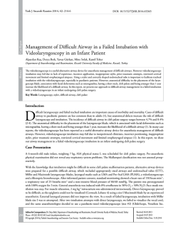
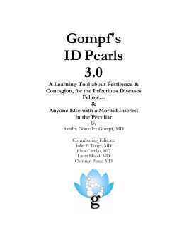
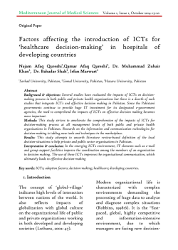
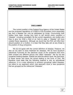
DOI: 10.5152/eurjrheum.2014.006
Review
Familial Mediterranean fever: An updated review
İsmail Sarı1, Merih Birlik1, Timuçin Kasifoğlu2
Abstract
Familial Mediterranean Fever (FMF) is a hereditary autoinflammatory disorder characterised by acute attacks of fever and serosal inflammation.
FMF primarily affects Jewish, Armenian, Turkish, and Arab populations. The disease is accompanied by a marked decrease in quality of life
due to the effects of attacks and subclinical inflammation in the attack-free periods. Untreated or inadequately treated patients run the risk of
amyloidosis, which is an important cause of morbidity and mortality. In this review, the current information available on FMF is summarised.
Key words: Familial Mediterranean fever, review, aetiology, amyloidosis, treatment
Introduction
Familial Mediterranean fever (FMF) is the most commonly seen fever syndrome and is significantly associated with ethnicity. It frequently occurs among Turks, Armenians, Jews and Arabs (1). Although it has been
known about for a long time, it was first mentioned in the literature in 1908 by Janeway and Mosenthal,
who reported recurrent fever and abdominal pain in a 6 year-old Jewish girl (2). Its first definition as a disease was based on a case report, published under the title “benign paroxysmal peritonitis” by the allergy
specialist Siegal from New York, as a compilation of Jewish patients with similar complaints. The periodical
fever definition was first used by Reimann in 1948, and Sohar et al. (2) defined the disease as FMF in 1955.
Prior to the use of colchicine, the disease was fatal, but a new era in the treatment of FMF began with the
introduction of colchicine in 1972. In a number of studies, it has been shown that this drug not only has an
effect on the symptoms, but also affects the development of amyloidosis (3, 4). In 1992, it was reported that
the abnormality associated with FMF is found on chromosome 16, and the gene responsible for the disease
was identified in 1997 (5, 6). The disease is accompanied by a marked decrease in quality of life due to the
effects of attacks and subclinical inflammation in the period between attacks (7). Untreated or inadequately
treated patients run the risk of amyloidosis, which is an important cause of morbidity and mortality (3). In
this review, the currently available information on FMF is summarised.
1 Department of Internal Medicine,
Division of Rheumatology, Dokuz
Eylül University Faculty of Medicine,
İzmir, Turkey
2 Department of Internal Medicine,
Division of Rheumatology, Osmangazi
University Faculty of Medicine,
Eskişehir, Turkey
Address for Correspondence:
İsmail Sarı, Department of Internal
Medicine, Division of Rheumatology,
Dokuz Eylül University Faculty of
Medicine, İzmir, Turkey
E-mail: [email protected]
Submitted: 10.01.2014
Accepted: 05.02.2014
Copyright 2014 © Medical Research and
Education Association
Epidemiology
Familial Mediterranean fever shows a marked ethnic distribution. The disease is most frequently observed in
Turkish, Armenian, Jewish and Arabic communities. Geographically, the disease is more commonly observed
among the nations of the Mediterranean region (1). Throughout the world, the disease is most frequently seen
in Turkey with a prevalence varying between 1:150 and 1:10,000 (8-13). The second most frequently affected
ethnic group is Armenians; studies carried out in Armenia report a prevalence of ca. 1:500 (14). In studies
conducted in Sephardic Jews, the prevalence of FMF is reported to be between 1:250 and 1:1000 (15). It has
been claimed that the rate is ca. 1:73,000 among Ashkenazi Jews (15). In studies conducted in Israel, it was
claimed that FMF incidence varies according to the ethnic group being studied (Ashkenazi or non-Ashkenazi
Jews), but that it is observed at a rate of 1:1000 on average (16). No precise information is available about the
prevalence of FMF among Arabs. On the other hand, recent studies conducted in countries such as Greece,
Cyprus and Italy indicate that this disease occurs more frequently than previously believed (17, 18). FMF has
also been identified in other countries. In Brazil, in a study assessing 102 cases with hereditary periodic syndrome, 17 patients with suspected FMF were reported and a mutation in both alleles was identified in three
of these patients (19). In a study carried out in countries in the Middle East and Eastern Europe, the incidence
of FMF among individuals under 19 years of age was reported to be ca. 2:1,000,000 (20). In a nationwide study
conducted in Japan in 2009, cases of clinical FMF were investigated. A number of hospitals did not participate
in this study; however, in participating centres, around 170 FMF cases were identified (21).
Aetiopathogenesis
Familial Mediterranean fever is a member of the group of autoinflammatory diseases. In these diseases,
the innate immune system is primarily affected (22). This disease group, including FMF, shows a genetical-
21
Sarı et al. An updated review of FMF
ly monogenic heredity, with mutations in the
affected genes causing the multiprotein complex called the inflammasomes to function
abnormally (23). Basically, the inflammasomes
activate the caspase-1 enzyme, which causes
interleukin (IL)-1β to be released. It is believed
that, in autoinflammatory diseases including
FMF, IL-1β release is uncontrolled (22, 24, 25).
FMF is an autosomal recessive hereditary disease and occurs as a result of point mutations
(single substitutions) in the Mediterranean Fever
(MEFV) gene on the short arm of chromosome
16. This gene encodes a protein called pyrine,
with a weight of 95 kDa (26). The pyrine protein
is essentially responsible for the regulation of
apoptosis, inflammation and cytokines, and is
mainly expressed in neutrophils, eosinophils,
dendritic cells and fibroblasts (26). At present,
no agreement exists about the physiological
role of the pyrine protein. However, it is assumed that the primary function of this molecule is to suppress the inflammatory response.
Structurally, the pyrine protein consists of the
following domains: (i) the pyrine domain (PYD)
located at the N-terminal end; (ii) the B box
zinc finger; (iii) alpha helix (coiled coil); and (iv)
B30.2 (PRYSPRY) located at the carboxy end
(26). Each of these domains has its own specific protein-protein interactions. The PYD domain at the terminal end has a death domain
fold (DDF) and forms homotypical bonds to
the apoptosis-associated speck-like protein
containing a CARD (ASC) protein, which plays
a role in apoptosis (27, 28). Under normal circumstances, this link activates the nuclear
factor kappa beta (NF-κβ) and pro-caspase-1,
while suppressing IL-1β production. It is presumed that the mutated pyrine molecule is
theoretically not able to suppress, and thus the
Eur J Rheum 2014; 1: 21-33
inflammatory response develops (Figure 1) (27,
28). B30.2 at the C-terminal end is an important
domain, and a significant number of mutations
in the MEFV gene occur here. At the same time,
this domain binds directly to caspase-1 (IL-1
transforming enzyme), independently of the
apoptosis-associated speck-like protein containing CARD (ASC) protein, and inhibits this
enzyme. Mutations in this domain are thought
to eliminate physiological caspase-1 inhibition
and cause uncontrolled IL-1β release (22).
MEFV Gene Mutations
The MEFV gene on the short arm of chromosome 16 consists of 10 exons. Most of the mutations identified occur in exon 10 (e.g., M680I,
M694V, M694I and V726A). Apart from this,
mutations in exon 2 (E148Q) and other exons
have also been identified (Figure 2). Most of
the mutations are point mutations, known as
missense mutations, and are characterised by
single-nucleotide changes. To date, 288 mutations have been identified in Infevers (http://
fmf.igh.cnrs.fr/ISSAID/infevers), which is an
online databank for mutations that play a role
in autoinflammatory diseases. The majority of
these mutations are rare, without causing any
clinical phenotype, and are mainly observed in
populations in which FMF is not prevalent. It
has been shown that the E148Q, M680I, M694V,
M694I and V726A mutations are responsible
for more than 80% of FMF cases in the Middle Eastern region (16). On the other hand,
because E148Q has a high carrier rate (>10%)
and does not cause an FMF phenotype, even
in homozygous cases, some researchers have
claimed that this should not be considered
a mutation, but rather a polymorphism (29).
Because of the autosomal recessive nature of
FMF, it is emphasised that individuals with clin-
Figure 1. Structure of the pyrine protein and molecular interactions
PYD:Pyrin domain; ASC: Apoptosis-associated speck-like protein containing a CARD
22
ical FMF should have two mutations (29, 30).
Nevertheless, studies have not demonstrated a
second allele in almost 30% of the individuals
with an FMF phenotype (30). The absence of
a second mutation in an autosomal recessive
disease has attracted the interest of many researchers, and a number of hypotheses have
been developed to account for this. The first
and simplest hypothesis is that the mutation cannot be identified using the presently
available laboratory methods (29, 30). Another explanation is that, even if there is a single
mutation in a patient, the other functional
allele is possibly not able to perform its function because of epigenetic mechanisms. The
existence of polymorphisms occurring in the
genes on the inflammatory cascade is claimed
to be another possible explanation. Another
important point in the occurrence of the disease phenotype in individuals with a single
mutation is the effect of environmental factors
on the abovementioned conditions (29). The
incidence of mutations in the MEFV gene may
differ between ethnic groups (Table 1). In the
Turkish population, the most commonly seen
mutation is M694V. This is followed by M680I
and V726A (31). To a lesser extent, other mutations such as E148Q, M694I, R761H, K695R,
E148V and P369S have been identified (31). Yilmaz et al. (32) reported the allele frequency in
FMF patients to be 51.1% for M694V and 9.22%
for M680I. Some studies have investigated the
MEFV gene mutation carrier state of healthy
individuals. In Turkey, the rate of carriers is reported to be 20% (32). This high rate is similar
to the carrier rate determined among the Ashkenazi and North African Jews. In Turkey, the
most frequent mutation determined in carriers
is E148Q; this is followed by the M680I, M694V
and V726A mutations (32).
The Phenotype-Genotype Relationship
Researchers have claimed that there is a relationship between FMF symptoms and the genotype.
According to some researchers, those individuals
who are homozygous for the M694V mutation
present with a more serious and severe disease
course (31, 34, 35). When considered from this
point of view, the most serious complication of
the disease, amyloidosis, is reported at a higher
incidence in homozygous M694V cases (35-37).
When patients are studied in terms of frequency
of fever and abdominal pain, which are the most
important clinical symptoms of FMF, studies provide contradictory data (31, 34, 38, 39). Whereas
some studies did not demonstrate such a relationship (31, 34), others determined that these
symptoms are more frequent in individuals with
an M694V or M680I genotype (38, 39). When the
age at onset of the disease is considered, some
studies claim that the disease develops earlier in
M694V mutation carriers (31, 40), whereas others
Eur J Rheum 2014; 1: 21-33
Sarı et al. An updated review of FMF
5'
R42W
3'
E148Q
P369S
R408Q
F479L
I591T
M694V
V726A
M680I (G/C)
M694I
M680I (G/A)
R761H
A744S
Figure 2. Distribution of MEFV gene mutations throughout the gene (33)
The MEFV gene consists of 10 exons. The mutations in this gene most frequently occur in exon 10. Nevertheless, mutations in other exons may also
be seen. Mutations emphasised in bold are the most commonly identified in FMF at present; those shown in a lighter colour are less frequently
observed. The majority of the mutations identified are not shown in this figure.
report that the disease occurs later in homozygous M694V patients (34). However, other studies could not determine a relationship between
genotype and the age at onset of the disease
(38). As a result, a considerable discrepancy exists between studies investigating the phenotype-genotype relationship. However, researchers seem to agree that the disease is more severe
in M694V homozygous patients.
Familial Mediterranean Fever Clinical Symptoms
Familial mediterranean fever is a disease that is
characterised by recurrent fever and serositis (e.g.,
peritonitis, pleuritis, synovitis) symptoms. Individual and ethnic differences may be seen in both the
frequency and course of the clinical symptoms.
While a single sign may sometimes accompany
high fever, at other times, more than one symptom can occur simultaneously. Even in the same
patients, clinical symptoms may differ over time
(1, 41). Attacks develop quite spontaneously and
continue for at least 12 hours. Most symptoms
resolve within 3-4 days, and the interval between
attacks is clinically relatively symptom-free. However, arthritis and myalgia may have a prolonged
course (1, 41). Disease onset is prior to 20 years
of age in 90% of cases and, in 60% of cases, the
age at onset is under 10 years. Nonetheless, the
disease may develop after the first years of life (2).
When the sex distribution of FMF is considered, a
slight male dominance can be noted (31).
The prodromal phase
In approximately half of FMF patients, various
constitutional and physical signs, including
restlessness at the site where the symptom is
about to occur, anxiety, irritability, increased
appetite and taste alterations, accompany the
onset of an attack. The period between the
prodromal signs and the onset of the attack
has been reported to average 20 hours (42).
Fever
High fever is the most important symptom
of FMF and one of the essential symptoms in
the diagnosis. Body temperature is generally
above 38°C. Fever typically emerges spontaneously, increases rapidly, and is followed by
a plateau and a rapid decrease, which completes the cycle. This course generally lasts 1-3
days. Non-specific findings such as weakness,
fatigue, myalgia, arthralgia, headache, lower
back and back pain commonly accompany
high fever (3). Most patients assess their fever
subjectively. According to one view, patients
who do not define high fever do not measure
their body temperature and therefore report
high fever as negative (2).
Gastrointestinal system symptoms
Peritonitis and related complications: Abdominal pain resulting from inflammation of the
peritoneal lining is the most frequent clinical
complaint in FMF. This is observed in more
than 90% of patients. On the other hand, abdominal pain attacks were reported to be the
most frequent baseline symptom of the disease in more than half of patients. Abdominal
pain starts in any region and quickly spreads
to the whole abdomen. In order to alleviate
the pain, the patient lies without moving in
the flexion position. Upon examination, there
are signs relating to peritoneal irritation (such
as abdominal swelling, tenderness, sensitivity,
stiffness, a decrease in bowel sounds, defence
and rebound), numerous air-fluid levels on
direct graphs and leucocytosis in laboratory
tests, and increased acute phase symptoms.
These symptoms resemble a surgical acute
abdominal picture. In some patients, haematuria may be determined in the urine, and this
finding may lead to inaccurate clinical interpretations in individuals that are not diagnosed
correctly. Although there is generally no gas or
stool excretion, about 10-20% of patients may
experience diarrhoea. Abdominal pain relief is
achieved within 6-12 hours; however, it generally takes 24-48 hours for it to resolve completely (2, 3, 31). Patients describing abdominal
attack experience various forms of pain on a
Table 1. The most frequently observed
mutations in the MEFV gene, by various
ethnic groups (16)
Turks
M694V, M680I, V726A, E148Q
Armenians
M694V, M680I, V726A, E148Q
Jews
North African M694V, E148Q
Iraq
V726A, M694V, E148Q, M680I
Ashkenazi
E148Q, V726A
Arabs
V726A, M680I, M694V, M694I,
E148Q
MEFV: Mediterranean fever
mild to severe range throughout their lives.
Some patients may undergo unnecessary
surgery, due to misinterpretation of the acute
abdominal clinical picture emerging during
the course of the disease. In a retrospective
study, it was found that abdominal surgeries
in FMF patients predominantly occurred (ca.
90%) prior to the FMF diagnosis and that, after
the diagnosis was made, this rate decreased to
around 10% (43). According to the same study,
whereas surgeries performed prior to the FMF
diagnosis were mainly done with the suspicion
of acute appendicitis, surgeries following FMF
diagnosis were more frequently performed
due to ileus (43). In a trial conducted in Turkey, it was determined that about one in five
FMF patients was operated on for a suspicion
of acute appendicitis (31). Although a number
of researchers claim that elective appendectomy may protect FMF patients against unnecessary examinations and surgical intervention,
this is not recommended and, moreover, can
increase adhesions of the peritoneal lining (3).
In previously performed studies, during an attack, hyperaemia in the peritoneal lining and,
in some patients, fluid accumulation in the
peritoneum was shown. This fluid was found
to be rich in fibrin and polymorphonuclear leukocytes and exhibits exudate characteristics.
On the other hand, in a computed tomography examination, peritoneal fluid may even be
detected during the asymptomatic period (44).
23
Sarı et al. An updated review of FMF
Recurrent accumulations of the exudative fluid
and its resolution may cause adhesions in the
peritoneal lining over time, and may thus lead
to complications such as mechanical bowel
obstruction, volvulus and strangulation (2). Too
many neutrophils in the peritoneal fluid are
believed to increase the risk of adhesion (45).
Studies have demonstrated that mechanical
bowel obstruction is seen more commonly in
FMF patients compared with the normal population (46). In FMF patients with no previous
surgery, the occurrence of intestinal obstruction is reported to be 3% (43). As a result of
recurrent attacks, some patients develop sclerosing peritonitis (encapsulated peritonitis),
which may lead to acid development (3).
Spleen and lymph node symptoms: Splenomegaly was reported in various series at different
rates, with an incidence between 10% and 60%
(3). In general, this occurs as a result of reactive changes secondary to the inflammation.
However, although rare, it may also occur as a
result of amyloid accumulation (47). In some
cases, peripheral and abdominal lymphadenopathy (LAP) secondary to the inflammatory
response was identified, with an incidence of
ca. 14%. Sometimes, cases undergoing laparotomy due to an enlargement of the mesenteric
lymph node have also been reported. The biopsy assessment of the lymph nodes revealed
non-specific lymphoid hyperplasia (44).
Hepatic symptoms: During the course of FMF,
hepatomegaly might occur, and this is identified to a lesser degree compared with splenomegaly (approximately 5%) (44, 48). A number
of observational studies have indicated that, as
cryptogenic hepatic cirrhosis is more frequently observed in FMF patients, there might be a
relationship between the diseases (49). Another study revealed that non-alcoholic steatohepatitis (NASH) is observed in FMF at a higher
rate than expected and associated this with
the effects of proinflammatory cytokines and
MEFV gene mutations on the liver. In the same
study, it was suggested that NASH might have
caused the increase in cryptogenic cirrhosis
mentioned earlier (50). Although an increase
in the incidence of cholelithiasis in FMF was
shown in some studies (3), others do not support this finding (47).
Mesothelioma: Observational studies produced
data indicating that mesothelioma is increased
in FMF. According to these researchers, over
time, chronic inflammation causes malignant
transformations in the serous membrane.
However, other observational studies do not
confirm the finding of increased mesothelioma in FMF (47).
24
Eur J Rheum 2014; 1: 21-33
Other gastrointestinal system symptoms: About
5% of FMF patients are reported to have irritable
bowel syndrome as an accompanying disease
(47). This might lead to abdominal pains that
are not related to the attacks. Another condition causing abdominal pain is colchicine intolerance, which may also be accompanied by
diarrhoea (47). Although rare, another condition
is diarrhoea and malabsorption due to the accumulation of amyloid in the small intestine (3).
Musculoskeletal system symptoms
Arthralgia and arthritis: Arthralgia and arthritis
are the most common clinical symptoms of
FMF. Arthritis generally develops in childhood,
with an incidence ranging between 20% and
70% (51, 52). In some cases (approximately
10%), it can be the baseline symptom of the
disease (53). Arthritis attacks are usually accompanied by fever; they demonstrate a monoarticular characteristic and manifest themselves
in the large joints of the lower extremities
(knee > ankle) (2). In arthritis cases, a rash is
frequently observed (2, 54). However, some
studies indicate that erysipelas-like erythema
(ELE) may also accompany up to 40% of cases
(55). Arthritis attacks tend to resolve within a
week (53). Upon examination of synovial fluid,
fluid accumulation that is more predominant
in neutrophils and with an inflammatory characteristic is detected. In some cases, leukocyte
numbers in the synovial fluid may increase dramatically, and this may be confused with septic
arthritis (56). Despite recurrent arthritis attacks,
erosion in the joints is not expected (51, 55).
Some 5-10% of FMF patients may experience
protracted arthritis attacks lasting more than a
month, sometimes even years (55-57). In protracted arthritis, mainly the knees and, to a lesser extent, the hip joints are affected (57). The inflammation in the knee joint is usually resolved
without sequelae, with massive fluid accumulation (57, 58). However, protracted hip arthritis
may exhibit destructive characteristics. In trials,
it has been shown that approximately 30% of
patients with hip manifestation required a total
hip arthroplasty operation (56, 57).
Clinical manifestations in the axial spine: Studies
indicate that the incidence of sacroiliitis is increased in FMF patients. As already known, sacroiliitis is a common characteristic of the spondyloarthropathy (SpA) disease category, and it
seems that FMF might also be in this spectrum.
Moreover, as arthritis in FMF tends to manifest
itself in the large joints of the lower extremities
and may cause manifestations in the root joint,
such as the hip joint, FMF is similar to the SpA
group of diseases in terms of the arthritis pattern. The association of FMF with SpA was first
pointed out in the 1960s, and this relationship
was further explored through case reports and
various other studies. In a study in which approximately 3000 FMF patients were evaluated,
SpA prevalence was reported to be 0.4% (59). In
a study conducted in Turkey, 503 FMF patients
were evaluated, and sacroiliitis was detected by
x-ray in up to 10.5% of these patients (60). When
FMF patients with sacroiliitis were assessed,
these patients are generally determined to be
HLA-B27 negative, whereas in HLA-B27-positive
cases, the axial manifestation symptoms were
observed to be more severe (61). In addition, it
is interesting to see that MEFV gene mutations
(particularly M694V) that play a role in FMF
pathogenesis are significantly increased in ankylosing spondylitis (AS) patients, with negative
FMF clinical symptoms (62, 63).
Muscle symptoms: During the course of FMF disease, various muscle symptoms may emerge,
and the incidence in this patient series is about
20-40% (31, 64). Types of myalgia that may occur during the course of FMF can be categorised as (i) muscle pain occurring in childhood
after exercising; (ii) disseminated muscle pain
due to fibromyalgia; (iii) protracted febrile
myalgia (PFM); (iv) muscle pain accompanying the attack; and (v) myalgia and myopathy
related to colchicine treatment. Post-exercise
muscle pains occur particularly in the lower
extremities. These are not accompanied by fever or acute phase response, last for 2-3 days
on average, may follow a severe course, and
usually begin in the evening (3). The incidence
of fibromyalgia syndrome, which causes generalised pain, has increased in both paediatric
and adult FMF patients. The basic cause of this
situation is that the pain threshold decreases
as a result of chronic illness. Acute phases and
muscle enzymes in myalgia resulting from this
are normal, and trigger points with pain can be
detected during examination (65, 66). Generalised muscle pain that accompanies fever and
other clinical findings during attacks is another
type of myalgia. This situation ends when the
attack ends (67). Another finding relevant to
muscles is myopathy, which develops due to
colchicine treatment. As is previously known,
colchicine is the gold standard treatment for
FMF. This risk increases in those with renal failure and who use cyclosporine. In colchicine
myopathy, in addition to weakness of proximal muscles, an increase in laboratory muscle
enzyme levels, a myopathic pattern in electromyographic (EMG) examination, and autophagic vacuoles histopathologically visible in
non-necrotic muscle fibrils, and lysosomal storage will probably be observed (68).
Another type of muscle pain is PFM. Its incidence has been stated to be 1-3% in various
Eur J Rheum 2014; 1: 21-33
case series (54, 69). It occurs particularly in the
lower extremities, continues with intense pain
and sensitivity, and seriously affects the patient’s
quality of life. Contrary to most FMF clinical findings, PFM can last for up to 6 weeks. Some authors have suggested that to be able to say that
a pain is PFM, it should last for at least 5 days (70).
PFM can occur before or after a serositis attack.
Approximately 70% of cases are accompanied
by fever (71). In PFM, muscle enzymes are at
normal levels, and no abnormality is detected in
muscle biopsy and EMG (3). However, in magnetic resonance imaging (MRI) examinations,
findings of oedema have been reported in the
involved muscles (72). A higher ratio of female
patients in the case series is noteworthy (64).
Studies of the genotype-phenotype relationship suggest that PFM is seen more frequently in
M694V homozygous individuals (73). Some authors suggest that Antistreptolysin O (ASO) titres
are high in these patients and, therefore, recent
streptococcal infection may trigger the condition (74). Because some cases are accompanied
by a skin rash, the existence of hyperglobulinaemia in laboratory tests, and symptoms that respond to high-dose (1 mg/kg) corticosteroids, it
has been suggested that autoimmunity may be
involved in the pathogenesis of PFM (3, 64). On
the other hand, the fact that the storage of immunoglobulin and complements is observed in
skin biopsies from some patients supports this
hypothesis (75).
Pulmonary findings: Pleural involvement occurs
in approximately 40% of FMF patients. Pain is
usually one-sided, gets worse with breathing
and can expand to the shoulder. Similar to the
other attacks, it lasts for 1-4 days. Sometimes
the right pleura and sometimes the left pleura
is affected. In a direct graphy of the patient, closure of the costophrenic sinus due to fluid can
be observed during attacks. Fluid analysis is exudative and neutrophils are dominant. In some
patients, thickening and adhesion have been
reported in the pleura as a result of repeated
attacks. Pleural inflammation can be accompanied by atelectasia and, where pleurisy and
fever co-exist, this situation can be diagnosed
as pneumonia by mistake. As with peritoneal
involvement, some authors have suggested an
increased risk of pleural mesothelioma; however, there is insufficient evidence to prove this.
In some patients, albeit rarely, pulmonary involvement due to amyloid has been reported,
and other organ involvement due to amyloid is
seen as well. On the other hand, some systemic
vasculites can be seen together with FMF, and
naturally, in such cases, pulmonary findings of
the disease can also accompany the clinical
picture. In some cases, especially in nephrotic
syndrome cases resulting from amyloidosis,
Sarı et al. An updated review of FMF
the risk of hypercoagulopathy increases, and
this situation, albeit rare, may cause pulmonary
thromboembolism (2, 31, 76).
Cardiovascular findings
Pericardial involvement: Inflammation of the
pericardial membrane in FMF is less frequent
compared with other serosal involvements.
Kees et al. (77) have reported the incidence
of pericarditis as 0.7% in a series of approximately 4000 patients. Pericarditis attacks last
for 1-14 days (average 4.2 days), and patients
often experience more than one pericarditis
attack. Pericardial inflammation and other serous membrane involvement can also be seen
during the course of the disease. Kees et al. (77)
have reported that pericardial attack follows
a benign course, and no sequelae remain in
any patients. Dabestani et al. (78) evaluated 30
patients whom they chose randomly among
their cohort of 210 patients, and reported that
the findings of pericarditis were found in eight
patients (27%) echographically. In this study,
the existence of pericardial thickening was
also observed in some patients. On the other
hand, the aforementioned study had various
methodological weaknesses, and the definition of pericarditis was based on effusion and/
or thickening findings determined by echocardiography. Therefore, various authors have
stated that this situation does not reflect the
actual incidence of pericarditis (77, 79). A prospective study investigated the incidence of
pericardial effusion during FMF attacks, and
reported that 3.6% of patients have pericardial effusion during attacks (79). It was observed
that the amount of effusion determined in this
study was around 5 mm on average (79). According to the current data, FMF involves the
pericardium less compared with other serosal
membranes, and this finding appears as a relatively later clinical finding of the disease may
cause pericardial thickening, and fluid is not
expected in massive amounts.
Other cardiovascular system findings: As is
known, inflammation causes atherogenic effects, and increased cardiovascular (CV) mortality due to early atherosclerosis is observed
in some inflammatory rheumatic diseases (80).
In recent years, various papers have been published on atherosclerosis in FMF patients. The
earliest step in atherogenesis is endothelial cell
damage (81). Various biomarker studies in FMF
have demonstrated findings of endothelial cell
damage both during and between attacks (82,
83). Additionally, some studies have pointed
out the existence of endothelial dysfunction
in ultrasonography during the subclinical period (84). However, some imaging studies have
reported that the existence of carotid plates,
which are used to show atherosclerotic load,
do not increase in FMF patients compared with
healthy control subjects (85, 86). According to
a very important and noteworthy study conducted by Langevitz et al. (87), in FMF patients
who regularly use colchicine, their CV mortality is lower than that of those control subjects
with another inflammatory disease. This study
also used another control group consisting of
relatives of FMF patients, and no difference in
terms of CV mortality was found between the
groups (87). As a result, although there are data
regarding endothelial damage in FMF disease,
this situation is not considered to cause an increase in the risk of CV mortality. The major reason for this is that, in contrast to the inflammation seen in rheumatoid arthritis and systemic
lupus erythematosus, inflammation in FMF follows a course in attacks, and there is the possible atheroprotective effect of colchicine (82).
In FMF, abnormalities in both atrial and ventricular transmission systems have been investigated, and no transmission system abnormality
has been detected in patients without amyloidosis (88-90). On the other hand, a study of
patients with amyloidosis reported that QT variability, which assesses the ventricular transmission abnormality, increases (91). Ventricle functions have been investigated in various studies.
Accordingly, while no abnormality is detected
in systolic functions, both right and left ventricle diastolic function has been detected in
periods between attacks in FMF patients, and
this function has been associated with inflammation (92-94). Some researchers have found
abnormalities in the variables of aortic stiffness
and arterial pulse wave velocity, which are parameters of atherosclerosis (93, 95). The clinical
significance of these findings is not yet known.
Renal findings other than amyloidosis: In 22% of
FMF patients, defined kidney problems other
than amyloidosis involvement have been defined. These include temporary or permanent
haematuria and/or proteinuria, recurring acute
pyelonephritis, tubulointerstitial nephritis and
glomerulonephritis (GN) (31, 96-98). However, vasculitic manifestations that accompany
FMF may also cause different types of kidney
involvement. Various types of GN such as diffuse proliferative, membranous and mesangioproliferative GN may occur during the course
of FMF (31, 96-98). Some researchers have
suggested that colchicine prevents glomerular
disease, and that the irregular use of this drug
triggers GN attacks (96).
Dermal findings: Various rashes have been defined during the course of FMF. The primary
dermal findings are urticaria, diffused erythema
on the palms and soles, subcutaneous nodules,
25
Sarı et al. An updated review of FMF
angioneurotic oedema, pyoderma, Raynaud’s
phenomenon, dermal findings related to accompanying vasculites and EBE (99). Among
these rashes, EBE is not considered to be typical
of the disease. This finding usually accompanies
fever or arthritis, and involves the front side of
the leg, ankle or dorsum of the feet. It is usually triggered by walking or standing for a long
time (99). The lesion is 15-50 cm in size, painful,
warm, swollen and has sharp boundaries, and
fades within approximately 1-3 days before disappearing (2). It is usually on one side, but it may
sometimes involve both extremities (99). No
specific anomaly has been defined in the histopathological examination of such lesions (99).
Other clinical findings: Inflammation of the tunica vaginalis, which is an extension of the peritoneum, may lead to acute scrotal attack. This
mostly occurs in children and young adults. It
is usually one-sided, and inflammation findings such as redness, pain and swelling can be
observed (3). Various nervous system findings
have been defined in the course of FMF. However, these findings are quite rare, and have
usually been reported as case reports. Some
observational studies have investigated the relationship between FMF and multiple sclerosis,
and have suggested that this condition is seen
2- to 4-fold more in FMF patients than in the
normal population (100-102). Involvement of
the eye is quite rare. There are publications, usually as case reports, of findings of eye involvement, scleritis, pre-uveitis, pan-uveitis, papillitis,
and objects similar to retinal colloid (103-105).
Another study has reported that ocular surface
anomalies are more common in FMF (106).
Amyloidosis in Familial Mediterranean Fever
The most important factors determining
the prognosis of FMF are the development
of amyloidosis and subsequent progression
to end-stage renal failure of the patient. The
most important factor in the development of
amyloidosis is the increase in production of
the SAA protein, which is synthesised in the
liver (107). Increased synthesis of this insoluble protein, and hence decreased elimination
of it, leads to accumulation of the molecule
in extracellular areas and the development of
amyloidosis (108). In the pre-colchicine period,
in FMF patients aged 40 years and above, the
incidence of amyloidosis has been reported
to be at quite high levels of 60-75% (109). The
incidence of amyloidosis has decreased markedly with the regular use of colchicine. Nevertheless, amyloidosis complications are still a
problem, especially in communities where the
disease is common. In two studies conducted
in Turkey on many patients, the frequencies of
amyloidosis have been reported as 12.9% (31)
26
Eur J Rheum 2014; 1: 21-33
and 8.6% (35). Although FMF carries a potential
risk of amyloidosis, not everyone with FMF develops amyloidosis. Various publications have
investigated the risk factors for amyloidosis.
The fact that the development of amyloidosis
is more common in Sephardic Jews compared
with Ashkenazi Jews indicates that the genetic background of patients is important in this
respect. The incidence of amyloidosis is also
very different between people of the same
ethnicity living in different geographic areas.
While the incidence of amyloidosis in Armenian FMF patients living in Armenia is 24%, the
incidence of amyloidosis in Armenians living in
California is 0% (110). Various publications have
reported the risk of amyloidosis in FMF patients
who are homozygous for the M694V mutation
in the MEFV gene (35, 37, 111). Kaşifoğlu et al.
(35) performed a study conducted on 2246 patients, and reported that FMF patients who are
M694V homozygotes carry a 6-fold risk of amyloidosis compared with FMF patients carrying
other MEFV gene mutations. Additionally, male
gender (35, 112), a family history of amyloidosis
(112), and the existence of a homozygous SAA
1.1/1.1 genotype are also other defined risk factors for amyloidosis.
Amyloid structures first accumulate in the
spleen, liver and kidneys, and then in various
tissues such as testicles, adrenal glands, the
gastrointestinal system and the nervous system (107). The development of nephrotic syndrome and renal failure due to damage to the
renal structure caused by amyloid fibrils is the
most common clinical course. While absorption failures and diarrhoea resulting from involvement of the gastrointestinal system with
amyloid can be seen, many patients can be asymptomatic. Storage of amyloid in the testicles
can cause azoospermia and infertility. Transmission anomalies and cardiac failure can be
seen as a result of cardiac storage. Storage in
joints can lead to amyloid arthropathy (2, 3, 15).
All patients followed up with the diagnosis of
FMF should also be regularly examined for amyloidosis by urinalysis. Tissue diagnosis can be
required in patients with proteinuria between
attacks. Renal biopsy is the most sensitive
method. An increased disposition to bleeding
due to amyloid storage can be limiting for renal biopsy. Although rectal biopsy is less sensitive, it is also a less invasive method, and can
therefore be considered an alternative. Some
studies have reported that the sensitivity of renal biopsy for diagnosis of amyloidosis is 88%,
and the sensitivity of rectal biopsy is 75%, and
gingival biopsy material is 19%. Other alternatives are bone marrow biopsy and fat tissue
biopsy. It has been reported that the sensitivity
of bone marrow biopsy is closer to the sensitivity of rectal biopsy for the diagnosis of amyloidosis, and that the sensitivity of subcutaneous
fat tissue is not very high for the diagnosis of
amyloidosis (15, 113, 114).
Clinical findings regarding amyloidosis are
usually seen at ages below 40 years. There is
another clinical subgroup called phenotype
II. In this clinical group, patients first have the
AA amyloidosis diagnosis confirmed by biopsy,
and the clinical manifestation of FMF occurs
after the diagnosis. In some cases with phenotype II, the clinical manifestation of FMF may
not occur; however, there are patients with
clinical manifestation of FMF in the families of
these cases (115).
Relationship between Familial Mediterranean
Fever and Vasculitis
Studies have found that vasculites such as
polyarteritis nodosa (PAN) and Henoch-Schönlein purpura (HSP) are more common compared with the normal population. According
to some researchers, Behçet’s disease (BD) is
also more common in FMF; however, this relationship is less common than that observed
with PAN and HSP.
Polyarteritis nodosa
Polyarteritis nodosa is a vasculitis that causes
necrotising inflammation in small and medium-sized arteries. There are various publications regarding the concurrence of PAN and
FMF. In a comprehensive study, the incidence
of PAN in FMF was reported to be 0.9%. If we
assume that the incidence of PAN in the normal population is 6 in 100,000, in that case,
the incidence of PAN in FMF has increased
approximately 200 times (31, 116). In cases of
PAN associated with FMF, first, the clinical manifestation of FMF occurs, and then the clinical
manifestation of PAN is added (116). In terms
of the symptoms, both diseases have common
findings such as abdominal pain, fever, muscle
pain, articular symptoms and skin rashes (67).
It has been reported that there are some differences between the PAN that accompanies
FMF and classic PAN. One of these is the age
at which the disease presents. Classic PAN
usually emerges at ages 40-60 years. However,
FMF-related PAN occurs at younger ages (116).
In another study, the average age at which
PAN accompanying FMF presents has been reported to be 14.9 years of age (117). Another
difference is the gender distribution among
patients. While classic PAN is usually seen in
men, FMF-related PAN is seen equally in men
and women (31). Another difference between
the groups is that spontaneous perirenal hematoma, which is rarely seen in classic PAN
Eur J Rheum 2014; 1: 21-33
cases, accompanies almost half of PAN cases
with FMF (117). The two groups are different
in terms of mortality. It has been reported that
FMF-related PAN cases follow a better course
(117). Another interesting point is that, in contrast to classic PAN cases, in the concurrent cases of FMF-PAN, glomerular involvement can be
seen (118, 119). Intensive myalgic complaints
are observed in patients in cases of PAN that
accompany FMF. Some clinicians have suggested that this could be a hint to suggest the
possibility of PAN; however, it should be kept
in mind that intensive muscle pain can also be
seen in FMF in PFM and colchicine-related myopathy (117).
Henoch-Schönlein purpura
Henoch-Schönlein purpura is a vasculitis of
small vessels that involves skin, kidneys, joints
and the gastrointestinal system, and is the
most common vasculitis of childhood. In various series, the incidence of HSP accompanying FMF has been given as 2.7-7.2% (31, 119).
In this respect, it is also the vasculitis that most
commonly accompanies FMF. Considering
that the incidence of HSP in childhood is 0.8%,
the incidence of this vasculitis has increased almost 4-fold (116). Researchers have stated that
classic HSP and FMF-related HSP are not clinically different from one another (119).
Behçet’s disease
Behçet’s disease is an idiopathic inflammatory
disease seen more commonly in the countries
on the historic Silk Road, which is defined as between the Mediterranean region and the Far East
(120). Although some studies have reported that
the incidence of BD increases in FMF, others have
not confirmed this (121, 122). It is suggested that
the clinical picture in FMF-related BD cases is not
different from that in classic cases (116).
Reproductive System and Pregnancy in Familial
Mediterranean Fever
As FMF especially affects individuals in fertile
age groups, it raises various concerns among
both patients and doctors regarding reproduction and pregnancy. The first of these concerns
is that FMF can cause infertility (123, 124). Another important concern is that it can lead to
peritonitis, and fever can lead to uterine contractions, thus leading to undesired complications such as abortion or premature birth (125,
126). Another concern is the possible teratogenic effect of colchicine, which is the gold
standard treatment for FMF (123).
Concerns about infertility
There are various studies on the relationship
between FMF and fertility. In a study conducted in Israel, infertility was reported in approx-
Sarı et al. An updated review of FMF
imately one-third of fertile women with FMF
not getting treatment for their disease, and
it was suggested that ovulatory dysfunction
could be responsible for this (127). Another
study was conducted on women who received
colchicine treatment for a long period of time,
and it was also reported in this group that infertility was more common than in the normal
population; this was because of ovulatory insufficiency (128). In another study conducted
in Italy, it was suggested that infertility in women with FMF was twice that observed in the
general population, and that ovary dysfunction
and peritoneal adhesions were the cause of
infertility (125). Another important conclusion
from this study was that the majority of infertile
women in this group consisted of women who
had not been receiving colchicine treatment
(125). Considering the literature data, particularly in untreated women, increased infertility
is seen as a result of ovulatory insufficiency and
peritoneal adhesions (because of attacks or
operations). Fertility can be achieved, and the
development of peritoneal adhesions can be
decreased with colchicine treatment (129). It
has been reported that artificial insemination
techniques such as in vitro fertilisation in infertile FMF patients are quite successful (126).
It has also been reported that male infertility
can be increased in FMF. The primary cause of
this is that the attacks occurring as a result of
inflammation of the tunica vaginalis can lead
to testicular insufficiency or amyloidosis, which
is a complication of FMF that can negatively affect spermatogenesis (123, 124). Sperm count
investigations may be suggested for men who
have amyloidosis and want to have children.
Concerns about pregnancy
Various studies have been conducted to investigate the risk of premature birth and
spontaneous abortion in FMF. According to
the literature data, the incidence of spontaneous abortions before colchicine treatment is
around 25-30% (128). This is probably because
FMF attacks (both peritonitis and fever) cause
or increase uterine contractions (123, 126).
Additionally, some studies have reported that
premature births and spontaneous abortions
are also increased in patients treated with colchicine (126). There are various studies on the
course of pregnancy in women with amyloidosis. According to these studies, pregnancy
in these patients can occur with spontaneous
abortion, stillbirth and worsening of kidney
functions (130, 131). Some authors suggest
that fertile women with amyloidosis complications should not become pregnant (123). Looking at the course of the disease during pregnancy, no definite remarks can be made on
this matter. According to some researchers, the
incidence of attacks increases during pregnancy; while, according to others, the incidence of
attacks decreases during pregnancy (129, 132).
Teratogenicity
At present, the gold standard treatment for FMF
is colchicine. This drug acts on the microtubular
structure of cells. Therefore, colchicine use may
affect various cellular processes such as mitosis.
Laboratory studies have reported that chromosome aberrations could be observed due to the
negative effect of colchicine on mitosis (133).
Demonstration of the passage of colchicine
through the placenta (134) and reports of trisomy in the children of women using colchicine in
some case presentations have resulted in serious
concerns on this issue (129). As a result, the manufacturing company has added a statement to
the package leaflet stating that it should not be
used during pregnancy (123, 129). As a result of
these concerns, physicians have recommended
the discontinuation of colchicine treatment for
at least 3 months before pregnancy (123, 129).
Some physicians have continued with this drug,
suggesting that the frequency of abortion increased due to the non-use of colchicine; however, they recommended amniocentesis and
karyotyping for their patients within the fifth
gestational month because of the possibility of
foetal malformation (123, 129). Subsequent animal studies have found that colchicine did not
result in any chromosome aberrations (129).
Analysis of large-scale monitoring data showed
that no difference in cytogenetic abnormalities was observed in patients taking colchicine
during pregnancy compared with those not taking colchicine (135-137). As amniocentesis is an
invasive procedure with a risk of miscarriage and
infection, some investigators have questioned
the need for this procedure and reported that
amniocentesis was not required in patients taking colchicine during pregnancy (124, 129, 135).
There are also some concerns about the negative impact of colchicine on spermatogenesis. This attracted attention because some
animal studies have reported the toxic effect
of colchicine on sperm (138). In fact, the colchicine dose used in these studies was 30- to
50-fold greater than the human dose (124). In
addition, various studies have reported that
there was no abnormality in the hormone values and sperm counts of patients on regular
colchicine treatment (123, 124). Some studies
evaluated the children of males on regular
colchicine treatment born to healthy women, and reported that there was no difference
between the groups in terms of abortion and
foetal malformation development compared
with control groups (123, 124, 139). Therefore,
27
Sarı et al. An updated review of FMF
the present opinion is that female patients
with FMF should continue taking the optimal
dose of colchicine during their pregnancy and
that there is no need for amniocentesis during
pregnancy. On the other hand, although data
from animal and laboratory studies suggest
that colchicine can affect sperm motility and
sperm count, human doses do not have any
reducing effect on sperm count and do not
result in foetal malformations.
Lactation and postpartum period
Colchicine may be excreted into breast milk.
Studies have reported that the drug reached
the maximum level in breast milk 1-2 hours
after administration. On the other hand, when
the amount passed to the child was analysed, it was observed to be only one-tenth
of the maternal dose (140). When children of
mothers taking colchicine were evaluated, no
side-effects were reported relating to the drug
(140, 141). Accordingly, it was suggested that
breastfeeding is safe in women taking colchicine. There is a limited number of studies relating to the course of disease in the postpartum
period. Available data show that breastfeeding
does not increase the frequency of episodes
(140, 142).
Diagnosis of Familial Mediterranean Fever
Familial Mediterranean fever disease is diagnosed clinically. People with an appropriate
ethnic origin, with recurrent findings and fever
accompanied by serositis ongoing for 1-4 days
are diagnosed with FMF (16). In the past, various criteria were suggested for diagnosis. One
of the most commonly used criteria includes
the Tel-Hashomer criteria (143) and diagnosis
sets recommended by Livneh et al. (144). According to Tel-Hashomer, two or more major
or one major plus one minor finding was sufficient for FMF diagnosis (Table 2) (143).
Both the sensitivity and the specificity of the diagnosis criteria adopted later, which were generated based on patient data in the records of
the Sheba Medical Center, were reported to be
99% (144). These diagnosis criteria suggested by
Livneh et al. (144) defined the episodes as typical and incomplete and also included supportive findings such as ethnicity and some laboratory data. As this set of criteria was very detailed,
the same working group recommended using a
simplified version (Table 3) (144).
In some cases, patients do not meet the clinical diagnosis criteria (144). Genetic diagnosis
tests can be used to detect MEFV mutations
that are frequently observed in patients with
symptoms suggesting FMF but without a certain diagnosis. In countries where the disease is
28
Eur J Rheum 2014; 1: 21-33
observed frequently with an accordingly high
rate of carriers, the presence of two mutations
in the MEFV gene is interpreted in favour of the
disease. However, the initiation of colchicine
treatment (1.5 mg/day; 6-12 months) is recommended for cases with no detected mutations
or with a single mutation. Then, colchicine is
discontinued and the response is evaluated in
these cases. Any response to treatment and occurrence (or increased occurrence) of episodes
after discontinuation is evaluated in favour of
FMF (16, 67). In some situations, there may be
cases without any clinical signs of FMF which
were studied for MEFV mutation analysis for
another reason. Concerning the cases without
clinical signs, the presence of a single or even
a double mutation in the MEFV gene does
not result in a treatment indication for these
patients. It is recommended to follow-up the
cases that are positive for two mutations concerning FMF clinical signs (67).
Laboratory Tests in Familial Mediterranean Fever
No laboratory tests specific to FMF are available
at present. Acute phase markers such as the
erythrocyte sedimentation rate (ESR), C-reactive protein, fibrinogen and serum amyloid A
(SAA) are frequently increased during episodes
(145, 146). Among these tests, CRP was reported to increase during almost every episode,
accompanied by an increase in ESR, fibrinogen
and leukocyte counts (90%, 60% and 50% of
cases, respectively) (145). However, it was reported that albumin, a negative acute phase
protein, remained unchanged during episodes,
and this was attributed to the short duration
of episodes (145). Similarly, it was reported that
there was no significant change in platelet levels in the acute episode period (145). On the
other hand, acute phase proteins may also remain higher in the inter-episode period, called
the subclinical period. The investigators reported that CRP levels were higher than normal in
nearly half of the cases within this period (145,
147). On the other hand, SAA levels, which are
considered to be related to amyloidosis, were
reported to be high in nearly 30% of cases in
the inter-episode period (146, 147). Interestingly, the level of acute phase proteins in healthy
control subjects with an MEFV gene mutation
was found to be higher than in healthy individuals with no mutation. This was explained by
the incorrect pyrine protein activating the cytokine pathway and triggering the acute phase
response (146, 148). Various cytokine levels
were studied in patients with FMF both during
episodes and in the inter-episode period. It was
reported that levels of IL-6, which stimulated
the production of acute phase proteins from
hepatocytes, increased during the episode.
However, data from the subclinical period were
controversial and reported to be high in some
studies and normal in others (149, 150). IL-1,
which has a major role in disease pathogenesis
at a molecular level, has been studied by various investigators; however, as the blood levels
of this cytokine could not be determined using
the routine enzyme-linked immunosorbent assay (ELISA), the investigators stated that more
sensitive assay methods were needed for this
Table 2. Tel-Hashomer diagnosis criteria (143)
Major criteria
Minor criteria
Recurrent febrile episodes with serositis (peritonitis, synovitis or pleuritis)
Amyloidosis of AA type without a predisposing disease Favourable response to regular colchicine treatment
Recurrent febrile episodes
Erysipelas-like erythema
FMF in a first-degree relative
Definitive diagnosis: 2 major or 1 major and 2 minor criteria. Probable diagnosis: 1 major and 1 minor criteria
Table 3. Simplified FMF diagnosis criteria suggested by Livneh et al. (144)
Major criteria
Typical attacks (1-4)
1- Generalised peritonitis
2- Unilateral pleuritis or pericarditis
3- Monoarthritis (hip, knee, ankle)
4- Fever alone
5- Incomplete abdominal attack
Minor criteria
Incomplete attacks involving either or both
of the following sites
1- Chest
2- Joint
3- Exertional leg pain
4- Favourable response to colchicine
FMF: familial Mediterranean fever
The requirements for the diagnosis of FMF have been defined as the presence of: at least 1 major; or at least 2 minor criteria.
Typical attacks must include all the following: recurrent (at least 3 episodes), febrile (rectal temperature ≥38°C) and short in duration (12
hours to 3 days). Incomplete attacks (must be recurrent) are defined as differing from typical attacks in 1 or 2 features as follows: (1) temperature <38°C; (2) attack duration longer or shorter than a typical attack (but no less than 6 hours and no more than 7 days); (3) no signs of
peritonitis during the attacks; (4) localised abdominal attacks; and (5) arthritis in a location other than the hip, knee, or ankle
Eur J Rheum 2014; 1: 21-33
purpose (149). Some investigators performed
studies on autoantibody frequencies. Thus,
the levels of rheumatoid factor, anti-nuclear
antibody, anti-CCP, and ENA in patients with
FMF showed no difference between patients
and healthy control groups (151, 152). Some
case presentations suggested that there was
an increase in bilirubin levels during FMF episodes. This was also confirmed in a subsequent
case-control study. Thus, it was reported that
there was a slight increase in both direct and
indirect bilirubin levels in nearly 25% of cases
during episodes, and this was reduced to normal limits in the subclinical period (153). In the
abovementioned study, transaminase measurements were performed and no difference
was observed (153). Urinalysis was performed
both during episodes and in the inter-episode
period in patients with FMF, and it was reported that transient haematuria and proteinuria
might occur during episodes (145). Ozdogan
et al. studied patients during episodes prospectively and detected the presence of occult
blood in faeces in nearly half of cases during
episodes. The authors attributed this finding
to an increase in vascular permeability during
episodes (119).
Treatment of Patients with Familial
Mediterranean Fever
The targets in the treatment of FMF patients
are: (i) treatment of acute episodes; (ii) prevention of episodes; (iii) suppression of subclinical
inflammation in the inter-episode period; (iv)
prevention of amyloidosis development and
stopping progression in amyloidosis cases; and
(v) treatment of other clinical findings accompanying FMF.
Colchicine has been used in the treatment
of FMF since the 1970s and still remains unrivalled in this respect. The efficacy of colchicine in preventing acute episodes has been
demonstrated in various randomized controlled studies (154-156). There is no consensus
for the optimal dose of colchicine. However, it
is recommended to treat adult patients with
a dose of at least 1 mg and increase the drug
dose to 1.5 and 2 mg for patients with ongoing
episodes on the previous dose. It is also recommended to give the total dose once and divide the dose in case of side-effects (157). In a
study performed with children, it was reported
that complete remission was achieved in twothirds of patients and partial remission was
achieved in one-third of cases with the use of
an appropriate colchicine dose, and that a lack
of response to colchicine was observed in only
5% of patients (158, 159). In other studies, the
lack of response to colchicine was also reported to be 5-10% (160). It was reported that no
Sarı et al. An updated review of FMF
amyloidosis complications developed in the
cases using regular and appropriate doses of
colchicine (161). In addition, in the cases who
developed amyloidosis but did not progress
to end-stage renal failure, disease progression
and the decrease in proteinuria was reported
with the use of colchicine at a dose of 1.5 mg
or higher (162, 163). In amyloidosis cases (also
referred to as high-risk cases) and in patients
with prior progression to end-stage renal failure due to FMF-related amyloidosis undergoing renal transplantation, it was recommended
to use 2 mg/day colchicine if tolerated by the
patient, without taking the clinical symptoms
into consideration (157). In cases with serious
renal failure (GFR<10 mL/min), the colchicine
dose should be reduced by 50% because of
possible colchicine toxicity (157). The first action in cases with a suspected lack of response
to colchicine is to evaluate drug compliance.
The studies performed reported that the majority of cases classified as lacking response to
the drug were in fact not taking colchicine regularly (164). In conclusion, colchicine is an indispensable drug for the treatment of this disease. Even if it does not reduce FMF episodes
to the desired level, the drug should not be
discontinued because of its protective effect
against amyloidosis.
Currently, there is no alternative treatment for
cases that are resistant to, or intolerant of, colchicine. However, there are several drugs being tested to fill this gap. The most promising
group of drugs appears to be the anti-IL-1 therapies. According to the data from case presentations, it was reported that anakinra (a human
IL-1 receptor antagonist binding competitively
to IL-1α and IL-1β) and canakinumab (an antiIL-1β monoclonal antibody) treatments had
a positive effect on both the development
and the intensity of episodes (167, 168). In a
randomised study with rilonacept (a dimeric
fusion protein consisting of the extracellular
domains of IL-1 type 1 receptor and IL-1 receptor accessory protein joined to the Fc region
of human Ig G1), this drug was reported to
significantly reduce episode frequency compared with placebo (169). In some case presentations, it was suggested that anti-tumour
necrosis factor (TNF) drugs were effective in
reducing episode frequency in patients unresponsive to colchicine, and another study reported that thalidomide, with TNF inhibition as
the main mechanism, also had positive effects
on episodes (170, 171). A significant point to
remember is that colchicine treatment should
be maintained whilst administering alternative
treatments to colchicine-resistant patients.
There are a limited number of actions to be
performed during FMF episodes. As there is no
effective therapy for acute episodes, supportive therapies such as bed rest, paracetamol or
non-steroidal anti-inflammatory drugs can be
administered. It was suggested that increasing the colchicine dose during an episode
did not provide any benefit and might even
lead to diarrhoea, resulting in the worsening
of gastrointestinal complaints (157). In several
studies with a limited number of cases, alternative therapies were tested for the treatment
of acute episodes. In a study by Erken et al.
(165), it was reported that a single intravenous
dose of 40 mg of methylprednisolone administered in the initial phase of an FMF episode
was significantly more efficient than placebo
in reducing symptoms. In some case studies,
although it was reported that interferon alpha
treatment administered in the early phase
of an episode had partial efficacy on clinical
symptoms, a randomised controlled study
demonstrated that this treatment was not superior to placebo in terms of attack symptoms
(166). In some studies with a limited number
of patients, it was suggested that blocking the
IL-1 cytokine, one of the molecules involved in
disease pathogenesis, could be effective in the
treatment of acute episodes in colchicine-resistant cases (167).
Informed Consent: N/A.
Ethics Committee Approval: N/A.
Peer-review: Externally peer-reviewed.
Author contributions: Concept - I.S.; Design - I.S.; Data
Collection&/or Processing - I.S., M.B., T.K.; Analysis&/
or Interpretation - I.S., M.B., T.K.; Literature Search - I.S.,
M.B., T.K.; Writing - I.S., T.K.; Critical Reviews - I.S., M.B.
Conflict of Interest: No conflict of interest was declared by the authors.
Financial Disclosure: The authors declared that this
study has received no financial support.
References
1.
Onen F. Familial Mediterranean fever. Rheumatol Int 2006; 26: 489-96. [CrossRef]
2. Sohar E, Gafni J, Pras M, Heller H. Familial Mediterranean fever. A survey of 470 cases and review of the literature. Am J Med 1967; 43: 22753. [CrossRef]
3. Livneh A, Langevitz P, Zemer D, Padeh S, Migdal
A, Sohar E, et al. The changing face of familial
Mediterranean fever. Semin Arthritis Rheum
1996; 26: 612-27.[CrossRef]
4. Mehmet T. Ailevi Akdeniz ateşinin tarihçesi
Dünya’da ve Türkiye’de ailevi Akdeniz ateşi.
Türkiye Klinikleri Dahili Tıp Bilimleri İmmünoloji
Romatoloji 2006; 2: 4-8.
5. French FMF Consortium. A candidate gene for
familial Mediterranean fever. Nat Genet 1997;
17: 25-31. [CrossRef]
6. The International FMF Consortium. Ancient
missense mutations in a new member of the
RoRet gene family are likely to cause familial
Mediterranean fever. Cell 1997; 90: 797-807.
[CrossRef]
29
Sarı et al. An updated review of FMF
7.
8.
9.
10.
11.
12.
13.
14.
15.
16.
17.
18.
19.
20.
21.
30
Ozcakar ZB, Yalcinkaya F, Yuksel S, Acar B, Gokmen D, Ekim M. Possible effect of subclinical
inflammation on daily life in familial Mediterranean fever. Clin Rheumatol 2006; 25: 149-52.
[CrossRef]
Cakir N, Pamuk ON, Dervis E, Imeryuz N, Uslu H,
Benian O, et al. The prevalences of some rheumatic diseases in western Turkey: Havsa study.
Rheumatol Int 2012; 32: 895-908. [CrossRef]
Cobankara V, Fidan G, Turk T, Zencir M, Colakoglu M, Ozen S. The prevalence of familial Mediterranean fever in the Turkish province of Denizli: a
field study with a zero patient design. Clin Exp
Rheumatol 2004; 22: S27-30.
Kisacik B, Yildirim B, Tasliyurt T, Ozyurt H, Ozyurt
B, Yuce S, et al. Increased frequency of familial
Mediterranean fever in northern Turkey: a population-based study. Rheumatol Int 2009; 29:
1307-9. [CrossRef]
Onen F, Sumer H, Turkay S, Akyurek O, Tunca
M, Ozdogan H. Increased frequency of familial
Mediterranean fever in Central Anatolia, Turkey.
Clin Exp Rheumatol 2004; 22: S31-3.
Ozdogan H, Ugurlu S, Hatemi G, Demirel Y,
Calli S, Ozgon G, et al. Prevalence of FMF and
Chronic Renal Failure due to Amyloidosis and
Frequency of MEFV Mutations in Zara, Turkey.
EULAR. London; 2011.
Ozen S, Karaaslan Y, Ozdemir O, Saatci U, Bakkaloglu A, Koroglu E, et al. Prevalence of juvenile
chronic arthritis and familial Mediterranean fever in Turkey: a field study. J Rheumatol 1998;
25: 2445-9.
Sarkisian T, Ajrapetian H, Beglarian A, Shahsuvarian G, Egiazarian A. Familial Mediterranean
Fever in Armenian population. Georgian Med
News 2008: 105-11.
Samuels J, Aksentijevich I, Torosyan Y, Centola M,
Deng Z, Sood R, et al. Familial Mediterranean fever at the millennium. Clinical spectrum, ancient
mutations, and a survey of 100 American referrals to the National Institutes of Health. Medicine
(Baltimore) 1998; 77: 268-97. [CrossRef]
Ben-Chetrit E, Touitou I. Familial mediterranean
Fever in the world. Arthritis Rheum 2009; 61:
1447-53. [CrossRef]
Deltas CC, Mean R, Rossou E, Costi C, Koupepidou P, Hadjiyanni I, et al. Familial Mediterranean
fever (FMF) mutations occur frequently in the
Greek-Cypriot population of Cyprus. Genet Test
2002; 6: 15-21. [CrossRef]
La Regina M, Nucera G, Diaco M, Procopio A,
Gasbarrini G, Notarnicola C, et al. Familial Mediterranean fever is no longer a rare disease in Italy. Eur J Hum Genet 2003; 11: 50-6. [CrossRef]
Jesus AA, Fujihira E, Watase M, Terreri MT, Hilario
MO, Carneiro-Sampaio M, et al. Hereditary autoinflammatory syndromes: a Brazilian multicenter study. J Clin Immunol 2012; 32: 922-32.
[CrossRef]
Toplak N, Dolezalova P, Constantin T, Sediva A,
Pasic S, Ciznar P, et al. Periodic fever syndromes
in Eastern and Central European countries: results of a pediatric multinational survey. Pediatr
Rheumatol Online J 2010; 8: 29. [CrossRef]
Migita K, Uehara R, Nakamura Y, Yasunami M,
Tsuchiya-Suzuki A, Yazaki M, et al. Familial Mediterranean fever in Japan. Medicine (Baltimore)
2012; 91: 337-43. [CrossRef]
Eur J Rheum 2014; 1: 21-33
22. Ozkurede VU, Franchi L. Immunology in clinic
review series; focus on autoinflammatory diseases: role of inflammasomes in autoinflammatory
syndromes. Clin Exp Immunol 2012; 167: 382-90.
[CrossRef]
23. Gattorno M, Galeotti C, Caorsi R, Hentgen V. Autoinflammatory syndromes. In: Bijlsma JWJ, ed.
EULAR Textbook on Rheumatic Diseases 2012.
p. 372-89.
24. Gross O, Thomas CJ, Guarda G, Tschopp J. The
inflammasome: an integrated view. Immunol
Rev 2011; 243: 136-51. [CrossRef]
25. Mankan AK, Kubarenko A, Hornung V. Immunology in clinic review series; focus on autoinflammatory diseases: inflammasomes: mechanisms of activation. Clin Exp Immunol 2012;
167: 369-81. [CrossRef]
26. Cantarini L, Rigante D, Brizi MG, Lucherini OM,
Sebastiani GD, Vitale A, et al. Clinical and biochemical landmarks in systemic autoinflammatory diseases. Ann Med 2012; 44: 664-73.
[CrossRef]
27. Chae JJ, Aksentijevich I, Kastner DL. Advances
in the understanding of familial Mediterranean
fever and possibilities for targeted therapy. Br J
Haematol 2009; 146: 467-78. [CrossRef]
28. Samuels J, Ozen S. Familial Mediterranean fever
and the other autoinflammatory syndromes:
evaluation of the patient with recurrent fever. Curr
Opin Rheumatol 2006; 18: 108-17. [CrossRef]
29. Ozen S. Changing concepts in familial Mediterranean fever: is it possible to have an autosomal-recessive disease with only one mutation?
Arthritis Rheum 2009; 60: 1575-7. [CrossRef]
30. Booty MG, Chae JJ, Masters SL, Remmers EF,
Barham B, Le JM, et al. Familial Mediterranean
fever with a single MEFV mutation: where is the
second hit? Arthritis Rheum 2009; 60: 1851-61.
[CrossRef]
31. Tunca M, Akar S, Onen F, Ozdogan H, Kasapcopur O, Yalcinkaya F, et al. Familial Mediterranean
fever (FMF) in Turkey: results of a nationwide
multicenter study. Medicine (Baltimore) 2005;
84: 1-11. [CrossRef]
32. Yilmaz E, Ozen S, Balci B, Duzova A, Topaloglu R,
Besbas N, et al. Mutation frequency of Familial
Mediterranean Fever and evidence for a high
carrier rate in the Turkish population. Eur J Hum
Genet 2001; 9: 553-5. [CrossRef]
33. Touitou I. The spectrum of Familial Mediterranean Fever (FMF) mutations. Eur J Hum Genet
2001; 9: 473-83. [CrossRef]
34. Yalcinkaya F, Cakar N, Misirlioglu M, Tumer N,
Akar N, Tekin M, et al. Genotype-phenotype
correlation in a large group of Turkish patients
with familial mediterranean fever: evidence for
mutation-independent amyloidosis. Rheumatology (Oxford) 2000; 39: 67-72. [CrossRef]
35. Kasifoglu T, Bilge SY, Sari I, Solmaz D, Senel S,
Emmungil H, et al. Amyloidosis and its related
factors in Turkish patients with familial Mediterranean fever: a multicentre study. Rheumatology (Oxford) 2013. [Epub ahead of print]
36.Dewalle M, Domingo C, Rozenbaum M,
Ben-Chetrit E, Cattan D, Bernot A, et al. Phenotype-genotype correlation in Jewish patients
suffering from familial Mediterranean fever
(FMF). Eur J Hum Genet 1998; 6: 95-7. [CrossRef]
37. Akpolat T, Ozkaya O, Ozen S. Homozygous
M694V as a risk factor for amyloidosis in Turkish
FMF patients. Gene 2012; 492: 285-9. [CrossRef]
38. Albayrak F, Selcuk NY, Odabas AR, Cetinkaya R,
Pirim I. Genotype-phenotype correlation in patients with familial Mediterranean fever in East
Anatolia (Turkey). Genet Test Mol Biomarkers
2010; 14: 325-8. [CrossRef]
39. Shinar Y, Livneh A, Langevitz P, Zaks N, Aksentijevich I, Koziol DE, et al. Genotype-phenotype
assessment of common genotypes among patients with familial Mediterranean fever. J Rheumatol 2000; 27: 1703-7.
40. Brik R, Shinawi M, Kepten I, Berant M, Gershoni-Baruch R. Familial Mediterranean fever: clinical and genetic characterization in a mixed pediatric population of Jewish and Arab patients.
Pediatrics 1999; 103: e70. [CrossRef]
41. Düzova A, Özen S. Ailevi Akdeniz ateşinin kliniği
ve tanısı. Türkiye Klinikleri Dahili Tıp Bilimleri İmmünoloji Romatoloji 2006; 2: 12-20.
42. Lidar M, Yaqubov M, Zaks N, Ben-Horin S, Langevitz
P, Livneh A. The prodrome: a prominent yet overlooked pre-attack manifestation of familial Mediterranean fever. J Rheumatol 2006; 33: 1089-92.
43. Kasifoglu T, Cansu DU, Korkmaz C. Frequency
of abdominal surgery in patients with familial Mediterranean fever. Intern Med 2009; 48:
523-6. [CrossRef]
44. Aharoni D, Hiller N, Hadas-Halpern I. Familial
Mediterranean fever: abdominal imaging findings in 139 patients and review of the literature.
Abdom Imaging 2000; 25: 297-300. [CrossRef]
45. Vural B, Canturk NZ, Esen N, Solakoglu S, Canturk Z, Kirkali G, et al. The role of neutrophils in
the formation of peritoneal adhesions. Hum
Reprod 1999; 14: 49-54. [CrossRef]
46. Berkun Y, Ben-Chetrit E, Klar A. Peritoneal adhesions and intestinal obstructions in patients
with familial Mediterranean fever--are they
more frequent? Semin Arthritis Rheum 2007;
36: 316-21. [CrossRef]
47. Mor A, Gal R, Livneh A. Abdominal and digestive system associations of familial Mediterranean fever. Am J Gastroenterol 2003; 98: 2594604. [CrossRef]
48. Ishak GE, Khoury NJ, Birjawi GA, El-Zein YR, Naffaa LN, Haddad MC. Imaging findings of familial Mediterranean fever. Clin Imaging 2006; 30:
153-9. [CrossRef]
49. Tweezer-Zaks N, Doron-Libner A, Weiss P, BenHorin S, Barshack I, Lidar M, et al. Familial Mediterranean fever and cryptogenic cirrhosis. Medicine (Baltimore) 2007; 86: 355-62. [CrossRef]
50. Rimar D, Rosner I, Rozenbaum M, Zuckerman
E. Familial Mediterranean fever: an association
with non-alcoholic fatty liver disease. Clin Rheumatol 2011; 30: 987-91. [CrossRef]
51. Brik R, Shinawi M, Kasinetz L, Gershoni-Baruch
R. The musculoskeletal manifestations of familial Mediterranean fever in children genetically
diagnosed with the disease. Arthritis Rheum
2001; 44: 1416-9. [CrossRef]
52. Jarjour RA, Dodaki R. Arthritis patterns in familial
Mediterranean fever patients and association
with M694V mutation. Mol Biol Rep 2011; 38:
2033-6. [CrossRef]
53. Lidar M, Kedem R, Mor A, Levartovsky D, Langevitz P, Livneh A. Arthritis as the sole episodic
manifestation of familial Mediterranean fever. J
Rheumatol 2005; 32: 859-62.
54. Majeed HA, Rawashdeh M. The clinical patterns
of arthritis in children with familial Mediterranean fever. QJM 1997; 90: 37-43. [CrossRef]
Eur J Rheum 2014; 1: 21-33
55. Ince E, Cakar N, Tekin M, Kendirli T, Ozkaya N, Akar
N, et al. Arthritis in children with familial Mediterranean fever. Rheumatol Int 2002; 21: 213-7.
[CrossRef]
56. Uthman I. The arthritis of familial Mediterranean
fever. J Rheumatol 2005; 32: 2278.
57. Sneh E, Pras M, Michaeli D, Shanin N, Gafni J. Protracted arthritis in familial Mediterranean fever.
Rheumatol Rehabil 1977; 16: 102-6. [CrossRef]
58. Younes M, Kahn MF, Meyer O. Hip involvement
in patients with familial Mediterranean fever. A
review of ten cases. Joint Bone Spine 2002; 69:
560-5. [CrossRef]
59. Langevitz P, Livneh A, Zemer D, Shemer J, Pras
M. Seronegative spondyloarthropathy in familial Mediterranean fever. Semin Arthritis Rheum
1997; 27: 67-72. [CrossRef]
60. Cefle A, Kamali S, Sayarlioglu M, Inanc M, Ocal L,
Aral O, et al. A comparison of clinical findings of
familial Mediterranean fever patients with and
without amyloidosis. Rheumatol Int 2005; 25:
442-6. [CrossRef]
61. Akkoc N, Gul A. Familial Mediterranean fever
and seronegative arthritis. Curr Rheumatol Rep
2011; 13: 388-94. [CrossRef]
62. Akkoc N, Sari I, Akar S, Binicier O, Thomas MG,
Weale ME, et al. Increased prevalence of M694V
in patients with ankylosing spondylitis: additional
evidence for a link with familial mediterranean fever. Arthritis Rheum 2010; 62: 3059-63. [CrossRef]
63. Cosan F, Ustek D, Oku B, Duymaz-Tozkir J, Cakiris
A, Abaci N, et al. Association of familial Mediterranean fever-related MEFV variations with
ankylosing spondylitis. Arthritis Rheum 2010;
62: 3232-6. [CrossRef]
64. Majeed HA, Al-Qudah AK, Qubain H, Shahin
HM. The clinical patterns of myalgia in children
with familial Mediterranean fever. Semin Arthritis Rheum 2000; 30: 138-43. [CrossRef]
65. Alayli G, Durmus D, Ozkaya O, Sen HE, Genc G,
Kuru O. Frequency of juvenile fibromyalgia syndrome in children with familial Mediterranean
fever: effects on depression and quality of life.
Clin Exp Rheumatol 2011; 29: S127-32.
66. Langevitz P, Buskila D, Finkelstein R, Zaks N,
Neuman L, Sukenik S, et al. Fibromyalgia in familial Mediterranean fever. J Rheumatol 1994;
21: 1335-7.
67. Livneh A, Langevitz P. Diagnostic and treatment concerns in familial Mediterranean fever.
Baillieres Best Pract Res Clin Rheumatol 2000;
14: 477-98. [CrossRef]
68. Sayarlioglu M, Sayarlioglu H, Ozen S, Erkoc R,
Gul A. Colchicine-induced myopathy in a teenager with familial Mediterranean fever. Ann
Pharmacother 2003; 37: 1821-4. [CrossRef]
69. Odabas AR, Cetinkaya R, Selcuk Y, Bilen H. Familial Mediterranean fever. South Med J 2002; 95:
1400-3. [CrossRef]
70. Kaplan E, Mukamel M, Barash J, Brik R, Padeh
S, Berkun Y, et al. Protracted febrile myalgia in
children and young adults with familial Mediterranean fever: analysis of 15 patients and suggested criteria for working diagnosis. Clin Exp
Rheumatol 2007; 25: S114-7.
71. Langevitz P, Zemer D, Livneh A, Shemer J, Pras
M. Protracted febrile myalgia in patients with
familial Mediterranean fever. J Rheumatol 1994;
21: 1708-9.
Sarı et al. An updated review of FMF
72. Kotevoglu N, Sahin F, Ozkiris SO, Bankaoglu M,
Sakiz D, Kuran B. Protracted febrile myalgia of familial Mediterranean fever. Clin Exp Rheumatol
2004; 22: S69-70.
73. Sidi G, Shinar Y, Livneh A, Langevitz P, Pras M,
Pras E. Protracted febrile myalgia of familial
Mediterranean fever. Mutation analysis and
clinical correlations. Scand J Rheumatol 2000;
29: 174-6. [CrossRef]
74. Soylu A, Kasap B, Turkmen M, Saylam GS, Kavukcu S. Febrile myalgia syndrome in familial Mediterranean fever. J Clin Rheumatol 2006; 12: 93-6.
[CrossRef]
75. Duru NS, Civilibal M, Karakoyun M, Payasli M,
Elevli M. Protracted febrile myalgia in two children with familial Mediterranean fever. Pediatr
Int 2010; 52: e137-40. [CrossRef]
76. Livneh A, Langevitz P, Pras M. Pulmonary associations in familial Mediterranean fever. Curr Opin
Pulm Med 1999; 5: 326-31. [CrossRef]
77. Kees S, Langevitz P, Zemer D, Padeh S, Pras M,
Livneh A. Attacks of pericarditis as a manifestation of familial Mediterranean fever (FMF). QJM
1997; 90: 643-7. [CrossRef]
78. Dabestani A, Noble LM, Child JS, Krivokapich
J, Schwabe AD. Pericardial disease in familial
Mediterranean fever: an echocardiographic
study. Chest 1982; 81: 592-5. [CrossRef]
79. Tutar E, Yalcinkaya F, Ozkaya N, Ekim M, Atalay S.
Incidence of pericardial effusion during attacks
of familial Mediterranean fever. Heart 2003; 89:
1257-8. [CrossRef]
80. Turesson C, Jacobsson LT, Matteson EL. Cardiovascular co-morbidity in rheumatic diseases.
Vasc Health Risk Manag 2008; 4: 605-14.
81. Ross R. The pathogenesis of atherosclerosis:
a perspective for the 1990s. Nature 1993; 362:
801-9. [CrossRef]
82. Sari I, Yuksel A, Kozaci D, Selcuk S, Gokce G,
Yildiz Y, et al. The effect of regular colchicine
treatment on biomarkers related with vascular
injury in newly diagnosed patients with familial
Mediterranean fever. Inflammation 2012; 35:
1191-7. [CrossRef]
83. Terekeci HM, Ulusoy ER, Kucukarslan NM, Nalbant
S, Oktenli C. Familial Mediterranean fever attacks
do not alter functional and morphologic tissue
Doppler echocardiographic parameters. Rheumatol Int 2008; 28: 1239-43. [CrossRef]
84. Akdogan A, Calguneri M, Yavuz B, Arslan EB,
Kalyoncu U, Sahiner L, et al. Are familial Mediterranean fever (FMF) patients at increased risk
for atherosclerosis? Impaired endothelial function and increased intima media thickness are
found in FMF. J Am Coll Cardiol 2006; 48: 2351-3.
[CrossRef]
85. Sari I, Karaoglu O, Can G, Akar S, Gulcu A, Birlik
M, et al. Early ultrasonographic markers of atherosclerosis in patients with familial Mediterranean fever. Clin Rheumatol 2007; 26: 1467-73.
[CrossRef]
86. Ugurlu S, Seyahi E, Cetinkaya F, Ozbakir F, Balci
H, Ozdogan H. Intima-media thickening in patients with familial Mediterranean fever. Rheumatology (Oxford) 2009; 48: 911-5. [CrossRef]
87. Langevitz P, Livneh A, Neumann L, Buskila D,
Shemer J, Amolsky D, et al. Prevalence of ischemic heart disease in patients with familial
Mediterranean fever. Isr Med Assoc J 2001; 3:
9-12.
88. Nussinovitch N, Livneh A, Katz K, Nussinovitch
M, Volovitz B, Lidar M, et al. P wave dispersion
in familial Mediterranean fever. Rheumatol Int
2011; 31: 1591-4. [CrossRef]
89. Nussinovitch U, Ben-Zvi I, Livneh A. QT variability in amyloidosis of familial Mediterranean fever. Isr Med Assoc J 2012; 14: 225-8.
90. Nussinovitch U, Kaminer K, Nussinovitch M,
Volovitz B, Lidar M, Nussinovitch N, et al. QT
interval variability in familial Mediterranean
fever: a study in colchicine-responsive and colchicine-resistant patients. Clin Rheumatol 2012;
31: 795-9. [CrossRef]
91. Nussinovitch U, Livneh A, Volovitz B, Nussinovitch M, Ben-Zvi I, Lidar M, et al. Normal QT
dispersion in colchicine-resistant familial Mediterranean fever (FMF). Clin Rheumatol 2012; 31:
1093-6. [CrossRef]
92. Sari I, Arican O, Can G, Akdeniz B, Akar S, Birlik M,
et al. Assessment of aortic stiffness and ventricular functions in familial Mediterranean fever.
Anadolu Kardiyol Derg 2008; 8: 271-8.
93. Tavil Y, Ozturk MA, Ureten K, Sen N, Kaya MG, Cemri M, et al. Assessment of aortic wall stiffness
in patients with Familial Mediterranean Fever.
Joint Bone Spine 2008; 75: 280-5. [CrossRef]
94. Kalkan GY, Bayram NA, Erten S, Keles T, Durmaz
T, Akcay M, et al. Evaluation of left ventricle
function by strain imaging in patients with familial Mediterranean fever. Echocardiography
2010; 27: 1056-60. [CrossRef]
95. Yildiz M, Masatlioglu S, Seymen P, Aytac E, Sahin B, Seymen HO. The carotid-femoral (aortic)
pulse wave velocity as a marker of arterial stiffness in familial Mediterranean fever. Can J Cardiol 2006; 22: 1127-31. [CrossRef]
96. Akpolat T, Akpolat I, Karagoz F, Yilmaz E, Kandemir B, Ozen S. Familial Mediterranean fever
and glomerulonephritis and review of the literature. Rheumatol Int 2004; 24: 43-5. [CrossRef]
97. Ozcakar ZB, Yuksel S, Ensari A, Ekim M, Yalcinkaya F. Acute renal failure in a patient with
familial Mediterranean fever. Rheumatol Int
2007; 27: 309-10. [CrossRef]
98. Tekin M, Yalcinkaya F, Tumer N, Cakar N, Kocak H, Ozkaya N, et al. Familial Mediterranean fever--renal involvement by diseases other than amyloid. Nephrol
Dial Transplant 1999; 14: 475-9. [CrossRef]
99. Barzilai A, Langevitz P, Goldberg I, Kopolovic J,
Livneh A, Pras M, et al. Erysipelas-like erythema
of familial Mediterranean fever: clinicopathologic correlation. J Am Acad Dermatol 2000; 42:
791-5. [CrossRef]
100.Akman-Demir G, Gul A, Gurol E, Ozdogan H,
Bahar S, Oge AE, et al. Inflammatory/demyelinating central nervous system involvement in
familial Mediterranean fever (FMF): coincidence
or association? J Neurol 2006; 253: 928-34.
[CrossRef]
101. Kalyoncu U, Eker A, Oguz KK, Kurne A, Kalan I,
Topcuoglu AM, et al. Familial Mediterranean fever and central nervous system involvement: a
case series. Medicine (Baltimore) 2010; 89: 75-84.
[CrossRef]
102. Yahalom G, Kivity S, Lidar M, Vaknin-Dembinsky
A, Karussis D, Flechter S, et al. Familial Mediterranean fever (FMF) and multiple sclerosis: an
association study in one of the world’s largest
FMF cohorts. Eur J Neurol 2011; 18: 1146-50.
[CrossRef]
31
Sarı et al. An updated review of FMF
103. Akalin T, Demirag MD, Tezcan ME, Ozturk MA.
Scleritis and sudden hearing loss associated
with familial Mediterranean fever. Clin Exp
Rheumatol 2010; 28: S103-4.
104. Berestizschevsky S, Weinberger D, Avisar I, Avisar R. Episcleritis associated with familial Mediterranean fever. Isr Med Assoc J 2008; 10: 318-9.
105. Yazici H, Pazarli H. Eye involvement in a patient
with familial Mediterranean fever. J Rheumatol
1982; 9: 644.
106. Karalezli A, Borazan M, Yilmaz S, Kiyici H, Akova YA.
Conjunctival impression cytology and tear-film
changes in patients with familial Mediterranean
fever. Acta Ophthalmol 2009; 87: 39-43. [CrossRef]
107. Obici L, Merlini G. Amyloidosis in autoinflammatory syndromes. Autoimmun Rev 2012; 12: 14-7.
[CrossRef]
108. van der Hilst JC, Simon A, Drenth JP. Hereditary
periodic fever and reactive amyloidosis. Clin
Exp Med 2005; 5: 87-98. [CrossRef]
109. Gafni J, Ravid M, Sohar E. The role of amyloidosis in familial mediterranean fever. A population
study. Isr J Med Sci 1968; 4: 995-9.
110. Schwabe AD, Peters RS. Familial Mediterranean
Fever in Armenians. Analysis of 100 cases. Medicine (Baltimore) 1974; 53: 453-62. [CrossRef]
111. Touitou I, Sarkisian T, Medlej-Hashim M, Tunca
M, Livneh A, Cattan D, et al. Country as the primary risk factor for renal amyloidosis in familial
Mediterranean fever. Arthritis Rheum 2007; 56:
1706-12. [CrossRef]
112. Saatci U, Ozen S, Ozdemir S, Bakkaloglu A, Besbas N, Topaloglu R, et al. Familial Mediterranean
fever in children: report of a large series and
discussion of the risk and prognostic factors
of amyloidosis. Eur J Pediatr 1997; 156: 619-23.
[CrossRef]
113. Sungur C, Sungur A, Ruacan S, Arik N, Yasavul U,
Turgan C, et al. Diagnostic value of bone marrow biopsy in patients with renal disease secondary to familial Mediterranean fever. Kidney
Int 1993; 44: 834-6. [CrossRef]
114. Tishler M, Pras M, Yaron M. Abdominal fat tissue
aspirate in amyloidosis of familial Mediterranean fever. Clin Exp Rheumatol 1988; 6: 395-7.
115.Soriano A, Manna R. Familial Mediterranean
fever: new phenotypes. Autoimmun Rev 2012;
12: 31-7. [CrossRef]
116.Aksu K, Keser G. Coexistence of vasculitides
with familial Mediterranean fever. Rheumatol
Int 2011; 31: 1263-74. [CrossRef]
117. Ozen S, Ben-Chetrit E, Bakkaloglu A, Gur H, Tinaztepe K, Calguneri M, et al. Polyarteritis nodosa in patients with Familial Mediterranean
Fever (FMF): a concomitant disease or a feature
of FMF? Semin Arthritis Rheum 2001; 30: 281-7.
[CrossRef]
118. Hatemi G, Masatlioglu S, Gogus F, Ozdogan H.
Necrotizing vasculitis associated with familial
Mediterranean fever. Am J Med 2004; 117: 516-9.
[CrossRef]
119. Ozdogan H, Arisoy N, Kasapcapur O, Sever L, Caliskan S, Tuzuner N, et al. Vasculitis in familial Mediterranean fever. J Rheumatol 1997; 24: 323-7.
120. Yazici Y, Yurdakul S, Yazici H. Behcet’s syndrome.
Curr Rheumatol Rep 2010; 12: 429-35. [CrossRef]
121. Schwartz T, Langevitz P, Zemer D, Gazit E, Pras
M, Livneh A. Behcet’s disease in Familial Mediterranean fever: characterization of the association between the two diseases. Semin Arthritis
Rheum 2000; 29: 286-95. [CrossRef]
32
Eur J Rheum 2014; 1: 21-33
122.Yurdakul S, Gunaydin I, Tuzun Y, Tankurt N,
Pazarli H, Ozyazgan Y, et al. The prevalence of
Behcet’s syndrome in a rural area in northern
Turkey. J Rheumatol 1988; 15: 820-2.
123. Ben-Chetrit E, Levy M. Reproductive system in
familial Mediterranean fever: an overview. Ann
Rheum Dis 2003; 62: 916-9. [CrossRef]
124. Mijatovic V, Hompes PG, Wouters MG. Familial
Mediterranean fever and its implications for
fertility and pregnancy. Eur J Obstet Gynecol
Reprod Biol 2003; 108: 171-6. [CrossRef]
125. Cerquaglia C, Verrecchia E, Fonnesu C, Giovinale
G, Marinaro A, de Socio G, et al. Female reproductive dysfunction in familial Mediterranean
fever patients with and without colchicine
treatment. Clin Exp Rheumatol 2010; 28: S101.
126. Ofir D, Levy A, Wiznitzer A, Mazor M, Sheiner E.
Familial Mediterranean fever during pregnancy:
an independent risk factor for preterm delivery.
Eur J Obstet Gynecol Reprod Biol 2008; 141:
115-8. [CrossRef]
127. Ismajovich B, Zemer D, Revach M, Serr DM, Sohar
E. The causes of sterility in females with familial
Mediterranean fever. Fertil Steril 1973; 24: 844-7.
128.Ehrenfeld M, Brzezinski A, Levy M, Eliakim M.
Fertility and obstetric history in patients with
familial Mediterranean fever on long-term colchicine therapy. Br J Obstet Gynaecol 1987; 94:
1186-91. [CrossRef]
129. Dotters-Katz S, Kuller J, Price T. The impact of familial Mediterranean fever on women’s health.
Obstet Gynecol Surv 2012; 67: 357-64. [CrossRef]
130. Cabili S, Livneh A, Zemer D, Rabinovitch O, Pras
M. The effect of pregnancy on renal function in
amyloidosis of familial Mediterranean fever. Am
J Reprod Immunol 1992; 28: 243-4. [CrossRef]
131. Livneh A, Cabili S, Zemer D, Rabinovitch O, Pras
M. Effect of pregnancy on renal function in
amyloidosis of familial Mediterranean fever. J
Rheumatol 1993; 20: 1519-23.
132. Nabil H, Zayed A, State O, Badawy A. Pregnancy
outcome in women with familial Mediterranean fever. J Obstet Gynaecol 2012; 32: 756-9.
[CrossRef]
133. Hansteen IL. Colchicine and chromosome aberrations. Lancet 1969; 2: 744-5. [CrossRef]
134. Amoura Z, Schermann JM, Wechsler B, Zerah X,
Goodeau P. Transplacental passage of colchicine in familial Mediterranean fever. J Rheumatol 1994; 21: 383.
135. Ben-Chetrit E, Ben-Chetrit A, Berkun Y. Pregnancy outcomes in women with familial Mediterranean fever receiving colchicine: is amniocentesis justified? Arthritis Care Res (Hoboken) 2010;
62: 143-8. [CrossRef]
136. Berkenstadt M, Weisz B, Cuckle H, Di-Castro M,
Guetta E, Barkai G. Chromosomal abnormalities
and birth defects among couples with colchicine treated familial Mediterranean fever. Am J
Obstet Gynecol 2005; 193: 1513-6. [CrossRef]
137.Diav-Citrin O, Shechtman S, Schwartz V,
Avgil-Tsadok M, Finkel-Pekarsky V, Wajnberg R,
et al. Pregnancy outcome after in utero exposure to colchicine. Am J Obstet Gynecol 2010;
203: 144.e1-6.
138. Andreu JM, Timasheff SN. Interaction of tubulin
with single ring analogues of colchicine. Biochemistry 1982; 21: 534-43. [CrossRef]
139. Ben-Chetrit E, Berkun Y, Ben-Chetrit A. The outcome of pregnancy in the wives of men with
familial mediterranean fever treated with colchicine. Semin Arthritis Rheum 2004; 34: 549-52.
[CrossRef]
140.Ben-Chetrit E, Scherrmann JM, Levy M. Colchicine in breast milk of patients with familial
Mediterranean fever. Arthritis Rheum 1996; 39:
1213-7. [CrossRef]
141. Milunsky JM. Breast-feeding during colchicine
therapy for familial Mediterranean fever. J Pediatr 1991; 119: 164. [CrossRef]
142.Makay B, Unsal E. Does breast-feeding affect
severity of familial Mediterranean fever? Clin
Rheumatol 2009; 28: 1389-93. [CrossRef]
143.Pras M. Familial Mediterranean fever: from
the clinical syndrome to the cloning of the
pyrin gene. Scand J Rheumatol 1998; 27: 92-7.
[CrossRef]
144. Livneh A, Langevitz P, Zemer D, Zaks N, Kees S,
Lidar T, et al. Criteria for the diagnosis of familial
Mediterranean fever. Arthritis Rheum 1997; 40:
1879-85. [CrossRef]
145. Korkmaz C, Ozdogan H, Kasapcopur O, Yazici H.
Acute phase response in familial Mediterranean
fever. Ann Rheum Dis 2002; 61: 79-81. [CrossRef]
146. Lachmann HJ, Sengul B, Yavuzsen TU, Booth DR,
Booth SE, Bybee A, et al. Clinical and subclinical
inflammation in patients with familial Mediterranean fever and in heterozygous carriers of
MEFV mutations. Rheumatology (Oxford) 2006;
45: 746-50. [CrossRef]
147. Duzova A, Bakkaloglu A, Besbas N, Topaloglu R,
Ozen S, Ozaltin F, et al. Role of A-SAA in monitoring subclinical inflammation and in colchicine dosage in familial Mediterranean fever.
Clin Exp Rheumatol 2003; 21: 509-14.
148. Tunca M, Kirkali G, Soyturk M, Akar S, Pepys MB,
Hawkins PN. Acute phase response and evolution of familial Mediterranean fever. Lancet
1999; 353: 1415. [CrossRef]
149. Manukyan GP, Ghazaryan KA, Ktsoyan Zh A, Tatyan
MV, Khachatryan ZA, Hakobyan GS, et al. Cytokine
profile of Armenian patients with Familial Mediterranean fever. Clin Biochem 2008; 41: 920-2. [CrossRef]
150.Oktem S, Yavuzsen TU, Sengul B, Akhunlar H,
Akar S, Tunca M. Levels of interleukin-6 (IL-6) and
its soluble receptor (sIL-6R) in familial Mediterranean fever (FMF) patients and their first degree
relatives. Clin Exp Rheumatol 2004; 22: S34-6.
151. Ceri M, Unverdi S, Altay M, Ureten K, Ozturk MA,
Gonen N, et al. Anti-cyclic citrullinated peptides
positivity rate in patients with familial Mediterranean fever. Clin Exp Rheumatol 2010; 28: S58-61.
152. Guler E, Kaptanoglu E, Sahin O, Candan F, Hayta
E, Elden H. Autoantibodies are not associated
with familial mediterranean fever. Acta Reumatol Port 2012; 37: 144-8.
153.Korkmaz C, Kasifoglu T. Changes in the liver
function tests during the attacks of familial Mediterranean fever. Rheumatol Int 2007; 27: 395-8.
[CrossRef]
154. Dinarello CA, Wolff SM, Goldfinger SE, Dale DC,
Alling DW. Colchicine therapy for familial mediterranean fever. A double-blind trial. N Engl J
Med 1974; 291: 934-7. [CrossRef]
155. Zemer D, Revach M, Pras M, Modan B, Schor S,
Sohar E, et al. A controlled trial of colchicine in
preventing attacks of familial mediterranean fever. N Engl J Med 1974; 291: 932-4. [CrossRef]
156. Goldstein RC, Schwabe AD. Prophylactic colchicine therapy in familial Mediterranean fever. A
Eur J Rheum 2014; 1: 21-33
controlled, double-blind study. Ann Intern Med
1974; 81: 792-4. [CrossRef]
157. Ozturk MA, Kanbay M, Kasapoglu B, Onat AM,
Guz G, Furst DE, et al. Therapeutic approach to
familial Mediterranean fever: a review update.
Clin Exp Rheumatol 2011; 29: S77-86.
158. Majeed HA, Barakat M. Familial Mediterranean
fever (recurrent hereditary polyserositis) in children: analysis of 88 cases. Eur J Pediatr 1989;
148: 636-41. [CrossRef]
159. Zemer D, Livneh A, Danon YL, Pras M, Sohar E.
Long-term colchicine treatment in children
with familial Mediterranean fever. Arthritis
Rheum 1991; 34: 973-7. [CrossRef]
160.Ben-Chetrit E, Ozdogan H. Non-response to
colchicine in FMF--definition, causes and suggested solutions. Clin Exp Rheumatol 2008; 26:
S49-51.
161. Kallinich T, Haffner D, Niehues T, Huss K, Lainka
E, Neudorf U, et al. Colchicine use in children
and adolescents with familial Mediterranean fever: literature review and consensus statement.
Pediatrics 2007; 119: e474-83. [CrossRef]
Sarı et al. An updated review of FMF
162.Livneh A, Zemer D, Langevitz P, Laor A, Sohar E,
Pras M. Colchicine treatment of AA amyloidosis of familial Mediterranean fever. An analysis
of factors affecting outcome. Arthritis Rheum
1994; 37: 1804-11. [CrossRef]
163. Zemer D, Livneh A, Langevitz P. Reversal of the
nephrotic syndrome by colchicine in amyloidosis of familial Mediterranean fever. Ann Intern
Med 1992; 116: 426. [CrossRef]
164. Ben-Chetrit E, Levy M. Familial Mediterranean
fever. Lancet 1998; 351: 659-64. [CrossRef]
165. Erken E, Ozer HT, Bozkurt B, Gunesacar R, Erken
EG, Dinkci S. Early suppression of familial Mediterranean fever attacks by single medium dose
methyl-prednisolone infusion. Joint Bone Spine
2008; 75: 370-2. [CrossRef]
166. Tunca M, Akar S, Soyturk M, Kirkali G, Resmi H,
Akhunlar H, et al. The effect of interferon alpha administration on acute attacks of familial
Mediterranean fever: A double-blind, placebo-controlled trial. Clin Exp Rheumatol 2004;
22: S37-40.
167. Ozen S, Bilginer Y, Aktay Ayaz N, Calguneri M.
Anti-interleukin 1 treatment for patients with
familial Mediterranean fever resistant to colchicine. J Rheumatol 2011; 38: 516-8. [CrossRef]
168. Meinzer U, Quartier P, Alexandra JF, Hentgen V,
Retornaz F, Kone-Paut I. Interleukin-1 targeting
drugs in familial Mediterranean fever: a case series and a review of the literature. Semin Arthritis Rheum 2011; 41: 265-71. [CrossRef]
169. Hashkes PJ, Spalding SJ, Giannini EH, Huang B,
Johnson A, Park G, et al. Rilonacept for colchicine-resistant or -intolerant familial Mediterranean fever: a randomized trial. Ann Intern Med
2012; 157: 533-41. [CrossRef]
170. Ozgocmen S, Akgul O. Anti-TNF agents in familial Mediterranean fever: report of three cases
and review of the literature. Mod Rheumatol
2011; 21: 684-90. [CrossRef]
171. Seyahi E, Ozdogan H, Celik S, Ugurlu S, Yazici H.
Treatment options in colchicine resistant familial Mediterranean fever patients: thalidomide
and etanercept as adjunctive agents. Clin Exp
Rheumatol 2006; 24: S99-103.
33
© Copyright 2026 Paperzz
![Keep Me Home If… (Guidelines for when your child is sick) [PDF]](http://s3.paperzz.com/store/data/003313584_1-1309463b5395897fd532a8cc91d91d14-250x500.png)