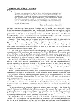
Turkish Journal of Medical Sciences http://journals.tubitak.gov.tr/medical/ Research Article Turk J Med Sci (2014) 44: 208-211 © TÜBİTAK doi:10.3906/sag-1203-72 A comparison of the generation of free radicals in saliva of active and passive smokers 1, 2 3 4 Murat DEMİRTAŞ *, Ünal ŞENEL , Sevda YÜKSEL , Mustafa YÜKSEL Department of Physiology, Faculty of Medicine, Bezmialem Vakıf University, İstanbul, Turkey 2 Department of Bioengineering, Faculty of Engineering, Çanakkale Onsekiz Mart University, Çanakkale, Turkey 3 Department of Child Development, School of Health Sciences, Turgut Özal University, Ankara, Turkey 4 Department of Audiology, School of Health Sciences, Turgut Özal University, Ankara, Turkey 1 Received: 20.03.2012 Accepted: 04.09.2012 Published Online: 15.01.2014 Printed: 14.02.2014 Aim: To reveal any correlation between cigarette smoke and malondialdehyde (MDA) values in the saliva fluid of subjects who are active smokers or nonsmoking subjects who are exposed to cigarette smoke in their environments. Materials and methods: Saliva samples were taken from 3 groups: a control group (group 1), a group inhaling smoke passively (group 2), and a test group (group 3) smoking 20 cigarettes per day; each group consisted of 20 members, giving a total sample of 60 people, aged 20 to 45 years. MDA, which is an indicator of lipid peroxidation, was measured via the colorimetric method. Results: Salivary MDA levels in smokers were found to be significantly higher compared to the control group and the group of passive smokers (P < 0.05). When compared with the control group, the MDA levels of passive smokers and active smokers were higher; when passive and active smokers were compared, the MDA levels of active smokers were higher. When all 3 groups were compared, the MDA levels in the control group (nonsmokers) were observed to be lower than the MDA level of the other 2 groups. Conclusion: It was observed that lipid peroxidation, which is an indicator used to determine oxidative stress, and MDA level, which is a product of this reactive chain, are significantly higher in individuals who are smokers. Such a high level of MDA in passive smokers indicates that smoking also affects nonsmokers negatively. Key words: Free radical, malondialdehyde, saliva, cigarette, passive smoking, thiobarbituric acid 1. Introduction Tobacco is a plant utilized in different ways throughout the world by using leaves of some of the strains of Nicotiana species, such as Nicotiana tabacum and Nicotiana rustica (1). Habitual smoking is the most significant threat to the world’s population today. Smoking causes 30% of deaths between the ages of 35 and 69. Cigarettes and tobacco products cause significant economic losses in society when the high cost of medical treatments for illnesses such as cancer and related maladies are taken into consideration, as well as consumer expenditures for the products themselves. It reduces workforce capacity by causing premature deaths and, therefore, impacts the economy at the individual and social levels (2). Passive smokers are people who are exposed to cigarette smoke although they do not smoke directly; they are as negatively affected by cigarette smoke as active smokers. Malondialdehyde (MDA) levels, which are an indicator of lipid peroxidation, were evaluated in this study. *Correspondence: [email protected] 208 Free radicals are produced continuously in the body as a result of various chemical activities. The degree of cell damage caused by free radicals is dependent upon the effectiveness of defense systems within the cell (3). Therefore, endogenous systems in the body have developed free radical preventative and scavenger features. In the case where the sensitive balance of oxidants and antioxidants moves toward oxidants, oxidative damage occurs (4). Since free radicals are potentially toxic, organisms have developed defense systems to eliminate them. These are called antioxidants (5). Cells inactivate enzymes, the peroxides of which are called the antioxidant defense system, and synthesize proteins that attract transition metals and scavenge free radicals, thus preventing the generation of free radicals and thereby limiting the damage caused by free radicals (6). MDA, as the final product of lipid peroxidation, is a chemically active molecule and may have harmful effects, particularly on proteins at the molecular level, by being DEMİRTAŞ et al. / Turk J Med Sci diffused easily in surrounding cells and tissues (7). MDA is a lipid peroxidation product that can be determined by thiobarbituric acid (TBA). Since other substances, such as bilirubin, react with TBA, this lipid peroxidation level is expressed as thiobarbituric acid reactive substances (TBARS) (8). Lipid peroxides produced as a result of lipid peroxidation are unstable structures and prone to secondary or tertiary reactions. One of the techniques to measure lipid peroxidation is the TBA test (9). This method is the most common and the easiest spectrophotometer method used for the measurement of lipid peroxide levels. MDA reacts with 2 molecules of TBA and creates a pink complex at 532 nm (10). There are many organs and tissues damaged by free radicals produced by substances contained in cigarette smoke or through the biotransformation of these substances. In light of this information, a study of this effect as observed in smokers is important in order to determine their exposure to free radicals and the damage they cause (11). The risk of lung cancer in smokers is 4–10 times higher than in nonsmokers; this figure is 15–30 times greater for heavy smokers. Retrospective studies carried out in the United States, England, and Canada showed that lung cancer risk increases proportionally with the number of cigarettes smoked (12). Each year 35,000 deaths in Turkey can be attributed to illnesses related to the use of tobacco products (13). The purpose of this study was to measure the MDA levels in the saliva fluids of active smokers and people exposed to cigarette smoke in their environment (passive smokers) and to then determine the degree of oxidative damage that developed due to cigarette smoke. There is abundant research showing that plasma lipid peroxidation increases in smokers. However, the current research was carried out due to insufficient prior research evaluating saliva MDA levels. 2. Materials and methods 2.1. Study groups Saliva samples were taken from 60 individuals forming 3 study groups. The nonsmoking group was defined as group 1 (control group); the passive smoking group was defined as group 2; and the group of active smokers, who consume at least 20 cigarettes a day, was defined as group 3. The individuals who gave saliva samples were all healthy males between the ages of 20 and 45 and not using medicine regularly, and those in group 3 had been smoking cigarettes for at least 1 year. The people who gave saliva samples rinsed their mouth with water prior to the collection of saliva samples and samples were placed in test tubes, where they were held for up to 5 min. Collected samples were kept at −20 °C until the analysis. 2.2. Laboratory measurements (determination of MDA level) Samples taken to measure MDA values in saliva fluid were processed with TBA and boiled for 30 min, and then were spectrophotometrically scanned at 532 nm. This method is based on dual boiling. In the initial heating, proteins were precipitated by being freed from bound MDA; in the second heating, total MDA entered into a reaction with TBA to create a colored complex in a hot and acidic environment. The concentration of MDA is calculated in direct proportion to the absorbance delivered by the color complex at 532 nm generated by MDA (14,15). In this analysis, 10% trichloroacetic solution (TCA; Ateks) and 0.675% TBA solution (Merck) were used. Next, 2.5 mL of TCA (10%) was put in control and sample tubes. Sample tubes received 0.5 mL of saliva, while control tubes received 0.5 mL of distilled water. Tubes were placed in a hot water bath filled with preheated water at 90 °C for 15 min after covering their tops tightly. Tubes were taken out of the water bath upon completion of the waiting period and cooled under running tap water. They were subjected to centrifugation for 10 min at 3000 rpm. A 2-mL sample from the supernatant on the top of each was introduced to other empty tubes. One milliliter of TBA solution (0.675%) was added to those tubes and they were similarly placed in a hot water bath at 90 °C for 15 min after covering their tops tightly. Tubes were taken out of the water bath upon completion of the waiting period and cooled under running tap water. Absorbance of the sample was measured by spectrophotometer at 532 nm against the blind samples. In the calculations, net absorbency was found by subtracting the absorbance of the blank from the absorbance of the sample. MDA concentrations were calculated in nmol/mL by taking into account the molar extinction coefficient of the TBA–MDA complex at 532 nm, i.e. 1.56 × 10⁵ M–1 cm–1, and by taking the dilution factor into consideration. 2.3. Statistical analysis Statistical analysis of data was carried out with SPSS 16.0 (SPSS Inc., Chicago, IL, USA). The Kolmogorov–Smirnov test was performed to examine the distribution of our data when comparing the values between groups, and Mann– Whitney U and Kruskal–Wallis tests were performed in an appropriate way. Significance level was accepted as P < 0.05. All data values are given as the mean ± standard deviation. 3. Results In this study, MDA levels as an indicator of lipid peroxidation were measured in saliva samples taken from active smokers, from passive smokers, and from nonsmokers (control); the results obtained were compared 209 DEMİRTAŞ et al. / Turk J Med Sci to the control group. In paired comparisons, a statistical increase at the P < 0.05 level was observed in MDA levels in parallel with the increase of smoking when comparing passive smokers with the control group, active smokers with the control group, and passive smokers with active smokers. In the Figure, the MDA values of the groups are specified. It was determined that the MDA levels of the nonsmoking group were lower than those of the other groups (P < 0.05). 4. Discussion It is known that cigarette and other tobacco products have extremely harmful effects on human health. It is not possible to cease production and consumption of tobacco and tobacco products. However, informing society through scientific studies can be helpful for human betterment. Therefore, the results of scientific studies with regard to the effects of cigarette smoking and the increasing threat to public health are important. Since the 8 Group 1 (Control) Malondialdehyde level nmol/mL 6.07 ± 2.33 * Group 2 7 Group 3 6 5 4.36 ± 0.68 3.47 ± 0.65 4 3 2 1 0 Figure. Levels of salivary MDA in group 1 (n = 20), group 2 (n = 20), and group 3 (n = 20). *: Salivary MDA levels in smokers were found to be significantly higher compared to the control group and the group of passive smokers (P < 0.05). change in biochemical parameters measured in the study pertaining to several diseases (primarily various cancers and cardiovascular diseases) is significant, the results of our study will be useful for evaluating the effects of smoking on public health. Cigarette smoke inhaled with normal respiration may impair the oxidant/antioxidant balance in the organism and cause the generation of free radicals (16). Free radicals in cigarette smoke or stimulated by smoking cause oxidative damage and may initiate the breakdown of lipids contained in the cell membrane (17,18). MDA, as the final product of lipid peroxidation, is a chemically active molecule and may have harmful effects, particularly on proteins at the molecular level, by being diffused easily in surrounding cells and tissues (19,20). It was reported that MDA levels measured in blood samples taken from rabbits exposed to cigarette smoke (for studying the effects of smoking on MDA levels) were higher than in the control group; smoking accelerates the production of free radicals and causes oxidative cell damage (21). It was also reported that the quantity of the smoking causes oxidative damage in smokers and leads to an increase in the MDA level, and thus increases the risk of coronary artery disease (22). Guentsch et al. (23) and Akalın et al. (24) reported that MDA levels increased in the saliva of smokers versus nonsmokers and these findings were in concordance with those reported by some previous studies in venous blood erythrocyte/serum/plasma (22,25–27). Our findings support these results. In conclusion, it was observed that lipid peroxidation, which is an indicator of oxidative stress, and MDA levels, which are a product of this reaction chain, are significantly higher in individuals who smoke. A high rate of this level in passive smokers indicates that smoking also affects nonsmokers negatively. This condition shows that the prohibition of smoking indoors is not sufficient. Therefore, it emphasizes that smoking outdoors where people are present should be prohibited and it is required that smoking shall be avoided in any environment where people younger than 18 years are present. Laws regulating the environments for smoking should be considered again and required regulations should be applied. References 1. Demir H. Tütün ve sigara içenlerde serum tiyosiyanat seviyeleri ile selenyum, C vitamini ve lipit peroksidasyonu seviyelerinin araştırılması. MSc, Harran University, Şanlıurfa, Turkey, 1998 (in Turkish). 2. Lesmes GR, Donofrio KH. Passive smoking: the medical and economic issues. Am J Med 1992; 93: 38–42. 210 3. Gianetti J, Pedrinelli R, Petrucci R. Inverse association between carotid intima-media thickness and the antioxidant lycopene in atherosclerosis. Am Heart J 2002; 143: 467–74. 4. Brent JA, Rumack BH. Role of free radicals in toxic hepatic injury. Free radical biochemistry. Clin Toxicol 1993; 31: 139– 71. DEMİRTAŞ et al. / Turk J Med Sci 5. Lohr JB. Oxygen radicals and neuropsychiatric illness. News and views. Arch Gen Psychiat 1991; 48: 1097–106. 6. De Zwart LL, Meerman JH, Commandeur JN, Vermeulen NP. Biomarkers of free radical damage applications in experimental animals and in humans. Free Radical Biol Med 1999; 26: 202– 26. 7. Parantainen J, Vapaatalo H, Hokkanen E. Clinical aspects of prostaglandins and leukotrienes in migraine. Cephalalgia 1986; 4: 95–101. 8. Knight JA, Pleper RK, Macellon L. Specificity of the thiobarbituric acid reaction: its use in studies of lipid peroxidaiton. Clin Chem 1988; 34: 2433–8. 9. Esterbauer H, Cheeseman KH, Dianzani MU, Poll G, Slater TF. Separation and characterization of the aldehydic products of ADP–Fe2+ stimulated lipid peroxidation in the rat liver microsomes. Biochem J 1982; 208: 129–40. 10. Gökpınar E. Maraş otunun (Ağızotu) Tükürük adenozin deaminaz, ksantin oksidaz aktiviteleri ile total sialik asit ve malondialdehit düzeylerine etkisinin araştırılması, MSc, Kahramanmaraş Sütçü İmam University, Kahramanmaraş, Turkey, 2008 (in Turkish). 11. Petruzelli S, Puntoni R, Mimotti P, Pulera N, Baliva F, Fornai E, Giuntini C. Plasma 3-nitrotyrosine in cigarette smokers. Am J Resp Crit Care 1997; 156: 1902–7. 12. Ertürk B. Akçiğer kanserli hastalarda malondialdehit (MDA) ve total antioksidan kapasite (TAOK) düzeyi ölçümü ile oksidan-antioksidan dengesinin araştırılması. Thesis, Republic of Turkey Ministry of Health, İstanbul, Turkey, 2006 (in Turkish). 13. Çavdar T, Ekim N. Akciğer kanseri. Multidisipliner yaklaşım. Toraks Kitapları 1999; 1: 17–53 (in Turkish). 14. Draper HH, Hadley M. Malondialdehyde determination as index of lipid peroxidation. Method Enzymol 1990; 186: 421– 30. 15. Hammode RMA, Khalil MMM, Salem A. Lipid peroxidation products in pleural fluid for separation of transudates and exudates. Clin Chem 1995; 41: 1315–24. 17. Church DF, Pryor WA. Free-radical chemistry of cigarette smoke and its toxicological implication. Environ Health Persp 1985; 64: 111–26. 18. Akkuş İ. Serbest Radikaller ve Fizyopatolojik Etkileri. Konya, Turkey: Mimoza Basım; 1995 (in Turkish). 19. Halliwell B. Drug antioxidant effects. A basis for drug selection? Drugs 1991; 42: 569–605. 20. Yılmaz G, Süer H, Üçler S, İnan L, Yücel D. Plasma nitric oxide metabolites in migraine with and without aura. Türk Biyokimya Dergisi 2005; 30: 236–40 (in Turkish with abstract in English). 21. Özaras R, Tahan V, Ünlü M, Demirci M, Bahçeci M, Uzun H, Aydın S, Delibaş N. Lp(a) levels and lipid peroxidation after smoke exposure in the rabbit. Free Radical Bio Med 1998; 25: 77. 22. Aliyev V. Sigara içenlerde oksitatif stres göstergelerinin değerlendirilmesi. MSc, Ankara University, Ankara, Turkey, 2006 (in Turkish). 23. Guentsch A, Jentsch H, Pfister W, Hoffmann T, Eick S. Moxifloxacin as an adjunctive antibiotic in the treatment of severe chronic periodontitis. J Periodontol 2008; 79: 1894–903. 24. Akalın FA, Baltacıoğlu E, Alver A, Karabulut E. Lipid peroxidation levels and total oxidant status in serum, saliva and gingival crevicular fluid in patients with chronic periodontitis. J Clin Periodontol 2007; 34: 558–65. 25. Solak ZA, Kabaroğlu C, Cok G, Parildar Z, Bayindir U, Ozmen, Bayindir O. Effect of different levels of cigarette smoking on lipid peroxidation, glutathione enzymes and paraoxonase 1 activity in healthy people. Clin Exp Med 2005; 5: 99–105. 26. Isik B, Ceylan A, Isik R. Oxidative stress in smokers and nonsmokers. Inhal Toxicol 2007; 19: 767–69. 27. Orhan H, Evelo CTA, Şahin G. Erythrocyte antioxidant defense response against cigarette smoking in humans - the glutathione S-transferase vulnerability. J Biochem Mol Toxic 2005; 19: 226–33. 16. Pryor WA, Prier DG, Church DF. Electron spin resonance study of mainstream and sidestream cigarette smoke: nature of free radicals in gas-phase smoke and in cigarette. Environ Health Persp 1993; 47: 345–55. 211
© Copyright 2026 Paperzz