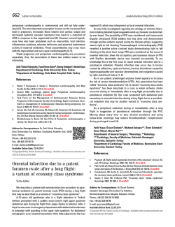
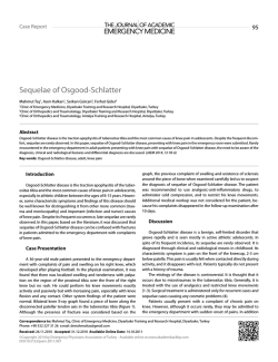
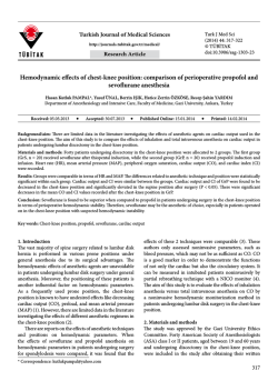
AĞRI 2014;26(1):34-38
CASE REPORT - OLGU SUNUMU
doi: 10.5505/agri.2014.30633
Ultrasound-guided psoas compartment block and
general anesthesia for arthroscopic knee surgery: a case report
Artroskopik diz cerrahisi için genel anestezi ve ultrason rehberliğinde
psoas kompartman blok uygulaması: Olgu sunumu
Emine Aysu ŞALVIZ,1 Yavuz GÜRKAN,2 Murat TEKİN,2 Levent BULUÇ3
Summary
Anesthetizing the lumbar plexus at its origin facilitates a more “complete” psoas compartment block compared to peripheral
approaches. It is usually performed using surface anatomical landmarks, and the site for local anesthetic injection is confirmed
by observing quadriceps muscle contraction to peripheral nerve stimulation. Ultrasound may provide guidance alone or together with the aid of nerve stimulation during nerve blocks. We present a 48-year-old male patient, American Society of
Anesthesiologists (ASA) physical status II, who refused spinal anesthesia, and underwent knee arthroscopy with ultrasoundguided psoas compartment block and general anesthesia. Following the standard monitoring and lateral decubitus positioning, the vertebral body, psoas, erector spinae, and quadratus lumborum muscles and hyperechoic nerve roots of the patient
were visualized at the level of L4-5 with curvilinear ultrasound probe. The needle was inserted with ultrasound guidance,
and correct tip position was confirmed with quadriceps contraction. Then, the mixture of 30 mL local anesthetic (10 mL 2%
lidocaine and 20 ml 5% levobupivacaine) was injected at the estimated position of the lumbar plexus (junction of the posterior
third and anterior two-thirds of the psoas muscle). He also received general anesthesia for the surgery. Anesthesia and surgical procedures were completed successfully without any additional anesthetic/analgesic requirement or complication. The
postoperative period was pain-free both at rest and during mobilization for 24 hours. This case report shows that ultrasoundguided psoas compartment block is feasible and efficient for peri- and postoperative analgesia during knee arthroscopy.
Key words: Knee arthroscopy; psoas compartment block; ultrasound.
Özet
Psoas kompartman bloğu, lumbar pleksusun kökler hizasında bloke edilmesi sebebiyle periferik yaklaşımlara göre daha tam bir bloktur. Genellikle yüzeyel anatomik işaret noktaları kullanılarak blok lokalizasyonu belirlenir ve lokal anestezik enjeksiyonu periferik
sinir stimülatörü ile kuadriseps kasının kontraksiyonu görülerek uygulanır. Ultrason sinir bloklarında tek başına veya sinir stimülatörü yardımı ile rehberlik sağlayabilir. Biz, bu yazıda spinal anestezi girişimini reddeden, genel anestezi ve ultrason rehberliğinde psoas
kompartman bloğu altında diz artroskopi cerrahisi geçiren, 48 yaşında, ASA II, erkek hasta ile ilgili deneyimimizi sunduk. Hastaya
standart monitorizasyon uygulandı ve lateral dekübit pozisyon verilmesi sonrasında vertebra, psoas, erektor spina and kuadratus
lumborum kasları L4-5 seviyesinde konveks prob ile gözlendi. İğne ultrason rehberliğinde ilerletildi ve iğne ucunun lokalizasyonu
kuadriseps kas kontraksiyonu ile doğrulandı. Otuz mL lokal anestezik karışımının (10 mL %2’lik lidokain ve 20 mL %5’lik levobupivakain) lumbar pleksus etrafına enjekte edilmesinin ardından hastaya genel anestezi uygulandı. Anestezi ve cerrahi işlem ek
anestezik/analjezik ihtiyacı veya komplikasyon olmaksızın başarıyla tamamlandı. Hastanın ameliyat sonrası dönemde istirahatte
ya da mobilizasyonda ilk 24 saatte ağrı şikayeti olmadı. Bu olgu sunumu, ultrason rehberliğinde psoas kompartman bloğunun diz
artroskopi cerrahisinde ameliyat esnası ve sonrası analjezi için uygun ve etkin bir yöntem olduğunu göstermektedir.
Anahtar sözcükler: Diz artroskopisi; psoas kompartman bloğu; ultrason.
Department of Anesthesiology and Reanimation, Şanlıurfa Training and Research Hospital, Sanliurfa;
Department of Anesthesiology and Reanimation, Kocaeli University Faculty of Medicine, Kocaeli;
3
Department of Orthopedics and Traumatology, Kocaeli University Faculty of Medicine, Kocaeli
1
Şanlıurfa Eğitim ve Araştırma Hastanesi, Anesteziyoloji ve Reanimasyon Kliniği, Şanlıurfa;
2
Kocaeli Üniversitesi Tıp Fakültesi, Anesteziyoloji ve Reanimasyon Anabilim Dalı, Kocaeli;
3
Kocaeli Üniversitesi Tıp Fakültesi, Ortopedi ve Travmatoloji Anabilim Dalı, Kocaeli
1
Current affiliation / Şimdiki kurumu:
2
1
Submitted (Başvuru tarihi) 24.02.2012
Accepted after revision (Düzeltme sonrası kabul tarihi) 15.06.2012
Department of Anesthesiology and Reanimation,
Istanbul University Istanbul Faculty of Medicine,
Istanbul;
1
İstanbul Ünivesitesi İstanbul Tıp Fakültesi,
Anesteziyoloji ve Reanimasyon Anabilim Dalı,
İstanbul
Correspondence (İletişim): Dr. Emine Aysu Şalvız. İstanbul Ünivesitesi, İstanbul Tıp Fakültesi, Anesteziyoloji ve Reanimasyon Anabilim Dalı, Çapa, İstanbul, Turkey.
Tel: +90 - 212 - 414 20 00 e-mail (e-posta): [email protected]
34
OCAK - JANUARY 2014
Ultrasound-guided psoas compartment block and general anesthesia for arthroscopic knee surgery
Introduction
The psoas compartment block (PCB) technique
is useful in anesthetizing the lumbar plexus at its
origin. Successfully placing local anesthetic (LA) at
the level of the lumbar plexus roots (junction of the
posterior third and the anterior two-thirds of the
psoas muscle) is a more “complete” method compared to peripheral approaches.[1-4] It provides the
blockade of the femoral, obturator and lateral cutaneous nerves of the thigh with a single injection.
[3-5]
Therefore, it may be used for anesthesia and analgesia of anterior, medial, and lateral procedures of
the thigh such as skin grafts and femoral osteotomy,
total knee replacement,[6] total hip replacement,[7,8]
hip fractures,[9,10] knee arthroscopy,[11] anterior and
posterior cruciate ligament reconstructions[12] and
chronic hip pain.[13]
The PCB technique is traditionally performed using
surface anatomical landmarks and observing quadriceps muscle contraction to peripheral nerve stimulation.[3] Lately, there has been a rise in the use of
ultrasound to visualize the sonoanatomy of lumbar
plexus and to guide the PCBs. Ultrasound-guided
regional anesthetic techniques of the lower limb can
be challenging because of the muscles and fat which
surround the nerves,[5,14] however technological improvements of ultrasound equipment have allowed
better visualization of the nerves and the other anatomical structures. The lumbar plexus and the posterior part of the psoas muscle, although deep, were
examined in cadavers and just in a few cases successfully.[1,3,15,16] We present a case, who underwent a
knee arthroscopy with successful ultrasound-guided
PCB and general anesthesia.
to provide a more complete anesthesia and analgesia for this patient, who refused to undergo spinal
anesthesia, an ultrasound-guided PCB and general
anesthesia was planned.
On arrival to the holding area where blocks were
performed, standard monitoring (pulse-oxymetry,
heart rate (HR), noninvasive blood pressure (NIBP)
and ECG) was applied. A 20-gauge intravenous
(IV) cannula was placed on the right hand of the
patient and a free running infusion of saline was
started. Oxygen 2 L/h was applied via mask. Sedation was provided with 3 mg IV midazolam after
the lateral decubitus position with the left operative side up, the hips and the knees flexed. After
disinfection and sterile draping, sterile gel was applied to the procedure area. A low frequency curvilinear ultrasound probe (1-8 MHz, Esaote My Lab
30, Genoa, Italy) was placed at the level of L4-5 in
the transverse plane (Figure 1). Vertebral body, articular process, psoas, erector spina, and quadratus
lumborum muscles were visualized (Figure 2). After
LA skin infiltration, a 22-gauge 100 mm insulated
needle (Pajunk, Geisingen, Germany) was inserted
lateral to the probe, using in-plane technique. The
correct tip position was confirmed with quadriceps
contraction at 8 cm by neurostimulator 0.5 mA.
Then, LA mixture of 30 mL (10 mL 2% lidocaine
and 20 mL 5% levobupivacaine) was injected incrementally in fractioned doses with frequent aspiration to detect any possible intravascular puncture.
The LA spread was observed on transverse axis scan.
Case Report
A 48-year-old male, ASA physical status II patient
with hypertension was scheduled for knee arthroscopy procedure. Patient history revealed that, the
patient had undergone appendectomy, nephrolitiazis operations under general anesthesia and knee
arthroscopy, hallux valgus, hemorrhoid operations
under spinal anesthesia. Moreover, he had had coronary angiography twice by the reason of chest pain
and hypertension, and got normal results. No abnormalities were noted on his preoperative blood
tests, chest X-ray and physical examination. In order
OCAK - JANUARY 2014
Figure 1.A low frequency curvilinear ultrasound probe was
placed at the level of L4-5 in the transverse plane while the
patient is placed in the right lateral decubitus position.
35
AĞRI
for these procedures.[18-21] Central neuraxial blocks
and general anesthesia provide effective anesthesia,
however neuraxial blocks cause nausea/emesis, urinary retention, post-dural puncture, hemodynamic
changes, motor weakness in the lower extremity and
on the other hand general anesthesia is reported with
high postoperative pain scores, and low satisfaction
ratings.[4,12] However, peripheral nerve blocks have
advantages of providing limb-specific anesthesia/analgesia without extended muscle weakness, allowing
early ambulation, enabling effective physiotherapy
and early discharge.[4,22,23]
Figure 2.The vertebral body, articular process, psoas, erector
spina, and quadratus lumborum muscles of the patient were
visualized with a low frequency curvilinear ultrasound probe.
Successful block was confirmed in 20 minutes by
decreased sensation in terminal nerves of the lumbar plexus (femoral, lateral femoral cutaneous, and
obturator) and quadriceps weakness.
Then, the patient was taken to the operating room.
Following the application of standard monitoring
and obtaining normal vital signs, general anesthesia was induced with propofol 2.5 mg kg-1 and
fentanyl 1 μg kg-1 iv. Laryngeal mask (number 4)
was placed for airway protection and anesthesia was
then maintained with 1.5-2% sevoflurane in N2O
and O2 (1:1 ratio). Knee arthroplasty and anesthesia
procedures were successfully completed within 50
minutes without any additional anesthetic/analgesic
requirement. Diclofenac 75 mg was ordered as a rescue analgesic that could be repeated every 6 h with
a maximum daily dose of 300 mg.
The patient was discharged on the next day. He was
very pleased because of pain free postoperative period both at rest and during mobilization, and his
analgesic consumption was 0 during the postoperative first 24 hours.
Discussion
Knee arthroscopy is one of the most commonly performed elective ambulatory orthopedic procedures
and associated with moderate to severe, often poorly controlled, postoperative pain.[4,17] Various types
of anesthesia including neuraxial blocks, general,
and peripheral nerve blocks have been performed
36
Although femoral nerve block is easy to perform,
the obturator and lateral femoral cutaneous nerves
are not consistently blocked with this technique.[24]
On the other hand, the lumbar plexus is made up of
the L1 through L5 spinal nerve roots. As the roots
of the lumbar plexus depart from their spinal nerves
and emerge from the intervertebral foramen, they
enter the posterior third or junction of the posterior third and the anterior two-thirds of the psoas
muscle.[1-4,25] In contrast to the femoral approach,
the PCB reliably produces blockade of the entire
lumbar plexus with high success rate.[14,24] Therefore, PCB with general anesthesia was chosen to
provide complete peri-/post-operative analgesia and
anesthesia during our clinical practice.
To reach the lumbar plexus by surface anatomical
landmarks is useful but can vary among patients.
Several techniques as loss of resistance, elicitation of
paresthesia, and quadriceps muscle contraction to
nerve stimulation have been described at different
levels using surface anatomical landmarks in the literature.[2,9,16,24,26,27] However, epidural spread of the
LA, systemic toxicity, renal hematoma, total spinal
anesthesia and circulatory cases have been reported
as complications of these approaches.[28-33] These
complications are usually related with failed or
multiple attempts, or inadvertent vascular injuries.
[33]
With the use of ultrasound for imaging of the
individual anatomy and real-time needle guidance,
the efficacy of the PCB might be increased and such
complications might be avoided.[16]
Ultrasound guidance during PCB may offer several
advantages compared to the traditional methods.
The related anatomy and target structures are clearly
OCAK - JANUARY 2014
Ultrasound-guided psoas compartment block and general anesthesia for arthroscopic knee surgery
identified, the advancement of the needle to the
posterior part of the psoas muscle and meanwhile
the spread of the LA during the injection can be
observed.[2,3,32] This may allow the anesthesiologist
to perform the PCB safely and successfully. Furthermore, two different techniques of ultrasound-guided PCB as transverse and longitudinal approaches
have been described.[1,3,16,17] Longitudinal technique
may cause bilateral spread and systemic LA toxicity because of the rapid absorption of large volumes
or inadvertent injection into one of the large paravertebral blood vessels.[5] Therefore, we preferred
transverse technique and were able to visualize the
kidney, vertebral body, spinous, articular, and transverse processes, psoas, erector spina and quadratus
lumborum muscles clearly in order to perform a
successful block and avoid the puncture.
Our observation of the lumbar plexus location in
the posterior part of the psoas muscle was coherent
with the previous data.[2,3] The location of the plexus
and the tip of the needle were confirmed both by
ultrasound and nerve stimulator. The block was successfully completed after one attempt. Paresthesia,
blood aspiration or injury to surrounding structures
were not noted during the procedure.
As a result, this case report describes that a successful ultrasound guided PCB technique provides a
pain free peri-/post-operative period without any
additional anesthetic or analgesic. This shows that
ultrasound-guided PCB is a feasible and efficient
technique for knee arthroscopy. Inspite of the impressive cadaver study and case report results, the
published prospective investigations are limited.
Further studies are necessary to evaluate the rate of
success, safety and efficacy of the ultrasound guided
PCB technique.
Conflict-of-interest issues regarding the authorship or article: None declared.
Peer-rewiew: Externally peer-reviewed.
References
1. Kirchmair L, Entner T, Wissel J, Moriggl B, Kapral S, Mitterschiffthaler G. A study of the paravertebral anatomy for ultrasound-guided posterior lumbar plexus block. Anesth Analg
2001;93(2):477-81. CrossRef
2. Farny J, Drolet P, Girard M. Anatomy of the posterior approach
OCAK - JANUARY 2014
to the lumbar plexus block. Can J Anaesth 1994;41(6):480-5.
3. Karmakar MK, Ho AM, Li X, Kwok WH, Tsang K, Ngan Kee WD.
Ultrasound-guided lumbar plexus block through the acoustic window of the lumbar ultrasound trident. Br J Anaesth
2008;100(4):533-7. CrossRef
4. Greengrass RA. Posterior lumbar plexus block. Tech Reg
Anesth Pain Manag 2003:7(1):3-7. CrossRef
5. Marhofer P, Harrop-Griffiths W, Willschke H, Kirchmair L. Fifteen years of ultrasound guidance in regional anaesthesia:
Part 2-recent developments in block techniques. Br J Anaesth 2010;104(6):673-83. CrossRef
6. Luber MJ, Greengrass R, Vail TP. Patient satisfaction and effectiveness of lumbar plexus and sciatic nerve block for total
knee arthroplasty. J Arthroplasty 2001;16(1):17-21. CrossRef
7. Capdevila X, Macaire P, Dadure C, Choquet O, Biboulet P,
Ryckwaert Y, et al. Continuous psoas compartment block
for postoperative analgesia after total hip arthroplasty: new
landmarks, technical guidelines, and clinical evaluation.
Anesth Analg 2002;94(6):1606-13. CrossRef
8. Türker G, Uçkunkaya N, Yavaşçaoğlu B, Yilmazlar A, Ozçelik S.
Comparison of the catheter-technique psoas compartment
block and the epidural block for analgesia in partial hip replacement surgery. Acta Anaesthesiol Scand 2003;47(1):306. CrossRef
9. Ho AM, Karmakar MK. Combined paravertebral lumbar
plexus and parasacral sciatic nerve block for reduction of
hip fracture in a patient with severe aortic stenosis. Can J Anaesth 2002;49(9):946-50. CrossRef
10.de Visme V, Picart F, Le Jouan R, Legrand A, Savry C, Morin
V. Combined lumbar and sacral plexus block compared with
plain bupivacaine spinal anesthesia for hip fractures in the
elderly. Reg Anesth Pain Med 2000;25(2):158-62. CrossRef
11.Jankowski CJ, Hebl JR, Stuart MJ, Rock MG, Pagnano MW,
Beighley CM, et al. A comparison of psoas compartment
block and spinal and general anesthesia for outpatient knee
arthroscopy. Anesth Analg 2003;97(4):1003-9. CrossRef
12.Klein SM, Greengrass RA, Grant SA, Higgins LD, Nielsen KC,
Steele SM. Ambulatory surgery for multi-ligament knee reconstruction with continuous dual catheter peripheral nerve
blockade. Can J Anaesth 2001;48(4):375-8. CrossRef
13.Goroszeniuk T, di Vadi PP. Repeated psoas compartment
blocks for the management of long-standing hip pain. Reg
Anesth Pain Med 2001;26(4):376-8. CrossRef
14.Saranteas T. Limitations in ultrasound imaging techniques
in anesthesia: obesity and muscle atrophy? Anesth Analg
2009;109(3):993-4. CrossRef
15.Kirchmair L, Entner T, Kapral S, Mitterschiffthaler G. Ultrasound guidance for the psoas compartment block: an imaging study. Anesth Analg 2002;94(3):706-10. CrossRef
16. Morimoto M, Kim JT, Popovic J, Jain S, Bekker A. Ultrasoundguided lumbar plexus block for open reduction and internal
fixation of hip fracture. Pain Pract 2006;6(2):124-6. CrossRef
17.O’Donnell BD, Iohom G. Regional anesthesia techniques
for ambulatory orthopedic surgery. Curr Opin Anaesthesiol
2008;21(6):723-8. CrossRef
18.Mulroy MF, Larkin KL, Hodgson PS, Helman JD, Pollock JE,
Liu SS. A comparison of spinal, epidural, and general anesthesia for outpatient knee arthroscopy. Anesth Analg
2000;91(4):860-4. CrossRef
19.Jacobson E, Forssblad M, Rosenberg J, Westman L, Weidenhielm L. Can local anesthesia be recommended for
routine use in elective knee arthroscopy? A comparison
between local, spinal, and general anesthesia. Arthroscopy
37
AĞRI
2000;16(2):183-90. CrossRef
20. Ilfeld BM, Le LT, Meyer RS, Mariano ER, Vandenborne K, Duncan PW, et al. Ambulatory continuous femoral nerve blocks
decrease time to discharge readiness after tricompartment
total knee arthroplasty: a randomized, triple-masked, placebo-controlled study. Anesthesiology 2008;108(4):703-13.
21.Gürkan Y, Kılıçkan L, Canatay H, Müezzinoğlu S, Toker K.
Artroskopik diz cerrahisinde farklı anestezi yöntemlerinin
karşılaştırılması. Türk Anesteziyoloji ve Reanimasyon
Cemiyeti Mecmuası 2002;30:408-12.
22.Capdevila X, Barthelet Y, Biboulet P, Ryckwaert Y, Rubenovitch J, d’Athis F. Effects of perioperative analgesic technique
on the surgical outcome and duration of rehabilitation after
major knee surgery. Anesthesiology 1999;91(1):8-15. CrossRef
23.Singelyn FJ, Deyaert M, Joris D, Pendeville E, Gouverneur
JM. Effects of intravenous patient-controlled analgesia with
morphine, continuous epidural analgesia, and continuous
three-in-one block on postoperative pain and knee rehabilitation after unilateral total knee arthroplasty. Anesth Analg
1998;87(1):88-92. CrossRef
24. Parkinson SK, Mueller JB, Little WL, Bailey SL. Extent of blockade with various approaches to the lumbar plexus. Anesth
Analg 1989;68(3):243-8. CrossRef
25.Di Benedetto P, Pinto G, Arcioni R, De Blasi RA, Sorrentino L,
Rossifragola I, et al. Anatomy and imaging of lumbar plexus.
38
Minerva Anestesiol 2005;71(9):549-54.
26.Chayen D, Nathan H, Chayen M. The psoas compartment
block. Anesthesiology 1976;45(1):95-9. CrossRef
27.Winnie AP, Ramamurthy S, Durrani Z, Radonjic R. Plexus
block for lower extremity surgery. Anesth Rev 1974;(1)1:11-6.
28. Huet O, Eyrolle LJ, Mazoit JX, Ozier YM. Cardiac arrest after injection of ropivacaine for posterior lumbar plexus blockade.
Anesthesiology 2003;99(6):1451-3. CrossRef
29.Breslin DS, Martin G, Macleod DB, D’ercole F, Grant SA. Central nervous system toxicity following the administration of
levobupivacaine for lumbar plexus block: A report of two
cases. Reg Anesth Pain Med 2003;28(2):144-7. CrossRef
30.Pousman RM, Mansoor Z, Sciard D. Total spinal anesthetic
after continuous posterior lumbar plexus block. Anesthesiology 2003;98(5):1281-2. CrossRef
31. Aida S, Takahashi H, Shimoji K. Renal subcapsular hematoma
after lumbar plexus block. Anesthesiology 1996;84(2):452-5.
32. Mullanu Ch, Gaillat F, Scemama F, Thibault S, Lavand’homme
P, Auffray JP. Acute toxicity of local anesthetic ropivacaine and
mepivacaine during a combined lumbar plexus and sciatic
block for hip surgery. Acta Anaesthesiol Belg 2002;53(3):2213.
33. Aveline C, Bonnet F. Delayed retroperitoneal haematoma after failed lumbar plexus block. Br J Anaesth 2004;93(4):58991. CrossRef
OCAK - JANUARY 2014
© Copyright 2026 Paperzz