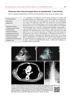
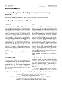
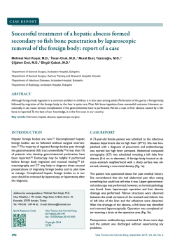
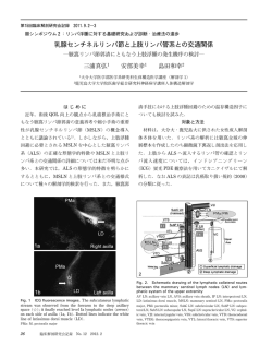
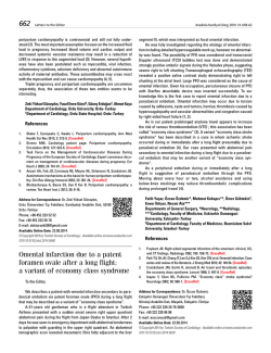
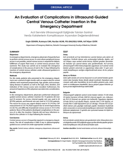
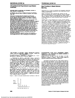
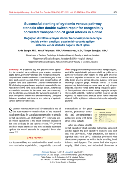
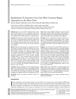
Türk Kardiyol Dern Arş - Arch Turk Soc Cardiol 2014;42(3):277-280 doi: 10.5543/tkda.2014.34356
277
Recurrent pulmonary embolism in an asthmatic patient who had
interrupted inferior vena cava with azygous continuation
Azigos ven ile devamlılığı olan kesintili vena kava inferiyora sahip
astımlı hastada tekrarlayıcı pulmoner emboli
Aylin Okur, M.D., Yavuz Selim İntepe, M.D.,# Halil İbrahim Serin, M.D.,
Uğur Yıldırım, M.D., Ertuğrul Mavili, M.D.*
Department of Radiology, Bozok University Faculty of Medicine, Yozgat;
#
Department of Chest Disease, Bozok University Faculty of Medicine, Yozgat;
*Department of Radiology, Erciyes University Faculty of Medicine, Kayseri
Summary– A 45-year-old woman with a history of recurrent
pulmonary embolism was admitted to the emergency clinic
with dyspnea, wheezing and tachypnea. Partial deep vein
thrombosis of the popliteal vein was seen on Doppler sonography. On the contrast-enhanced thorax computed tomography (CT) scan, a clot was detected in the right main pulmonary
artery and its major descending branch. Moreover, the azygos
vein was prominently dilated. Abdominal multi-slice computed
tomography (MSCT) scan revealed absence of the hepatic
segment of the inferior vena cava (IVC) with continuation of
the IVC as a dilated right-sided azygos vein. The hepatic veins
were draining directly into the right atrium. Thus, we discuss
herein this rare anatomic variant presented with recurrent pulmonary embolism, together with the findings on MSCT.
I
nterruption of the Abbreviations:
inferior vena cava CT Computed tomography
(IVC) is a congeni- CTA Computed tomography angiography
tal anomaly. The ve- IVC Inferior vena cava
MSCT Multi-slice computed tomography
nous drainage of the PTE Pulmonary thromboembolism
lower extremities is
achieved through a compensatory dilated vena azygos
system. Computed tomography (CT) and magnetic
resonance imaging (MRI) are non-invasive imaging techniques that can be used to detect these vascular anomalies. Awareness of the existence of IVC
anomalies is important for preoperative planning.
These anomalies are rarely associated with thrombosis of the iliac and femoral veins, especially in young
Özet– Tekrarlayıcı pulmoner emboli hikayesi olan 45 yaşında kadın hasta dispne, hırıltılı solunum ve taşipne şikayetleri ile acil kliniğine başvurdu. Doppler ultrasonografide,
popliteal vende kısmi trombüs saptandı. Kontrastlı toraks
bilgisayarlı tomografisinde (BT), sağ ana pumoner ven ve
büyük dallarında trombüs mevcuttu. Ayrıca, azigos ven belirgin olarak genişlemişti. Abdominal çok kesitli bilgisayarlı
tomografide (ÇKBT), vena kava inferiyor’un (VKİ) hepatik
segmentinin olmadığı ve VKİ’nin genişlemiş azigos ven
olarak devam ettiği saptandı. Hepatik venler, doğrudan sağ
atriyuma boşalmaktaydı. Böylelikle, tekrarlayan pulmoner
emboli ile ortaya çıkışı nadir olan bu anatomik varyasyonu,
ÇKBT bulguları ile tartışmayı amaçladık.
patients.[1] Here, we report a patient with interrupted
IVC anomaly who presented with deep vein thrombosis of the femoral veins and recurrent pulmonary
thromboembolism (PTE).
CASE REPORT
A 45-year-old woman who had been followed since
2009 presented to the emergency department with
dyspnea, wheezing and tachypnea in January 2010.
She was admitted to the hospital and received asthma
medication but did not recover. Thus, she underwent
thorax CT scan, which revealed a thrombus in the
right pulmonary artery (Figure 1). Lower extrem-
Received: May 20, 2013 Accepted: October 02, 2013
Correspondence: Dr. Aylin Okur. Bozok Üniversitesi Tıp Fakültesi, Radyoloji Anabilim Dalı, Yozgat.
Tel: +90 354 - 212 60 70 e-mail: [email protected]
© 2014 Turkish Society of Cardiology
Türk Kardiyol Dern Arş
278
Figure 1. CT scan shows the thrombus in the right main
pulmonary artery (arrow).
ity color Doppler sonography revealed deep venous
thrombosis. She had no evident risk factors for thromboembolic disease, such as trauma, surgery, obesity,
or history of a long journey (longer than 6 hours), or
any other acquired risk factor. Standard therapy with
heparin was given for six months without complication. The follow-up CT scan revealed resolution of the
thrombus. One year later, the patient presented with
symptoms of dyspnea, hemoptysis and swelling of the
right lower extremity. Partial deep vein thrombosis of
the lower extremity (popliteal vein) was seen on DopA
pler sonography. Genetic tests were performed since
she had a history of recurrent PTE. Tests for thrombophilia such as protein S, protein C, antithrombin III,
homocysteine, and antiphospholipid antibody were
found to be normal. Mediastinal widening was seen on
the X-ray evaluation. On the contrast-enhanced thorax
CT scan, a clot was detected in the right main pulmonary artery and its major descending branch. The CT
scan revealed a prominently dilated azygos vein (Figure 2a, b). An abdominal CT scan was performed for
the evaluation of the distal vascular structures. On the
64-slice multi-slice computed tomography (MSCT)
and computed tomography angiography (CTA), absence of the hepatic segment of the IVC was noted.
The IVC continued as a dilated, right-sided azygos
vein. The hepatic veins were draining directly into the
right atrium (Figure 3). The left renal vein was retroaortic. No additional congenital anomalies, including
heart disease or polysplenia, were seen. The patient
was treated using unfractionated heparin and oral anticoagulants. A standard regimen was used including
initial intravenous bolus of 80 IU/kg heparin followed
by continuous infusion with 18 IU/kg/h. We measured
activated partial thromboplastin time value every six
hours to determine whether the therapeutic threshold
was reached. Warfarin was added and maintained after 24 hours of heparin therapy. At the time of the first
attack, warfarin therapy had been continued for nine
months. After a second attack, a new therapeutic plan
B
Figure 2. (A) Axial and (B) coronal CT scans demonstrate the enlarged azygos vein at the hepatic level (arrow).
Recurrent pulmonary embolism in an asthmatic patient
279
of 0.6%. It is often associated with congenital heart
disease, polysplenia, and less commonly, asplenia.
[5]
Isolated occurrence without heart disease may be
encountered incidentally during radiological imaging
studies or vascular interventions. Our patient had no
detectable additional congenital anomalies.
Figure 3. Coronal CT scan shows the hepatic veins draining
directly into the right atrium (arrow).
PARS HEPATICA
DIAPHRAGMATIC
CRUS
AORTA
Figure 4. Schematic drawing shows lack of contiguity between the pre-renal segment of the IVC (arrow) and the hepatic segment (ref. 5).
was established. She has had no recurrent symptoms.
DISCUSSION
The embryology of the IVC is complex.[2] The normal
IVC consists of four main segments: hepatic, suprarenal, renal, and infrarenal.[3] Hepatic segment anomalies occur when the right subcardinal vein fails to converge with hepatic sinusoids.[3] Thus, blood is shunted
from the suprasubcardinal anastomosis through the
retrocrural azygos vein.[4] Azygos continuation of the
IVC is defined as absence of the hepatic segment of
the IVC with azygos continuation (Figure 4).[4] This
anomaly is a rare clinical condition, with a prevalence
The azygos vein generally originates below the
diaphragm at the level of the first two lumbar vertebrae by confluence of the ascending lumbar vein and
the right subcostal vein.[6] A normal azygos vein is
about 3-7 mm in width and cannot be seen on conventional radiography. When azygos continuation of
the IVC is present, the visceral and lower extremity
blood returns to the right atrium through the azygos
vein. The enlarged azygos vein may manifest as mediastinal widening on chest radiography, and it can
be mistakenly interpreted as right-sided paratracheal
adenopathy and mediastinal mass.[7] In the presented
case, widening of the right mediastinum was present
on chest radiography; however, the final diagnosis
was established with CT.
Knowledge of the presence of an interrupted IVC
may be important for patients undergoing right heart
catheterization, electrophysiological studies, cardiopulmonary bypass surgery, femoral vein catheter insertion, IVC filter placement, and temporary pacing
via the transfemoral route. If unidentified, it can lead
to life-threatening complications during abdominal
surgery.[5]
Inferior vena cava (IVC) anomalies have been recognized as possible risk factors for deep vein thrombosis. Ruggeri et al.[8] showed IVC anomalies in four
of 75 patients with a first- attack deep vein thrombosis. In another study, 31 patients with iliofemoral deep
vein thrombosis were prospectively evaluated by venography and MR angiography, and five anomalous
IVCs were found in that study.[2] An interrupted IVC
may cause increased blood pressure in the lower extremity veins in the presence of insufficient collateral
vessels. Eventually, venous stasis, which predisposes
to deep vein thrombosis and indirectly to PTE, as in
our case, may occur. PTE in patients with interrupted
IVC with azygos continuation is reported rarely, and
most of the publications are case reports.[3]
Another important feature of our case is the presence of asthma, which is a chronic pulmonary disease
characterized by reversible airflow obstruction. Dyspnea, wheezing, cough, and chest tightness are com-
280
mon symptoms. Pulmonary infections, congestive
heart failure, bronchiectasis, upper airway obstructions, and other airway diseases such as chronic obstructive pulmonary disease (COPD), pulmonary embolism, and gastroesophageal reflux disease should
be considered in the differential diagnosis. Bronchoconstriction, the main feature of asthma, can also be
encountered in pulmonary embolism. Bronchospasm
was seen after experimental thromboembolism in dog
studies.[9] In particular, wheezing and dyspnea could
interfere with the diagnosis of pulmonary embolism
in asthmatic patients. Recently, studies have shown
that asthmatic patients have activated coagulation in
the airway.[10] Inflammation affects procoagulant and
fibrinolytic activities, and there is endothelial dysfunction in the airway circulation in asthmatic patients, which is a primary factor in the pathogenesis of
pulmonary embolism. Studies have shown that asthmatic patients have local vascular inflammation and
increased risk for pulmonary embolism.[10]
Türk Kardiyol Dern Arş
REFERENCES
1. Obernosterer A, Aschauer M, Schnedl W, Lipp RW. Anomalies of the inferior vena cava in patients with iliac venous
thrombosis. Ann Intern Med 2002;136:37-41. CrossRef
2. Bass JE, Redwine MD, Kramer LA, Harris JH Jr. Absence
of the infrarenal inferior vena cava with preservation of the
suprarenal segment as revealed by CT and MR venography.
AJR Am J Roentgenol 1999;172:1610-2. CrossRef
3. Bass JE, Redwine MD, Kramer LA, Huynh PT, Harris JH Jr.
Spectrum of congenital anomalies of the inferior vena cava:
cross-sectional imaging findings. Radiographics 2000;20:63952. CrossRef
4. Ginaldi S, Chuang VP, Wallace S. Absence of hepatic segment
of the inferior vena cava with azygous continuation. J Comput
Assist Tomogr 1980;4:112-4. CrossRef
5. Ovalı GY, Örgüç Ş, Serter S, Göktan C, Pekindil G.
Bilgisayarlı tomografide vena kava inferior anomalileri. Türk
Göğüs Kalp Damar Cer Derg 2006;14:169-71.
6. Sadler TW. Cardiovascular system. In: Langman’s medical
embryology. 6th ed. Baltimore: Williams&Wilkins; 1990. p.
217-20.
7. Lee SY, Kuo HT, Peng MJ, Lin FJ, Shih SC, Sheu CY, et al.
Azygos vein varix mimicking mediastinal mass in a patient
with liver cirrhosis: a case report. Chest 2005;127:661-4. CrossRef
8. Ruggeri M, Tosetto A, Castaman G, Rodeghiero F. Congenital
absence of the inferior vena cava: a rare risk factor for idiopathic deep-vein thrombosis. Lancet 2001;357:441. CrossRef
9. Windebank WJ, Boyd G, Moran F. Pulmonary thromboembolism presenting as asthma. Br Med J 1973;1:90-4. CrossRef
10.Majoor CJ, Kamphuisen PW, Zwinderman AH, Ten Brinke A,
Amelink M, Rijssenbeek-Nouwens L, et al. Risk of deep vein
thrombosis and pulmonary embolism in asthma. Eur Respir J
2013;42:655-61. CrossRef
In conclusion, we recommend that patients with
recurrent PTE or deep vein thrombosis should be
evaluated for IVC anomalies. The presence of a dilated azygos vein may be an indicator of interrupted
IVC; therefore, additional images should be obtained
to identify associated vascular anomalies. Interrupted IVC or asthma can be a risk factor for recurrent
PTE. MSCT and CTA imaging are the most helpful
techniques in the evaluation of vascular abnormalities. When complex congenital vascular anatomy is
suspected, CTA is currently an excellent choice as the
first-line diagnostic imaging modality.
Key words: Asthma; azygos vein/radiography; diagnosis, differential;
pulmonary embolism; venae cavae abnormalities.
Conflict-of-interest issues regarding the authorship or
article: None declared.
Anahtar sözcükler: Astım; azigos ven/radyografi; tanı, ayırıcı; pulmoner emboli; vena kava anormalliği.
© Copyright 2026 Paperzz