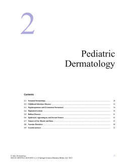
Letter to the Editor
DOI: 10.4274/tjh.2014.0037
Hemophagocytic Lymphohistiocytosis Syndrome
Associated with Gaucher Disease Type 2
Tip 2 Gaucher Hastalığı ile İlişkili Hemafagositik Lenfohistiositozis
Sendromu
Gholamreza Bahoush 1, Ghasem Miri-Aliabad2
1Iran
University of Medical Sciences, Ali Asghar Children’s Hospital, Pediatric Hematology-Oncology, Tehran, Iran
University of Medical Sciences, Children and Adolescent Health Research Center, Department of Pediatric Hematology-Oncology,
Zahedan, Iran
2Zahedan
To the Editor,
We present the case of a 5.5-month-old female infant
hospitalized because of fever, lethargy, pallor, poor feeding,
convulsion, and developmental delay. She was the first
child of the family and her parents were relatives. Physical
examination revealed hepatosplenomegaly, opisthotonus,
and pitting edema. Laboratory studies showed pancytopenia
(WBC: 3500/mm3, Hb: 6 g/dL, Plt: 40.000/mm3),
coagulopathy (PT: 19.4 s, aPTT: 41 s, fibrin degradation
products: 1.8 mg/mL, D-dimer: 4 mg/L), AST of 132 U/L,
ALT of 107 U/L, ALP of 313 U/L, triglycerides of 280 mg/
dL, and albumin of 2.7 g/dL. Analysis of the cerebrospinal
fluid (CSF) revealed lymphocytic pleocytosis. Serum ferritin
level was 1020 ng/mL and fibrinogen level was 150 mg/dL.
Other test results, including renal function tests, electrolytes,
serum and urine amino acid chromatography, TORCH study,
and virology studies (HIV, HCV, HBV, HAV, EBV), were
normal. Blood, urine, stool, CSF, and bone marrow cultures
were all negative. Bone marrow examination revealed
hemophagocytosis caused by macrophages (Figure 1). It
also showed an increase in the number of histiocytic cells,
which was not typical of Gaucher disease. However, dried
blood spot testing showed a drastic reduction of 8.45 pmol/
spot (normal range: 200-2000 pmol/spot in 20 h) in the betaglucosidase enzyme activity and confirmed the diagnosis of
Gaucher disease. Since 6 of the 8 criteria of hemophagocytic
lymphohistiocytosis (HLH) syndrome were observed in the
Figure 1. A) A macrophage that has phagocytosed a band
cell and a lymphocyte B) A macrophage that has engulfed
many normoblast.
patient, she was subjected to treatment with the HLH 2004
Protocol [2], broad-spectrum antibiotics, and anticonvulsant
drugs while supportive measures were also taken. Despite
the aforementioned efforts, the patient’s clinical condition
gradually worsened and she died 10 days after the beginning
of the treatment. It was not possible to study the mutations
associated with primary HLH or to measure the level of sIL2R
and the activity of NK cells. Informed consent was obtained.
This case is important because it is an example of HLH
syndrome associated with Gaucher disease type 2. Gaucher
disease is a lysosomal storage disorder that is caused by the
deficiency of beta-glucocerebrosidase enzyme. Gaucher
disease has 3 types. Type 2, which is known as the acute
infantile form, is characterized with neurological symptoms
and death in early childhood [1]. HLH syndrome is a rare and
rapidly progressive disease that has a primary (familial) and a
Address for Correspondence: Ghasem MIRI-ALİABAD, M.D.,
Zahedan University of Medical Sciences, Children and Adolescent Health Research Center, Department of Pediatric Hematology-Oncology, Zahedan, Iran
Phone: +985413411250 E-mail: [email protected]
Received/Geliş tarihi : January 26, 2014
Accepted/Kabul tarihi : February 10, 2014
307
Turk J Hematol 2014;31:307-308
secondary (acquired) form. If this disorder is left untreated,
it can quickly lead to death [2]. Diagnosis of primary HLH
is confirmed by studying the related genetic mutations, such
as STX11, UNC13D, and PRF1. The secondary type of HLH
also develops after infections or with rheumatologic diseases,
Chediak–Higashi syndrome, Griscelli syndrome, X-linked
lymphoproliferative disease, malignancies, and inborn errors
of metabolism. This potentially fatal disease is caused by
uncontrolled over-production of inflammatory cytokines
by cytotoxic T lymphocytes and histiocytes [2,3]. HLH
syndrome is rarely associated with Gaucher disease. It was
Lee et al. that first found erythrophagocytosis in Gaucher
cells [4]. Hibbs et al. [5] and Bitton et al. [6] also reported
erythrophagocytic activity in Gaucher cells. In another study,
similar clinical symptoms were seen in patients with Gaucher
disease type 2 and patients with HLH [7]. Approximately 15%
of Gaucher cells were found to have abnormal morphologies
or significant erythrophagocytic activity [8]. Sharpe et
al. reported a female infant with Gaucher disease type 2
manifesting as severe HLH [9]. Only that study addressed
the association of these 2 diseases, while other reports have
only found erythrophagocytosis in Gaucher cells.
Although it was not possible to study the genetic mutations
of HLH in this patient, on the basis of the familial background
of her parents, the progressive trend of her disease, and no
response of disease to treatment, it can be concluded that the
patient possibly suffered from primary HLH. Measurement
of the activity of beta-glucosidase enzyme confirmed the
diagnosis of Gaucher disease. Due to the presence of severe
and rapidly progressive neurologic symptoms, the patient
was diagnosed with Gaucher disease type 2. These 2 diseases
have many overlapping clinical symptoms. Therefore, in
similar occasions, a careful evaluation of the presence of
storage diseases such as Gaucher disease is recommended.
Conflict of Interest Statement
The authors of this paper have no conflicts of interest,
including specific financial interests, relationships, and/
or affiliations relevant to the subject matter or materials
included.
308
Gholamreza B, et al: Hemophagocytic Syndrome Associated with Gaucher Disease
Key Words: Gaucher disease, Hemophagocytic
lymphohistiocytosis, Hepatosplenomegaly
Anahtar Sözcükler: Gaucher hastalığı, Hemafagositik
lenfohistiositozis, Hepatosplenomegali
References
1. Oski FA, Brugnara C, Nathan DG. Storage disease of the
reticuloendothelial system. In: d’Azzo A, Kolodny EH,
Bonton E, Annunziata I. Nathan and Oski’s Hematology
of Infancy and Childhood. 7th ed. Philadelphia, Saunders,
2009.
2. Henter JI, Horne A, Aricó M, Egeler RM, Filipovich AH,
Imashuku S, Ladisch S, McClain K, Webb D, Winiarski J,
Janka G. HLH-2004: diagnostic and therapeutic guidelines
for hemophagocytic lymphohistiocytosis. Pediatr Blood
Cancer 2007;48:124-131.
3. Kliegman R, Stanton B, St Geme J, Schor NF, Behrman RE
(eds). Nelson Text Book of Pediatrics. 19th ed. Philadelphia,
Saunders Elsevier, 2011.
4. Lee RE, Balcerzak SP, Westerman MP. Gaucher’s disease: a
morphologic study and measurements of iron metabolism.
Am J Med 1967;42:891-898.
5. Hibbs RG, Ferrans VJ, Cipriano PR, Tardiff KJ. A
histochemical and electron microscopic study of Gaucher
cells. Arch Pathol 1970;89:137-153.
6. Bitton A, Etzell J, Grenert JP, Wang E. Erythrophagocytosis
in Gaucher cells. Arch Pathol Lab Med 2004;128:11911192.
7. Mignot C, Doummar D, Maire I, De Villemeur TB; French
Type 2 Gaucher Disease Study Group. Type 2 Gaucher
disease: 15 new cases and review of the literature. Brain Dev
2006;28:39-48.
8. Machaczka M, Klimkowska M, Regenthal S, Hagglund H.
Gaucher disease with foamy transformed macrophages and
erythrophagocytic activity. J Inherit Metab Dis 2011;34:233235.
9. Sharpe LR, Ancliff P, Amrolia P, Gilmour KC, Vellodi A.
Type II Gaucher disease manifesting as haemophagocytic
lymphohistiocytosis. J Inherit Metab Dis 2009;32 Suppl
1:107-110.
© Copyright 2026 Paperzz