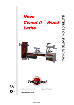
Reproductive BioMedicine Online (2014) xxx, xxx– xxx
www.sciencedirect.com
www.rbmonline.com
ARTICLE
Rapid warming increases survival
of slow-frozen sibling oocytes: a step towards
a single warming procedure irrespective
of the freezing protocol?
Lodovico Parmegiani a,*, Carla Tatone b, Graciela Estela Cognigni a, Silvia
Bernardi a, Enzo Troilo a, Alessandra Arnone a, Antonio Manuel Maccarini a,
Giovanna Di Emidio b, Maurizio Vitti b, Marco Filicori a
a
Reproductive Medicine Unit, GynePro Medical Centers, Bologna, Italy;
Environmental Sciences, University of L’Aquila, L’Aquila, Italy
b
Department of Life, Health, and
* Corresponding author. E-mail address: [email protected] (L Parmegiani).
After completing a Master’s degree in biology in 1996 at the University of Bologna, Italy, Lodovico Parmegiani
completed his postgraduate specialization in biochemistry and clinical chemistry in 2000 at the University of
Modena and Reggio Emilia, Italy. In 2008, he received certification as a senior clinical embryologist from the
European Society for Human Reproduction and Embryology. In his career he has developed and optimized
devices and methods for clinical embryology. His current research interests are cryobiology, gamete selection
and micromanipulation. He is currently the Laboratory Director at the Reproductive Medicine Unit, GynePro
Medical Centers in Bologna, Italy.
Abstract Nowadays, human oocytes/embryos are cryopreserved via slow freezing or vitrification. The aim of this study was to eval-
uate a rapid warming protocol for slow-frozen human oocytes based on the standard warming procedure for vitrification. This was a
prospective study on 216 sibling oocytes randomized for either conventional rapid thawing or rapid warming with vitrification warming solution. The primary endpoint was morphological assessment of survival at 2 h. Surviving oocytes were divided into two subgroups: (i) parthenogenetically activated; and (ii) fixed and observed for spindle/chromosome configuration. Secondary
endpoints were parthenogenetic development and spindle/metaphase configuration. Survival rate with rapid warming was higher
(92/102, 90.2%) than with rapid thawing (85/114, 74.6%; P = 0.005), and after 3 d of culture the rapidly warmed parthenotes had
more blastomeres compared with those rapidly thawed (P = 0.042). Meiotic spindle and chromosomal configuration were not significantly influenced by rapid warming or rapid thawing. The finding of this study allows IVF centres to increase the efficiency of oocyte
slow freezing, enabling survival rates comparable to vitrification protocols, and potentially to optimize costs by using the same
warming protocol for both slow-frozen and vitrified reproductive cells. RBMOnline
ª 2014, Reproductive Healthcare Ltd. Published by Elsevier Ltd. All rights reserved.
KEYWORDS: oocyte slow freezing, oocyte vitrification, parthenogenetic activation, rapid thawing, universal warming protocol, warming
http://dx.doi.org/10.1016/j.rbmo.2014.01.015
1472-6483/ª 2014, Reproductive Healthcare Ltd. Published by Elsevier Ltd. All rights reserved.
Please cite this article in press as: Parmegiani, L et al. Rapid warming increases survival of slow-frozen sibling oocytes: a step towards a single
warming procedure irrespective of the freezing protocol?. Reproductive BioMedicine Online (2014), http://dx.doi.org/10.1016/j.rbmo.2014.01.015
2
L Parmegiani et al.
Introduction
Materials and methods
Nowadays, in assisted reproduction laboratories, human
oocytes and embryos are cryopreserved by two main methods: vitrification or slow freezing.
During vitrification, the cells are converted to a glassy
state without ice-crystal formation by using a viscous
medium with a very high concentration of cryoprotectants
(Rall and Fahy, 1985; Yavin and Arav, 2007). However, during slow freezing due to the progressive permeation of
some cryoprotectants as the ice crystals form in the cryopreservation solution, a glassy/vitrified state is also
obtained within the cell throughout the cooling procedure
(Vanderzwalmen et al., 2013). For this reason, since both
vitrified and slow-frozen oocytes have a vitrified cytoplasm, it can be postulated that the same warming protocol can be used for both. Furthermore, since for any given
concentration of cryoprotectant, the warming rates are
much faster than the critical cooling rates (Fahy et al.,
1987), the minimal concentration of cryoprotectant to prevent crystallization during warming must be higher than
during cooling (Vanderzwalmen et al., 2012). In fact, in
most vitrification protocols or commercial kits, the cryoprotectant concentration in the first warming solution is
approximately 1 mol/l (The Alpha Consensus Meeting,
2012), which is higher than the cryoprotectant concentration in both the freezing solution and the first thawing
solution for rapid thawing of slow-frozen oocytes or
embryos (e.g. for oocytes: 0.2–0.3 mol/l in freezing
solution, 0.3–0.5 mol/l in thawing solution; Bianchi
et al., 2007; Boldt et al., 2006; Fabbri et al., 2001; Parmegiani et al., 2008a). This suggests that vitrification
warming solution could be used for thawing slow-frozen
oocytes/embryos.
For a number of reasons, but most of all for the guaranteed high survival rate, vitrification is increasingly used
worldwide, especially for oocytes (Edgar and Gook, 2012).
Nevertheless, to date, perhaps millions of slow-frozen
oocytes/embryos have already been stored in IVF cryobanks. Furthermore, IVF centres that have completely
switched their cryopreservation programme from slow
freezing to vitrification may still receive slow-frozen
oocytes/embryos from other centres, cryopreserved by
various different protocols. Nowadays, regulations recommend the use of FDA/CE-marked thawing media approved
for human IVF; because the shelf life of these media is
usually short (some months), it becomes expensive to
keep available the appropriate thawing solution for any
slow freezing protocol. In this scenario, the possibility
of using a ‘universal medium’ to thaw any cell/tissue irrespective of the freezing protocol may simplify the management of thawing procedures. For all these reasons,
the aim of this study is to evaluate a rapid warming protocol for slow-frozen human oocytes based on the standard warming procedure for vitrification, to optimize
the survival rate and reduce costs by using the same solutions for both slow freezing and vitrification warming. The
rapid warming protocol proposed in this study is potentially applicable also to slow-frozen embryos at any stage
of cleavage.
Study population
Since April 2004, all patients undergoing an IVF treatment at
the GynePro Medical Centres, Bologna, Italy have had the
option of cryopreserving their surplus oocytes not inseminated during the fresh cycle. This study was performed
between December 2012 and January 2013 on 216 sibling
oocytes obtained from 40 patients and cryopreserved via
slow freezing from April 2004 to November 2008. Patient
age (mean ± standard error) at time of freezing was
33.70 ± 0.77 years. The number of oocytes thawed/warmed
per patient was 5.40 ± 0.53.
Ethical approval
All the women included in this cryopreservation programme
were informed of the procedure and written consent was
obtained from each at the time of oocyte freezing. The
written consent included the option of oocyte donation
for research purposes before destruction, should the patient
decide to discontinue the cryostorage. A further agreement
was obtained from any patient before starting the study.
This study was approved by the Institutional Review Board
of the clinic (reference no. 15.10.2012, approved 3 December 2012).
Ovarian stimulation, oocyte retrieval and selection,
study design, and endpoints
Ovarian stimulation and oocyte retrieval and selection
before cryopreservation were performed as previously
described (Parmegiani et al., 2008a). For this study,
slow-frozen oocytes were randomized for either conventional rapid thawing or rapid warming via vitrification
warming solutions. Randomization was performed by a
different embryologist to the operator who performed
oocyte thawing/warming using a specific software tool
(http://www.randomizer.org): for example, for six straws
containing slow-frozen oocytes, a randomized set, 1, 2, 6,
was generated, so oocytes in straws 1, 2 and 6 were thawed
by conventional rapid thawing and oocytes in straws 3, 4 and
5 were rapidly warmed. The primary outcome measure was
morphological assessment of survival at 2 h. Secondary outcome measures were parthenogenetic development and
spindle/metaphase configuration.
Cryopreservation
The cryopreservation protocol consisted of a slow-cooling
method (Fabbri et al., 2001). Oocyte freezing solutions
(OocyteFreeze; Origio, Denmark) contained Dulbecco’s
phosphate-buffered saline (PBS) supplemented with human
serum albumin, alpha- and beta-globulins and 1,2-propanediol (PROH) and sucrose as cryoprotectants.
After washing in a PBS solution (Vial 1–OocyteFreeze;
Origio), oocytes were equilibrated for 10 min at room
temperature in 1.5 mol/l 1,2-PROH (Vial 2–OocyteFreeze,
Please cite this article in press as: Parmegiani, L et al. Rapid warming increases survival of slow-frozen sibling oocytes: a step towards a single
warming procedure irrespective of the freezing protocol?. Reproductive BioMedicine Online (2014), http://dx.doi.org/10.1016/j.rbmo.2014.01.015
Rapid warming of slow-frozen human oocytes
Origio) and then transferred into the loading solution of
1.5 mol/l 1,2-PROH and 0.3 mol/l sucrose (Vial 3–OocyteFreeze; rigio). Between one and three oocytes were loaded
in plastic straws (Paillette Cristal 133 mm; Cryo Bio System,
France) and transferred into an automated biological vertical freezer (Kryo 360–1.7; Planer, UK). The cooling process
was initiated, reducing the chamber temperature from 20C
to 7C at a rate of 2C/min. Ice nucleation was induced
manually at 7C (seeding). After a hold time of 10 min at
7C, the straws were cooled slowly to 30C at a rate of
0.3C/min and then rapidly to 150C at a rate of 50C/min.
After 10–12 min at stabilization temperature, the straws
were transferred into liquid nitrogen and stored for later
use.
Rapid thawing
The straws were air-warmed for 30 s and then immersed in a
30C water bath for 40 s. The cryoprotectant was removed
at room temperature by stepwise dilution of PROH in the
thawing solutions: the contents of the straws were expelled
in 1.0 mol/l 1,2-PROH and 0.3 mol/l sucrose solution (Vial
1–OocyteThaw) and the oocytes were equilibrated for
5 min. The oocytes were then transferred into 0.5 mol/l
1,2-PROH and 0.3 mol/l sucrose solution (Vial 2–OocyteThaw) for 5 min and then into 0.3 mol/l sucrose solution
(Vial 3–OocyteThaw) for 10 min before final dilution in
PBS solution (Vial 4–OocyteThaw) for 20 min (10 min at
room temperature and 10 min at 37C). The oocytes were
finally cultured at 37C in ISM1 medium (Origio) in an atmosphere of 6% CO2 in air for 2 h before assessment of survival.
Rapid warming
The straws were air-warmed for 10 s and then immersed in a
30C water bath until the external ice had melted (but not
for longer than 10 s because this is a critical step). Each
straw was opened and the content was immediately
expelled in 4 ml warming solution containing 1 mol/l
sucrose at 37C (Sucrose Warming Solution; Sage, USA).
Then, the oocytes were incubated at room temperature
for 3 min first in 0.5 mol/l Sucrose Warming Solution and
subsequently in 0.25 mol/l of 1:1 (v/v) of 0.5 mol/l Sucrose
Warming Solution and MOPS Solution (Sage) and finally
washed for 4 min in basic medium (MOPS Solution) before
culture. The oocytes were finally cultured at 37C in ISM1
medium in an atmosphere of 6% CO2 in air for 2 h before
assessment of survival.
Survival evaluation and oocyte destiny
After a 2-h post-thaw/post-warming culture, survival was
evaluated by inverted microscopy with Hoffman modulation
contrast. Oocytes were considered to have survived in the
absence of negative characteristics: dark or contracted
ooplasm, vacuolization, cytoplasmic leakage, abnormal perivitelline space, cracked zona pellucida. Some of these surviving oocytes were then studied either for their
developmental competence or for their spindle/metaphase
configuration, whereas 33 sibling oocytes taken from seven
patients were observed only for survival. The remaining
3
oocytes were randomly assigned to parthenogenesis or
spindle study by a sealed envelope procedure: 96 sibling
oocytes taken from 26 patients were parthenogenetically
activated and 48 sibling oocytes taken from seven patients
were fixed and observed for spindle/chromosome
configuration.
Parthenogenetic activation
Two hours after thawing/warming, oocytes were exposed to
calcium-ionophore (GM508 Cult Active Mediuml Gynemed,
Germany) for 15 min at 37C with 6% CO2 in air then washed
twice and subsequently incubated in 2 mmol/l 6-dimethylaminopurine (Sigma–Aldrich, Italy) in ISM1 medium for
3 h at 37C with 6% CO2 in air. Oocytes were then washed
in fresh ISM1 medium, placed in 50-ll microdrops of the
same medium under oil (Ferticult Mineral oil; Fertipro, Belgium) and cultured at 37C with 6% CO2 in air. After 18–20 h
(with exposure to calcium-ionophore as time 0) oocytes
were evaluated for signs of activation. Oocytes showing
one enlarged pronucleus and no extrusion of the second
polar body were considered activated (Loi et al., 1998). Parthenotes were kept in culture in ISM1 for 3 d, followed by a
further 2 d of culture in Blast Assist Medium (Origio). Parthenote development was assessed as previously described
(Paffoni et al., 2007), by counting the number of blastomeres at 42–44 h (day 2) and at 66–68 h (day 3) after activation; blastocyst formation was evaluated at 118–120 h
(day 5) after activation.
Assessment of meiotic spindle and chromosome
configuration
Two hours after thawing/warming, surviving oocytes were
fixed in 4% formaldehyde and used for the study of spindle
and chromatin organization as previously described (Antczak and Van Blerkom, 1997; Cobo et al., 2008a; Messinger
and Albertini, 1991) but with a slightly modified procedure.
Briefly, the oocytes were washed with a solution of PBS (Life
Technologies, USA) with 3 mg/ml polyvinylpyrrolidone
(Sigma–Aldrich) and 0.01% Tween 20 (Bio-Rad Laboratories,
USA) and was successively treated with pronase type XIV
(Sigma–Aldrich) for 20 min at 37C for zona pellucida
removal. Permeabilization was performed with a PBS/PVP
solution
supplemented
with
0.5%
Triton-X-100
(Sigma–Aldrich) for 1 h at 37C, followed by washing in
blocking solution consisting of PBS/PVP solution supplemented with 1.5% bovine serum albumin (Sigma–Aldrich)
for 2 h at 37C. Oocytes were further processed for immunostaining through overnight incubation with a monoclonal
primary anti-a tubulin antibody (Santa Cruz Biotechnology,
USA) diluted 1:100 in blocking solution, followed by incubation for 2 h at room temperature with secondary antibodies
conjugated with Alexa 488 (Life Technologies) diluted 1:500
in blocking solution; after each antibody incubation,
oocytes were washed in blocking solution. Chromatin
staining was performed by using 5 lg/ml Hoechst 33342
(Sigma–Aldrich) for 8 min at room temperature. After
washing, the oocytes were mounted with a mixture of glycerol/PBS (I:I, v/v) and analysed by confocal microscopy (TCS
SP5 II; Leica Microsystems, Germany).
Please cite this article in press as: Parmegiani, L et al. Rapid warming increases survival of slow-frozen sibling oocytes: a step towards a single
warming procedure irrespective of the freezing protocol?. Reproductive BioMedicine Online (2014), http://dx.doi.org/10.1016/j.rbmo.2014.01.015
4
Meiotic spindle configurations of the oocytes were classified as follows: (i) normal, if microtubules formed two
opposite poles; (ii) slightly aberrant, if microtubule structures displayed reduced dimensions of the spindle and lost
normal poles; (iii) aberrant, if disorganized microtubule
patterns were observed; and (iv) absent, if the spindle
was not found. Based on chromosomal distribution in the
metaphase plate, oocytes were classified as follows: (i) normal, in the case of normal distribution; (ii) misaligned, if
the plate showed some disorganization, with one or two
chromosomes separated from the plate; (iii) displaced, in
the case of scattered and decondensed chromosomes or disorganized chromosomal plate; and (iv) activated, if sister
chromatid separation was observed.
Statistical analysis
Calculation of sample size was based on this group’s previous study performed on 437 slow-frozen oocytes, with a survival rate after thawing of 75% (Parmegiani et al., 2008a).
This analysis revealed that at least 174 oocytes would be
necessary to obtain a confidence level of 95% and a margin
of error (confidence interval) of 5%. For this reason, the
study was performed on more than 200 oocytes. Continuous
variables are presented as mean ± standard error. Categorical variables are presented as percentage. Normality of
distribution of continuous variables was assessed with a
Kolmogorov–Smirnov test (with Lillefor correction).
Between-group differences of normally distributed continuous variables were assessed with parametric statistic
(Student’s t-test), whereas non parametric statistics
(Mann–Whitney rank sum test) were employed when the
normality test was not passed. Between-group differences
in noncontinuous variables were assessed using chi-squared
test with Yates correction if needed or with Fisher’s exact
test. Difference was considered significant when a P-value
was <0.05.
Results
Survival
The mean number of oocytes warmed/thawed per group
was comparable: 2.55 ± 0.29 in the rapid warming group
versus 2.85 ± 0.30 in the rapid thawing group. Survival rate
was 90.2% (92/102) in the rapid warming group versus 74.6%
(85/114) in the rapid thawing group (P = 0.005).
An interesting observation was that, during warming,
slow-frozen oocytes in the rapid warming group displayed
the behaviour typical of vitrified oocytes: as soon as the
oocytes were expelled from the straw, they tended to float
and appear vitreous in 1 mol/l sucrose solution; they subsequently shrank in 0.5 mol/l sucrose, and then in 0.25 mol/l
sucrose and the washing solution they progressively recovered their original shape. (Parmegiani in The Alpha Consensus Meeting, 2012). This similarity is due to the ideal sucrose
concentration (1 mol/l) and temperature (37C) in the first
warming solution, which both determine better osmolarity
conditions for propanediol dilution compared with the conventional slow-freezing thawing solution used in the rapid
thawing group (0.3 mol/l sucrose, 1 mol/l propanediol;
L Parmegiani et al.
room temperature). For this reason it was critical to expel
the oocytes into the first warming solution at 37C as soon
as the external ice around the straw had melted to avoid
or minimize fluid exchanges between oocytes and melted
holding medium inside the straw. Rapidly warmed oocyte
were expelled in 1 mol/l sucrose at 37C still shrunken and
rehydration occurred within 1 min (Figure 1A, B); subsequently, the oocytes went through the same recovery process as vitrified oocytes (Figure 1C–E). Full recovery of
the original cell shape is obtained after 10 min from expulsion from the straw, compared with 30 min for rapidly
thawed oocytes (Figure 1F–J). The rapid warming in
1 mol/l sucrose at 37C (which may better protect against
the risk of water recrystallization) and the faster dilution
of intracellular cryoprotectants may both contribute to
the significantly better performance of rapidly warmed
oocytes compared with rapidly thawed oocytes.
Parthenogenetic development
A total of 96 sibling oocytes from 26 patients were activated
for parthenogenesis. The patient age was 33.69 ±
0.94 years. In the rapid warming and the rapid thawing
groups, the parthenogenetic activation rate was comparable (90.4% versus 81.8%, respectively). Parthenogenetic
development was comparable at day 2 (2.67 ± 0.42 blastomeres versus 2.00 ± 0.00 blastomeres, respectively); however, at day 3, the rapidly warmed parthenotes had
5.00 ± 0.47 blastomeres versus 3.56 ± 0.26 blastomeres for
the rapidly thawed parthenotes (P = 0.042). Blastocyst
development was comparable (10.6% versus 5.6%, respectively). The results for parthenogenetic development are
summarized in Table 1.
Spindle/metaphase configuration
A total of 48 sibling oocytes taken from seven patients were
fixed and observed for meiotic spindle/metaphase configuration. More specifically, in 35 sibling oocytes taken from
six patients, the meiotic spindle together with the metaphase asset was studied, and in 13 sibling oocytes taken
from one patient, only the metaphase asset was studied.
The mean patient age was 34.17 ± 2.50 years.
In the rapid warming and the rapid thawing groups, the
meiotic spindle was present in the majority of the oocytes
observed (94.4% and 88.2%, respectively; not significant).
No significant differences were observed between the two
groups in normal and abnormal spindle and metaphase plate
configurations. Nevertheless, a better trend in spindle restoration and normal spindle/chromosomal pattern was
observed in the rapid warming group. The results are summarized in Table 2.
Discussion
Human reproductive cells can be cryopreserved either via
vitrification or via slow freezing. Especially for embryos
and oocytes, vitrification is increasingly used due to the high
survival rate obtainable, and this procedure is overtaking
slow freezing in human assisted reproduction treatment
(Edgar and Gook, 2012).
Please cite this article in press as: Parmegiani, L et al. Rapid warming increases survival of slow-frozen sibling oocytes: a step towards a single
warming procedure irrespective of the freezing protocol?. Reproductive BioMedicine Online (2014), http://dx.doi.org/10.1016/j.rbmo.2014.01.015
Rapid warming of slow-frozen human oocytes
5
Figure 1 Rapid warming and rapid thawing of slow-frozen sibling oocytes. Slow-frozen sibling oocytes from the same patient were
warmed via rapid warming or via a conventional rapid thawing procedure. (A–E) A rapidly warmed oocyte was expelled into 1 mol/l
sucrose at 37C still shrunken (A) and its rehydration occurred within 1 min (B); then, it was moved to 0.5 mol/l sucrose, where it
shrank again (C); subsequently, in 0.25 mol/l sucrose (D) and washing solution (E), the oocyte completely recovered its original cell
shape. (F–J) A rapidly thawed oocyte was expelled in 0.3 mol/l sucrose at room temperature when intra/extracellular fluids (water
and cryoprotectants) had already been exchanged inside the cryostraw; for this reason, its shape was already partially recovered (F,
G); the oocyte remained partially rehydrated throughout the third step of thawing, in 0.3 mol/l sucrose solution (H–I) and fully
recovered its original shape only in the washing solution, at 30 min after its release from the straw (J).
It is important to note that, during both these procedures, despite the different behaviour of the cryopreservation medium when cooled below the solidification point
(vitrification medium appears ‘glassy’ whereas slow freezing
medium is ‘iced’ due to the presence of ice crystals), the
cell cytoplasm itself is converted from liquid to solid state
without formation of ice crystals. In other words, slow
freezing is just a method of intracellular vitrification with
ice being present in the extracellular compartment (Katkov
et al., 2012). In fact, intracellular formation of ice crystals
has a detrimental effect on cellular organelles and membranes and must therefore be avoided during both cooling
and thawing phases (Leibo et al., 1978; Luyet, 1937; Mazur
et al., 2005). This is ensured in different ways during
vitrification or slow freezing, but at the end of both these
procedures the cell is vitrified. ‘Vitrification’ is defined as
the conversion of a superviscous liquid into a glassy state
when it is cooled below its glass transition temperature
Please cite this article in press as: Parmegiani, L et al. Rapid warming increases survival of slow-frozen sibling oocytes: a step towards a single
warming procedure irrespective of the freezing protocol?. Reproductive BioMedicine Online (2014), http://dx.doi.org/10.1016/j.rbmo.2014.01.015
6
L Parmegiani et al.
Table 1
Parthenogenetic development.
Parameter
Rapid warming (n = 58)
Rapid thawing (n = 60)
P value
Survival rate
Activated oocytes
Parthenogenetic activation rate
Blastomeres on day 2 (42–44 h)
Blastomeres one day 3 (66–68 h)
Blastocyst rate
52 (89.7)
52 (2.00 ± 0.16)
47 (90.4)
2.67 ± 0.42
5.00 ± 0.47
5 (10.6)
44 (73.3)
44 (1.69 ± 0.22)
36 (81.8)
2.00 ± 0.00
3.56 ± 0.26
2 (5.6)
0.041
NS
NS
NS
0.042
NS
Values are n (%), n (mean ± standard error) or mean ± standard error.
NS = not statistically significant.
Table 2
Spindle/metaphase configuration.
Parameter
Rapid warming
Rapid thawing
Meiotic spindle
No. of oocytes observed
Oocytes displaying meiotic spindle
at 2 h from thawing/warming
xxx
18
17 (94.4)
17
15 (88.2)
x
xxxx
Type of meiotic spindle
Normal
Slightly aberrant
Aberrant
Absent
xxx
8 (44.4)
4 (22.2)
5 (27.8)
1 (5.6)
xxxx
xxx
23
10 (43.5)
5 (21.7)
8 (34.8)
0 (0)
x
Chromosomal distribution
No. of oocytes observed
Normal
Misaligned
Displaced
Activated
4
6
5
2
(23.5)
(35.3)
(29.4)
(11.8)
25
9 (36.0)
6 (24.0)
8 (32.0)
2 (8.0)
Values are n (%) unless otherwise stated. There were no statistically significant differences between the groups.
(Fahy et al., 1987). Any material can vitrify as long as the
cooling rate is fast enough to prevent the formation of
organized crystals; the probability of obtaining a glassy
state depends on the relationship between viscosity, cooling
rate and sample volume (Vajta and Kuwayama, 2006; Yavin
and Arav, 2007).
During vitrification for cryopreservation of reproductive
cells, cells are converted to the glassy state by using a viscous medium with very high concentration of cryoprotectants (Rall and Fahy, 1985). Two types of cryoprotectants
are involved in cell cryopreservation: intracellular, which
can permeate the cell membrane (e.g. ethylene glycol,
propanediol, dimethylsulphoxide) and extracellular, which
cannot permeate inside the cell (e.g. sucrose). During vitrification, high concentrations of cryoprotectants in the holding medium (about 4.5–6.5 mol/l for intracellular and
about 0.5–1 mol/l for extracellular cryoprotectants) encapsulate the cell in a vitrifying sheath and, at the same time,
determine a viscous environment inside the cell (Vanderzwalmen et al., 2013). The intracellular cryoprotectants permeate the cell membrane and replace the inner water
which has drained out from the cell due to the very high
osmotic gradient generated during the consecutive steps
of vitrification (in equilibration and vitrification solutions).
Subsequently the cells (loaded in specifically designed carriers which allow fast cooling rates) vitrify by direct contact
with liquid nitrogen or exposure to nitrogen vapours
(Kuwayama et al., 2005; Parmegiani, 2011; Vajta and
Kuwayama, 2006; Yavin and Arav, 2007).
On the other hand, during slow freezing, the cryoprotectant concentration in the cryopreservation medium is lower
than in the vitrification medium (about 1–2 mol/l for intracellular and about 0.1–0.3 mol/l for extracellular cryoprotectants). During slow freezing, the cells are exposed to
this moderate concentration of extracellular cryoprotectants which establish an osmotic gradient to facilitate a
progressive movement of water out of the cell (cell dehydration), while intracellular cryoprotectants enter the cell,
replacing the intracellular water drawn out. At the end of
this dehydration process, intracellular cryoprotectants are
estimated to be approximately 1.5–1.7 mol/l (McGrath,
2009; Son and Tan, 2009). However, it must be pointed
out that, at the moment of cytoplasm solidification, intracellular cryoprotectant concentration is postulated to be
Please cite this article in press as: Parmegiani, L et al. Rapid warming increases survival of slow-frozen sibling oocytes: a step towards a single
warming procedure irrespective of the freezing protocol?. Reproductive BioMedicine Online (2014), http://dx.doi.org/10.1016/j.rbmo.2014.01.015
Rapid warming of slow-frozen human oocytes
higher than this. This is explained by the ‘solution effect’:
as the solution is cooled to lower and lower subzero temperatures, more of the water freezes and the solution becomes
increasingly concentrated (Leibo, 2008). Thus, as ice crystals form and the osmolarity rises in the cryopreservation
solution, intracellular cryoprotectants continue to progressively permeate the cell and further dehydration, shrinkage
and increase in intracellular solute concentration occur
(Leibo and Pool, 2011; Son and Tan, 2009). This happens
in the programmable freezer chamber during the seeding
of ice nucleation along the length of the straw/vial containing the cell; after this, the viscous cell cytoplasm is supercooled and converted to a glassy state (McGrath, 2009).
By using computerized predictive models, McGrath (2009)
calculated an intracellular cryoprotectant concentration
(1,2-propanediol) of 2.7 mol/l for human oocytes slow
cooled via a 0.3 mol/l sucrose protocol (Fabbri et al., 2001)
after 10 min holding time after ice nucleation seeding. At
this point the oocyte is postulated to be close to equilibrium, the freezer chamber temperature slightly decreases
at 0.3C/min from 7C to 20C and cell vitrification
occurs. In a recent paper, Vanderzwalmen et al. (2013)
demonstrated that cryoprotectant concentrations at the
moment of solidification inside slow-frozen mouse oocytes
are actually higher than inside vitrified oocytes (approximately 2.14 mol/l). This may be one of the reasons why
slow-frozen oocytes/embryos can survive the low thawing
rates (about 600C/min when straws/vials are air warmed
at 22–25C) during conventional slow freeze–thaw protocols. In fact, warming speeds play an even more important
role for cell survival than cooling rates (Seki and Mazur,
2008, 2009). This is well accepted in the case of vitrification, where a high warming speed (about 20,000C/min) is
critical to prevent cell lysis; if the warming speed is not fast
enough, the supercooled liquid is rapidly transformed into
ice crystals because intracellular cryoprotectant concentration is too low to prevent recrystallization (Vanderzwalmen
et al., 2012, 2013).
During the thawing/warming process, extracellular cryoprotectants are used to generate an osmotic gradient which
favours cell rehydration and drains intracellular cryoprotectants. Since for any given concentration of cryoprotectants,
the warming rates are much faster than the critical cooling
rates (Fahy et al., 1987), the minimum concentration of
cryoprotectant to prevent crystallization during warming
must be higher than during cooling (Vanderzwalmen et al.,
2012). This means that, when thawing slow-frozen
oocytes/embryos, vitrification warming solution could be
used to optimize cell survival because extracellular cryoprotectant concentration in the first warming solution in any
vitrification protocol is usually 1 mol/l, which is higher than
in slow-freezing thawing solution (e.g. 0.3–0.5 mol/l for
human oocytes). For this reason, this high extracellular
cryoprotectant concentration in the medium should better
protect against the risk of the recrystallization of the cellular water during warming. This was the rationale of the
present study, which tested the vitrification warming protocol on slow-frozen human oocytes, which are well known to
be the most sensitive reproductive cells during cryopreservation/thawing process. Furthermore, predictive models
have suggested that there may be opportunities for optimizing the thawing protocol for oocytes slow frozen with
7
0.3 mol/l sucrose (McGrath, 2009). As far as is known, this
study is the first to investigate the possibility of using vitrification warming solution for thawing human oocytes.
Interestingly, the survival rate was significantly improved
when the slow-frozen oocytes were rapidly warmed (90.6%)
rather than thawed with the conventional procedure
(75.0%). In the rapid thawing group, the survival rate was
comparable to the survival previously reported in clinical
studies on oocytes slow frozen with 0.3 or 0.2 mol/l sucrose
(Bianchi et al., 2012; Parmegiani et al., 2008a,b, 2009a,b);
in fact, evidence from widespread practice suggests that a
plateau has been reached at 70–80% with currently available slow freezing/rapid thawing methodology (Edgar and
Gook, 2012; Gook and Edgar, 2007). On the other hand,
the rapidly warmed oocyte survival rate in the current study
(about 90%) is comparable with survival obtained in the
majority of vitrification studies (Edgar and Gook, 2012;
Kuwayama et al., 2005), particularly in those performed
on infertile patients (Parmegiani et al., 2011; Practice Committees of American Society for Reproductive Medicine and
Society for Assisted Reproductive Technology, 2013; Rienzi
et al., 2010; The Alpha Consensus Meeting, 2012).
Furthermore, rapidly warmed oocytes showed a better
development after parthenogenetic activation. Parthenogenesis is a tool for the preclinical evaluation of experimental procedures, because the developmental competence of
activated oocytes is similar to fertilized oocytes (Paffoni
et al., 2007). The mean number of blastomeres in day 3
was significantly higher in rapid warming than in rapidly
thawed parthenotes, and this was comparable with that of
parthenotes derived from fresh oocytes in the study by Paffoni et al. (2007), as was the blastulation rate (about 10%).
These findings seem to demonstrate that it is possible to
increase the efficiency of oocyte slow freezing simply by
improving the thawing procedure.
This study also analysed the oocytes by confocal microscopy in order to observe the meiotic spindle rearrangement
dynamics (Bromfield et al., 2009). The human metaphase-II
oocyte has its chromosomes equatorially located on a
spindle, a temporary and dynamic structure of microtubules
which is very sensitive to cooling (Pickering et al., 1990). It
is already known that prolonged chilling at temperatures
about 0C is highly detrimental and causes oocyte meiotic
spindle disassembly (Zenzes et al., 2001). However, in
oocytes cryopreserved via slow freezing, the spindle should
be protected by intracellular cryoprotectants (George
et al., 1996; Gook et al., 1993; Van der Elst et al., 1988;
Yang et al., 2010). Nevertheless, spindle alterations and disassembly have been displayed during the rapid thawing procedure (Coticchio et al., 2006; Rojas et al., 2004; Wu et al.,
2006). In studies with polarization microscopy, oocyte spindles were identified immediately after thawing, disappeared during the subsequent removal of cryoprotectants
and finally reappeared after 3–5 h of culture (Bianchi
et al., 2005; Rienzi et al., 2004). On the contrary, during vitrification protocols, the meiotic spindle and chromosome
alignment are both maintained throughout the processes
of vitrification, warming and post warming (Chang et al.,
2011; Larman et al., 2007). Through the use of a vitrification
warming protocol on slow-frozen oocytes, the present study
might give interesting information on the mechanism of
spindle disruption/recovery during slow freezing. Thus,
Please cite this article in press as: Parmegiani, L et al. Rapid warming increases survival of slow-frozen sibling oocytes: a step towards a single
warming procedure irrespective of the freezing protocol?. Reproductive BioMedicine Online (2014), http://dx.doi.org/10.1016/j.rbmo.2014.01.015
8
spindle and chromosome dynamics were studied at 2 h from
thawing/warming in order to discover if different protocols
might influence spindle recovery. The presence of spindles
were observed in the majority of the oocytes; this is in
agreement with previous studies showing time-dependant
spindle repolymerization (Bianchi et al., 2005; Mandelbaum
et al., 2004; Pickering et al., 1990; Rienzi et al., 2004). Furthermore, both normal spindle and chromosomal configuration were comparable between the two study groups
(Table 2). Although slightly better figures for spindle presence and normal spindle/chromosome pattern may suggest
a better dynamic in the rapid warming group, the spindle
repolymerization does not seem to be influenced by the
choice of thawing/warming protocol, and it is at least clear
that the quick removal of intracellular cryoprotectants during rapid warming does not negatively affect meiotic spindle
reformation.
Nowadays, human oocyte cryopreservation has moved
from bench-to-bedside and it is no longer considered an
experimental technique but a valuable clinical tool (ASRM
Practice Committee, 2012); this is due particularly to studies on vitrification demonstrating that vitrified oocytes perform as well as their fresh counterparts (Cobo et al., 2008b;
Parmegiani et al., 2011; Rienzi et al., 2010). The present
study demonstrates that it is possible to increase the survival of slow-frozen oocytes by changing the thawing protocol and to potentially obtain results comparable to
vitrification; furthermore, this study demonstrates that
the fast removal of intracellular cryoprotectants during
rapid warming does not negatively affect meiotic spindle
reformation. Even though oocyte slow freezing has been
optimized in the last 5 years (Bianchi et al., 2007, 2012; Parmegiani et al., 2008a, 2009a), the oocyte still remains the
most fragile human cell with the highest risk of ice crystal
formation and cryoinjury during freezing/thawing, due to
an unfavourable nucleus/cytoplasm ratio and elevated
intracellular water concentration. Since the rapid warming
procedure proposed in this study is viable for oocytes, it is
also potentially applicable to other less fragile reproductive
cells frozen via slow freezing, such as zygotes or embryos at
any stage of cleavage (from 2 cells to blastocyst). A recent
prospective study performed on 359 slow-frozen pronuclear
embryos in Japan confirmed the positive effect on embryo
development of the rapid warming protocol: the blastocyst
formation rate was significantly increased when the
embryos were warmed with vitrification warming media
rather than with conventional rapid thawing (Kojima
et al., 2012). Furthermore, since in any protocol or commercial kit, vitrification warming solutions have a concentration of 1 mol/l (first warming step) and 0.5 mol/l
(second warming step) of extracellular cryoprotectants,
these solutions can be used for the warming of any reproductive cell irrespective of the freezing protocols, thus
streamlining laboratory activity and potentially reducing
costs.
In conclusion, this is a pilot study that need confirmation
in large-scale clinical studies but, if these preliminary data
are confirmed, this finding will redefine the scenario of slow
freezing and vitrification, as it emphasizes the pivotal role
of warming and indicates that the results obtained until
now with slow freezing/rapid thawing can be improved.
The application of this warming protocol increased the
L Parmegiani et al.
efficiency of the oocyte/embryo slow freezing procedure,
enabling survival rates comparable with those reported in
vitrification studies. This could be the biggest breakthrough
in human oocyte cryopreservation since the introduction of
vitrification. Furthermore, this finding demonstrates that it
is possible to optimize the management of cryopreservation
cycles in the IVF laboratory by using a single warming protocol for both slow-frozen and vitrified oocytes and potentially other reproductive cells.
Acknowledgements
Thawing and warming solutions were kindly supplied by
Origio. The authors wish to thank Ms Maggie Baigent for
revising the manuscript and Dr Azzurra Rastellini and Dr Sara
Lanzilotti (GynePro Medical Centres) for their valuable
contribution in thawing/warming and parthenogenesis
experiments.
References
Alpha Scientists in Reproductive Medicine, 2012. The Alpha
consensus meeting on cryopreservation key performance indicators and benchmarks: proceedings of an expert meeting.
Reprod. Biomed. Online 25, 146–167.
Antczak, M., Van Blerkom, J., 1997. Oocyte influences on early
development: the regulatory proteins leptin and STAT3 are
polarized in mouse and human oocytes and differentially
distributed within the cells of the preimplantation stage
embryo. Mol. Hum. Reprod. 3, 1067–1086.
Bianchi, V., Coticchio, G., Fava, L., Flamigni, C., Borini, A., 2005.
Meiotic spindle imaging in human oocytes frozen with a slow
freezing procedure involving high sucrose concentration. Hum.
Reprod. 20, 1078–1083.
Bianchi, V., Coticchio, G., Distratis, V., Di, G.N., Flamigni, C.,
Borini, A., 2007. Differential sucrose concentration during
dehydration (0.2 mol/l) and rehydration (0.3 mol/l) increases
the implantation rate of frozen human oocytes. Reprod. Biomed.
Online 14, 64–71.
Bianchi, V., Lappi, M., Bonu, M.A., Borini, A., 2012. Oocyte slow
freezing using a 0.2–0.3 M sucrose concentration protocol: is it
really the time to trash the cryopreservation machine? Fertil.
Steril. 97, 1101–1107.
Boldt, J., Tidswell, N., Sayers, A., Kilani, R., Cline, D., 2006.
Human oocyte cryopreservation: 5-year experience with a
sodium-depleted slow freezing method. Reprod. Biomed. Online
13, 96–100.
Bromfield, J.J., Coticchio, G., Hutt, K., Sciajno, R., Borini, A.,
Albertini, D.F., 2009. Meiotic spindle dynamics in human oocytes
following slow-cooling cryopreservation. Hum. Reprod. 24,
2114–2123.
Chang, C.C., Lin, C.J., Sung, L.Y., Kort, H.I., Tian, X.C., Nagy, Z.P.,
2011. Impact of phase transition on the mouse oocyte spindle
during vitrification. Reprod. Biomed. Online 22, 184–191.
Cobo, A., Perez, S., de los Santos, M.J., Zulategui, J., Domingo, J.,
Remohi, J., 2008a. Effect of different cryopreservation protocols on the metaphase II spindle in human oocytes. Reprod.
Biomed. Online 17, 350–359.
Cobo, A., Kuwayama, M., Perez, S., Ruiz, A., Pellicer, A., Remohi,
J., 2008b. Comparison of concomitant outcome achieved with
fresh and cryopreserved donor oocytes vitrified by the Cryotop
method. Fertil. Steril. 89, 1657–1664.
Coticchio, G., De, S.L., Rossi, G., Borini, A., Albertini, D.,
Scaravelli, G., Alecci, C., Bianchi, V., Nottola, S., Cecconi, S.,
2006. Sucrose concentration influences the rate of human
Please cite this article in press as: Parmegiani, L et al. Rapid warming increases survival of slow-frozen sibling oocytes: a step towards a single
warming procedure irrespective of the freezing protocol?. Reproductive BioMedicine Online (2014), http://dx.doi.org/10.1016/j.rbmo.2014.01.015
Rapid warming of slow-frozen human oocytes
oocytes with normal spindle and chromosome configurations
after slow-cooling cryopreservation. Hum. Reprod. 21,
1771–1776.
Edgar, D.H., Gook, D.A., 2012. A critical appraisal of cryopreservation (slow cooling versus vitrification) of human oocytes and
embryos. Hum. Reprod. Update 18, 536–554.
Fabbri, R., Porcu, E., Marsella, T., Rocchetta, G., Venturoli, S.,
Flamigni, C., 2001. Human oocyte cryopreservation: new
perspectives regarding oocyte survival. Hum. Reprod. 16,
411–416.
Fahy, G.M., Levy, D.I., Ali, S.E., 1987. Some emerging principles
underlying the physical properties, biological actions, and utility
of vitrification solutions. Cryobiology 24, 196–213.
George, M.A., Pickering, S.J., Braude, P.R., Johnson, M.H., 1996.
The distribution of alpha- and gamma-tubulin in fresh and aged
human and mouse oocytes exposed to cryoprotectant. Mol.
Hum. Reprod. 2, 445–456.
Gook, D.A., Edgar, D.H., 2007. Human oocyte cryopreservation.
Hum. Reprod. Update 13, 591–605.
Gook, D.A., Osborn, S.M., Johnston, W.I., 1993. Cryopreservation
of mouse and human oocytes using 1,2-propanediol and the
configuration of the meiotic spindle. Hum. Reprod. 8,
1101–1109.
Katkov, I.I., Bolyuk, V.F., Chernetsov, O.A., Dudin, P.I., Grigoriev,
A.Y., Isachenko, V., Isachenko, E., Lulat, A.G.M., Moskovtsev,
S.I., Petrushko, M.P., Pinyaev, V.I., Sokol, K.M., Sokol, Y.I.,
Sushko, A.B., Yakhnenko, I., 2012. Kinetic vitrification of
spermatozoa of vertebrates: what can we learn from nature?
In: Katkov, I.I. (Ed.), Current Frontiers in Cryobiology. InTech,
New York, pp. 3–40.
Kojima, E., Fukunaga, N., Nagai, R., Kitasaka, H., Ohno, H., Asada,
Y., 2012. The vitrification method is significantly better for
thawing of slow-freezing embryos. Fertil. Steril. 98, S124.
Kuwayama, M., Vajta, G., Kato, O., Leibo, S.P., 2005. Highly
efficient vitrification method for cryopreservation of human
oocytes. Reprod. Biomed. Online 11, 300–308.
Larman, M.G., Minasi, M.G., Rienzi, L., Gardner, D.K., 2007.
Maintenance of the meiotic spindle during vitrification in
human and mouse oocytes. Reprod. Biomed. Online 15,
692–700.
Leibo, S.P., 2008. Cryopreservation of oocytes and embryos:
optimization by theoretical versus empirical analysis. Theriogenology 69, 37–47.
Leibo, S.P., Pool, T.B., 2011. The principal variables of cryopreservation: solutions, temperatures, and rate changes. Fertil.
Steril. 96, 269–276.
Leibo, S.P., McGrath, J.J., Cravalho, E.G., 1978. Microscopic
observation of intracellular ice formation in unfertilized mouse
ova as a function of cooling rate. Cryobiology 15, 257–271.
Loi, P., Ledda, S., Fulka Jr., J., Cappai, P., Moor, R.M., 1998.
Development of parthenogenetic and cloned ovine embryos:
effect of activation protocols. Biol. Reprod. 58, 1177–1187.
Luyet, B.J., 1937. The vitrification of organic colloids and of
protoplasm. Biodynamica 1, 1–14.
Mandelbaum, J., Anastasiou, O., Levy, R., Guerin, J.F., de
Larouzie
`re, V., Antoine, J.M., 2004. Effects of cryopreservation
on the meiotic spindle of human oocytes. Eur. J. Obstet.
Gynecol. Reprod. Biol. 113, S17–S23.
Mazur, P., Seki, S., Pinn, I.L., Kleinhans, F.W., Edashige, K., 2005.
Extra- and intracellular ice formation in mouse oocytes. Cryobiology 51, 29–53.
McGrath, J.J., 2009. Predictive models for the development of
improved cryopreservation protocols for human oocytes. In:
Borini, A., Coticchio, G. (Eds.), Cryopreservation of Human
Oocytes. Informa, London, pp. 62–82.
Messinger, S.M., Albertini, D.F., 1991. Centrosome and microtubule
dynamics during meiotic progression in the mouse oocyte. J. Cell
Sci. 100, 289–298.
9
Paffoni, A., Brevini, T.A., Somigliana, E., Restelli, L., Gandolfi, F.,
Ragni, G., 2007. In vitro development of human oocytes after
parthenogenetic activation or intracytoplasmic sperm injection.
Fertil. Steril. 87, 77–82.
Parmegiani, L., 2011. Vitrification with UV-sterilized supercooled
air. Fertil. Steril. 95, e43.
Parmegiani, L., Cognigni, G.E., Bernardi, S., Ciampaglia, W.,
Infante, F., Pocognoli, P., de Fatis, C.T., Troilo, E., Filicori,
M., 2008a. Freezing within 2 h from oocyte retrieval increases
the efficiency of human oocyte cryopreservation when using a
slow freezing/rapid thawing protocol with high sucrose concentration. Hum. Reprod. 23, 1771–1777.
Parmegiani, L., Fabbri, R., Cognigni, G.E., Bernardi, S., Pocognoli,
P., Filicori, M., 2008b. Blastocyst formation, pregnancy, and
birth derived from human oocytes cryopreserved for 5 years.
Fertil. Steril. 90, 2014.e7–2014.e10.
Parmegiani, L., Garello, C., Granella, F., Guidetti, D., Bernardi, S.,
Cognigni, G.E., Revelli, A., Filicori, M., 2009a. Long-term
cryostorage does not adversely affect the outcome of oocyte
thawing cycles. Reprod. Biomed. Online 19, 374–379.
Parmegiani, L., Bertocci, F., Garello, C., Salvarani, M.C., Tambuscio, G., Fabbri, R., 2009b. Efficiency of human oocyte slow
freezing: results from five assisted reproduction centres.
Reprod. Biomed. Online 18, 352–359.
Parmegiani, L., Cognigni, G.E., Bernardi, S., Cuomo, S., Ciampaglia, W., Infante, F.E., Tabarelli de, F.C., Arnone, A., Maccarini,
A.M., Filicori, M., 2011. Efficiency of aseptic open vitrification
and hermetical cryostorage of human oocytes. Reprod. Biomed.
Online 23, 505–512.
Pickering, S.J., Braude, P.R., Johnson, M.H., Cant, A., Currie, J.,
1990. Transient cooling to room temperature can cause irreversible disruption of the meiotic spindle in the human oocyte.
Fertil. Steril. 54, 102–108.
Practice Committees of American Society for Reproductive Medicine and Society for Assisted Reproductive Technology, 2013.
Mature oocyte cryopreservation: a guideline. Fertil. Steril. 99,
37–43.
Rall, W.F., Fahy, G.M., 1985. Ice-free cryopreservation of mouse
embryos at
196 degrees C by vitrification. Nature 313,
573–575.
Rienzi, L., Martinez, F., Ubaldi, F., Minasi, M.G., Iacobelli, M.,
Tesarik, J., Greco, E., 2004. Polscope analysis of meiotic spindle
changes in living metaphase II human oocytes during the freezing
and thawing procedures. Hum. Reprod. 19, 655–659.
Rienzi, L., Romano, S., Albricci, L., Maggiulli, R., Capalbo, A.,
Baroni, E., Colamaria, S., Sapienza, F., Ubaldi, F., 2010. Embryo
development of fresh ‘versus’ vitrified metaphase II oocytes
after ICSI: a prospective randomized sibling-oocyte study. Hum.
Reprod. 25, 66–73.
Rojas, C., Palomo, M.J., Albarracin, J.L., Mogas, T., 2004.
Vitrification of immature and in vitro matured pig oocytes:
study of distribution of chromosomes, microtubules, and actin
microfilaments. Cryobiology 49, 211–220.
Seki, S., Mazur, P., 2008. Effect of warming rate on the survival of
vitrified mouse oocytes and on the recrystallization of intracellular ice. Biol. Reprod. 79, 727–737.
Seki, S., Mazur, P., 2009. The dominance of warming rate over
cooling rate in the survival of mouse oocytes subjected to a
vitrification procedure. Cryobiology 59, 75–82.
Son, W.Y., Tan, S.L., 2009. Comparison between slow freezing and
vitrification for human embryos. Expert. Rev. Med. Devices 6,
1–7.
Vajta, G., Kuwayama, M., 2006. Improving cryopreservation systems. Theriogenology 65, 236–244.
Van der Elst, J., Van den Abbeel, E., Jacobs, R., Wisse, E., Van,
S.A., 1988. Effect of 1,2-propanediol and dimethylsulphoxide on
the meiotic spindle of the mouse oocyte. Hum. Reprod. 3,
960–967.
Please cite this article in press as: Parmegiani, L et al. Rapid warming increases survival of slow-frozen sibling oocytes: a step towards a single
warming procedure irrespective of the freezing protocol?. Reproductive BioMedicine Online (2014), http://dx.doi.org/10.1016/j.rbmo.2014.01.015
10
Vanderzwalmen, P., Zech, N.H., Ectors, F., Stecher, A., Lejeune,
B., Vanderzwalmen, S., Wirleitner, B., 2012. Blastocyst transfer
after aseptic vitrification of zygotes: an approach to overcome
an impaired uterine environment. Reprod. Biomed. Online 25,
591–599.
Vanderzwalmen, P., Connan, D., Grobet, L., Wirleitner, B., Remy,
B., Vanderzwalmen, S., Zech, N., Ectors, F.J., 2013. Lower
intracellular concentration of cryoprotectants after vitrification
than after slow freezing despite exposure to higher concentration of cryoprotectant solutions. Hum. Reprod. 28, 2101–2110.
Wu, C., Rui, R., Dai, J., Zhang, C., Ju, S., Xie, B., Lu, X., Zheng, X.,
2006. Effects of cryopreservation on the developmental competence, ultrastructure and cytoskeletal structure of porcine
oocytes. Mol. Reprod. Dev. 73, 1454–1462.
Yang, D., Winslow, K.L., Nguyen, K., Duffy, D., Freeman, M.,
Al-Shawaf, T., 2010. Comparison of selected cryoprotective
L Parmegiani et al.
agents to stabilize meiotic spindles of human oocytes during
cooling. J. Exp. Clin. Assist. Reprod. 7, ii.
Yavin, S., Arav, A., 2007. Measurement of essential physical
properties of vitrification solutions. Theriogenology 67, 81–89.
Zenzes, M.T., Bielecki, R., Casper, R.F., Leibo, S.P., 2001. Effects
of chilling to 0 degrees C on the morphology of meiotic spindles
in human metaphase II oocytes. Fertil. Steril. 75, 769–777.
Declaration: LP holds an international patent: ‘Device and method
for sterilizing liquid nitrogen by ultraviolet radiation’. The other
authors report no financial or commercial conflicts of interest.
Received 22 August 2013; refereed 22 January 2014; accepted 22
January 2014.
Please cite this article in press as: Parmegiani, L et al. Rapid warming increases survival of slow-frozen sibling oocytes: a step towards a single
warming procedure irrespective of the freezing protocol?. Reproductive BioMedicine Online (2014), http://dx.doi.org/10.1016/j.rbmo.2014.01.015
© Copyright 2026 Paperzz