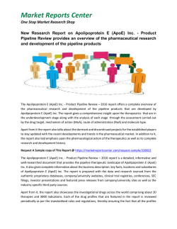
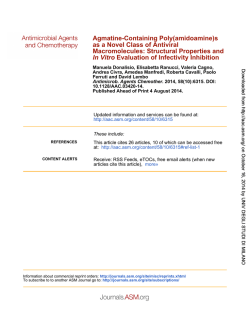
ORIGINAL RESEARCH ARTICLE
published: 23 April 2014
doi: 10.3389/fnagi.2014.00061
AGING NEUROSCIENCE
Physical activity reduces hippocampal atrophy in elders at
genetic risk for Alzheimer’s disease
J. Carson Smith 1 , Kristy A. Nielson 2,3 , John L. Woodard 4 , Michael Seidenberg 5 , Sally Durgerian 3 ,
Kathleen E. Hazlett 2 , Christina M. Figueroa 2 , Cassandra C. Kandah 5 , Christina D. Kay 5 ,
Monica A. Matthews 5 and Stephen M. Rao 6*
1
2
3
4
5
6
Department of Kinesiology, School of Public Health, University of Maryland, College Park, MD, USA
Department of Psychology, Marquette University, Milwaukee, WI, USA
Department of Neurology, Medical College of Wisconsin, Milwaukee, WI, USA
Department of Psychology, Wayne State University, Detroit, MI, USA
Department of Psychology, Rosalind Franklin University of Medicine and Science, North Chicago, IL, USA
Cleveland Clinic, Schey Center for Cognitive Neuroimaging, Neurological Institute, Cleveland, OH, USA
Edited by:
Emil C. Toescu, Birmingham
University, UK
Reviewed by:
José M. Delgado-García, University
Pablo de Olavide, Seville, Spain
Diego Ruano, University of Sevilla,
Spain
Kirk I. Erickson, University of
Pittsburgh, USA
*Correspondence:
Stephen M. Rao, Cleveland Clinic,
Schey Center for Cognitive
Neuroimaging, Neurological
Institute, 9500 Euclid Ave./U10,
Cleveland, OH 44195, USA
e-mail: [email protected]
We examined the impact of physical activity (PA) on longitudinal change in hippocampal
volume in cognitively intact older adults at varying genetic risk for the sporadic form of
Alzheimer’s disease (AD). Hippocampal volume was measured from structural magnetic
resonance imaging (MRI) scans administered at baseline and at an 18-month follow-up
in 97 healthy, cognitively intact older adults. Participants were classified as High or
Low PA based on a self-report questionnaire of frequency and intensity of exercise.
Risk status was defined by the presence or absence of the apolipoprotein E-epsilon 4
(APOE-ε4) allele. Four subgroups were studied: Low Risk/High PA (n = 24), Low Risk/Low
PA (n = 34), High Risk/High PA (n = 22), and High Risk/Low PA (n = 17). Over the 18
month follow-up interval, hippocampal volume decreased by 3% in the High Risk/Low PA
group, but remained stable in the three remaining groups. No main effects or interactions
between genetic risk and PA were observed in control brain regions, including the caudate,
amygdala, thalamus, pre-central gyrus, caudal middle frontal gyrus, cortical white matter
(WM), and total gray matter (GM). These findings suggest that PA may help to preserve
hippocampal volume in individuals at increased genetic risk for AD. The protective effects
of PA on hippocampal atrophy were not observed in individuals at low risk for AD. These
data suggest that individuals at genetic risk for AD should be targeted for increased levels
of PA as a means of reducing atrophy in a brain region critical for the formation of episodic
memories.
Keywords: cognitive aging, Alzheimer’s disease, volumetric MRI, association studies in genetics, physical activity,
exercise
INTRODUCTION
Possessing an apolipoprotein-E ε4 (APOE-ε4) allele increases the
risk for developing the sporadic form of Alzheimer’s disease (AD)
(Bird, 2008). We have previously shown that APOE-ε4 status
in healthy elders, in combination with measures of hippocampal atrophy obtained at study entry, can predict future cognitive
decline after as short an interval as 18 months (Woodard et al.,
2010). Such a finding is consistent with the commonly held view
that the neuropathology of AD begins decades prior to the clinical diagnosis (Jack, 2012). Yet not all individuals with an APOE-ε4
allele will develop AD (Bird, 2008), suggesting that other genetic,
and possibly lifestyle, factors may offer protection from neurodegeneration that ultimately leads to cognitive decline and a
diagnosis of clinical AD.
Exercise training and physical activity (PA) are associated
with the preservation cognitive function (Etnier et al., 2006;
Angevaren et al., 2008) and hippocampal volume (Erickson et al.,
2011) in healthy older adults. Whether APOE-ε4 status interacts
with PA to slow the longitudinal course of neurodegeneration
Frontiers in Aging Neuroscience
is unknown. Cross-sectional studies have reported differences
between high and low PA on cognitive outcomes (Woodard
et al., 2012; Smith et al., 2013), amyloid burden (Head et al.,
2012), and brain function (Smith et al., 2011) in APOE-ε4 carriers (although see Lindsay et al., 2002). One such study (Schuit
et al., 2001) reported reduced odds for experiencing cognitive
decline in physically active older male APOE-ε4 carriers compared to sedentary carriers. Another study (Rovio et al., 2005)
reported reduced odds of receiving an AD diagnosis in physically active male and female APOE-ε4 carriers compared to
inactive carriers. Importantly, in both of these epidemiological
studies, the positive effects of PA were not apparent in noncarriers. An additional epidemiological study (Lindsay et al.,
2002) reported that engaging in regular PA reduced the odds
of being diagnosed with AD after a 5 year follow-up period;
in this study, however, the effects of PA were not modified by
APOE-ε4 inheritance. Etnier and colleagues reported that greater
cardiorespiratory fitness was associated with better neurocognitive test performance in healthy older women who were APOE-ε4
www.frontiersin.org
April 2014 | Volume 6 | Article 61 | 1
Smith et al.
Physical activity reduces hippocampal atrophy
homozygotes, but the same relationship was not observed in
APOE-ε4 heterozygotes or non-carriers. We (Woodard et al.,
2012) have previously reported, in a sub-sample of the current
study, that physically active APOE-ε4 carriers had significantly
reduced odds of cognitive decline over 18 months compared to
physically inactive carriers. However, it is not known if this pattern of neuroprotection among APOE-ε4 carriers also occurs in
the hippocampus.
The aim of this study was to track the 18-month changes
in hippocampal volume, a proxy for neurodegeneration, in four
groups of cognitively intact older adults that varied on the basis
of PA (low vs. high) and genetic risk (APOE-ε4 carriers vs.
non-carriers). We hypothesized that high PA would result in
preservation of hippocampal volume in APOE-ε4 carriers compared to low PA carriers. In contrast, the protection offered by PA
would be less apparent in APOE-ε4 non-carriers.
MATERIALS AND METHODS
PARTICIPANTS
Healthy adults between the ages of 65 and 89 were recruited
from newspaper advertisements. A telephone screen was
administered initially to 459 individuals to determine eligibility based on inclusion/exclusion criteria. Potential participants were excluded if they reported a history of cognitive
deterioration and/or dementia, neurological disease (cerebral
ischemia, vascular headache, carotid artery disease, cerebral
palsy, epilepsy, brain tumor, chronic meningitis, multiple sclerosis, pernicious anemia, normal-pressure hydrocephalus, HIV
infection, Parkinson’s disease, and Huntington’s disease), medical illnesses (untreated hypertension, glaucoma, and chronic
obstructive pulmonary disease), major psychiatric disturbance
or substance abuse generally consistent with DSM-IV Axis I
criteria or a Geriatric Depression Scale (GDS) score greater
than 15. Participants were allowed to take cardiovascular
drugs.
To enrich the sample with a higher percentage of APOE-ε4 carriers, half the sample was recruited based on a family history of
dementia, since the APOE-ε4 allele is more common in individuals with a family history of dementia than among those without
such a history (Sager et al., 2005). Family history was defined as
a report of a clear clinical diagnosis of AD or a reported history
of gradual decline in memory and other cognitive functions, confusion, or judgment problems without a formal diagnosis of AD
prior to death in a first-degree relative.
Of those who met criteria, 112 agreed to undergo APOE genotyping from blood samples, complete a PA questionnaire, and be
administered a brief neurobehavioral assessment and a structural
magnetic resonance imaging (MRI) scan. All participants were
invited to undergo a repeat MRI scan 18 months later. Followup MRI scans were obtained from 99 of 112 (88.4%) participants
using the identical scanner and pulse sequence (see below). Of the
13 participants who did not undergo follow-up scanning, nine
withdrew from the study, two had excessive head motion at the
baseline scan, and two were lost to follow-up. Of the 99 participants who completed both scan sessions, two were excluded
due to excessive head motion during follow-up scanning. From
the final pool of 97 participants, four subgroups were formed
Frontiers in Aging Neuroscience
based on the absence/presence of one or both APOE-ε4 alleles
(Low Risk vs. High Risk) and self-reported amounts of leisuretime PA (Low PA vs. High PA). The mean (±SD) inter-scan
interval for all participants was 554 (±41) days (range 489–763);
the mean inter-scan interval did not differ between groups
(p = 0.92).
This study was approved by the institutional review board
at the Medical College of Wisconsin and conducted in accordance with the Helsinki Declaration. Written informed consent was obtained and all participants received modest financial
compensation.
PHYSICAL ACTIVITY
Frequency and intensity of leisure time PA were measured using
the Stanford Brief Activity Survey (SBAS) (Taylor-Piliae et al.,
2006). SBAS scores have demonstrated validity for assessing
habitual PA (Taylor-Piliae et al., 2006, 2007). Participants who
endorsed one of the two items indicating two or fewer days
of low intensity PA (ranging from no PA to slow walking or
light chores) were classified as physically inactive (Low PA).
Participants endorsing one of the remaining three items describing moderate to vigorous intensity PA three or more days per
week (ranging from brisk walking, jogging or swimming for
15 min or more, or moderately difficult chores for 45 min, to regular jogging, running, bicycling or swimming for 30 min or more,
or playing sports such as handball or tennis for an hour or more)
were classified as physically active (High PA).
GENETIC TESTING
APOE genotype was determined using a polymerase chain reaction method (Saunders et al., 1996). Deoxyribonucleic acid was
isolated with Gentra Systems Autopure LS for Large Sample
Nucleic Acid Purification. Participants with one or both APOEε4 alleles were classified as High Risk for developing AD and the
remainder classified as Low Risk. APOE genotype results for the
four groups were as follows: Low Risk/High PA (n = 24: three
ε2/ε3 and 21 ε3/ε3), Low Risk/Low PA (n = 34: six ε2/ε3 and 28
ε3/ε3), High Risk/High PA (n = 22: 20 ε3/ε4 and two ε4/ε4), and
High Risk/Low PA (n = 17: two ε2/ε4 and 15 ε3/ε4).
NEUROBEHAVIORAL TESTING
Participants were administered the Mini-Mental State
Examination (Folstein et al., 1975), Mattis Dementia Rating
Scale 2 (DRS-2) (Jurica et al., 2001), GDS (Yesavage, 1988), and
Lawton Activities of Daily Living (ADL) (Lawton and Brody,
1969) at study entry.
STRUCTURAL MRI ACQUISITION
MR imaging was conducted at baseline and 18-month followup on a General Electric (Waukesha, WI) Signa Excite 3T short
bore scanner equipped with a quad split quadrature transmit/receive head coil. High-resolution, 3-dimensional spoiled
gradient-recalled at steady state (SPGR) anatomic images were
acquired [TE 3.9 ms; repetition time (TR) 9.5 ms; inversion recovery preparation time 450 ms; flip angle 12◦ ; number of excitations 2; slice thickness 1.0 mm; FOV 24 cm; resolution 256 ×
224]. Foam padding was used to reduce head movement.
www.frontiersin.org
April 2014 | Volume 6 | Article 61 | 2
Smith et al.
Physical activity reduces hippocampal atrophy
VOLUMETRIC ANALYSES
The hippocampus was selected a priori as the key structure
for examining the longitudinal volumetric effects of PA. In
addition, we measured changes in volumes of the thalamus,
caudate, amygdala, caudal middle frontal gyrus, pre-central
gyrus, total gray matter (GM), and cortical white matter (WM)
to determine if PA effects were specific to the hippocampus.
Volumetric measurements were obtained from parcellation of
anatomical T1 images at baseline and at 18-months using the
longitudinal method of FreeSurfer software (v. 5.1) (Fischl et al.,
2004). The first stage consisted of identifying the GM/WM
boundary (Dale et al., 1999) using both intensity and continuity
information from the entire 3D volume. Maps were then created
using spatial intensity gradients across tissue classes. Using an
automated labeling system, GM and WM were subdivided into
distinct volumes of interest per hemisphere (Desikan et al., 2006).
Homologous left and right hemisphere volumes were summed,
as these volumes were highly correlated and we did not have a
hypothesis regarding hemispheric differences. In addition, total
intracranial volume (ICV) was calculated to account for interindividual differences in head size. Each volume of interest was
expressed as a percentage of total ICV (%ICV). Change in volume
was achieved by subtracting baseline and follow-up %ICV and
then expressed as a percent change from baseline. This change
score was subjected to a 2 (High Risk vs. Low Risk) × 2 (High PA
vs. Low PA) analysis of variance (ANOVA; SPSS 21, Chicago, IL).
Significant effects were followed by post-hoc group comparisons.
RESULTS
BASELINE ANALYSES
The four participant groups did not differ in age, education, or sex
at baseline (see Table 1). There were no differences in the number
of participants in each group based on PA and Risk classification
(p > 0.2). At baseline, the High Risk groups performed slightly
worse on the MMSE than the Low Risk groups, but all study
participants performed within the normal range indicating intact
cognitive abilities. No significant group differences were observed
on a measure of depression (GDS) and all participants had intact
activities of daily living. At baseline, no significant main or interaction effects were observed for %ICV for the hippocampus,
thalamus, caudate, caudal middle frontal gyrus, and cortical WM.
A significant PA effect was observed for the amygdala, pre-central
gyrus, and total GM (Low PA > High PA).
demonstrated that the rate of hippocampal atrophy can be influenced by the extent of leisure-time PA. The protective effect
of PA on hippocampal volume, however, was specific to persons at genetic risk for AD and was not observed in individuals
at low genetic risk. Remarkably, the protective effect of PA in
high risk individuals was seen over a relatively brief 18 month
follow-up interval. Our findings were found to be specific to
the hippocampus, since similar PA effects were not observed in
the thalamus, caudate, amygdala, caudal middle frontal gyrus,
pre-central gyrus, total GM, or cortical WM volumes.
This study is the first to demonstrate the protective effects
of PA on hippocampal atrophy in persons at genetic risk for
AD. Our results are compatible with epidemiological studies
that have examined the joint effects of PA and APOE-ε4 inheritance on the extent of cognitive decline and the diagnosis of
AD (Smith et al., 2013). Our findings are also consistent with
evidence from other brain imaging modalities. Head and colleagues, using [11 C] Pittsburgh Compound B (PiB) as a measure
of amyloid mean cortical binding potential, reported less brain
amyloid in a physically active compared to a physically inactive
group of APOE-ε4 carriers, an effect not observed in non-carriers.
Deeny et al. (2012) demonstrated that among ε4 carriers, activation in the left inferior temporal cortex during performance of the
Sternberg working memory task was greater in those with higher
levels of fitness. Finally, in a task-activated fMRI study conducted
on a sub-sample of this study, we (Smith et al., 2011) demonstrated that semantic memory activation was greatest in high PA
APOE-ε4 carriers relative to low PA carriers and non-carriers. We
have also demonstrated that increased semantic memory activation, along with APOE-ε4 non-carrier status, is protective against
future cognitive decline (Woodard et al., 2010). In a sub-sample
of the current study, physically active APOE-ε4 carriers had significantly reduced odds of cognitive decline over 18 months
compared to physically inactive carriers (Woodard et al., 2012);
as in the current study of hippocampal volume, the protective
effect of PA was not observed in APOE-ε4 non-carriers. Other
factors, such as the presence of depressive symptoms, may also
lead to reductions in cognitive function and hippocampal volume in older adults (Sexton et al., 2013). However, in our sample
depression scores were below the clinical range, did not differ
between the groups at baseline, and did not significantly change
at the 18-month follow-up. Thus, these effects do not appear
to be related to differences in symptoms of depression between
the groups.
LONGITUDINAL CHANGE IN BRAIN VOLUMES
The percent change from baseline to 18-month follow-up scans
are shown in Table 2 (see Table 3 for raw volumetric data).
A significant interaction was observed between Risk and PA for
the hippocampus. Hippocampal volume decreased 3% in the
High Risk/Low PA group (see Figure 1), whereas the volumetric changes in the remaining three groups were negligible. No
significant main or interaction effects were observed for the thalamus, caudate, amygdala, caudal middle frontal gyrus, pre-central
gyrus, total GM, and cortical WM volumes.
DISCUSSION
Atrophy of the hippocampus is a hallmark of AD progression
and, to a lesser extent, occurs as part of healthy aging. We have
Frontiers in Aging Neuroscience
POTENTIAL MECHANISMS FOR PHYSICAL ACTIVITY AND APOE-ε4
INTERACTIONS
The precise neurophysiological mechanisms by which PA might
protect human APOE-ε4 carriers from cognitive decline and ADrelated neuropathology are less well understood (Smith et al.,
2013). Animal studies would suggest that PA counteracts the
physiologic impact of the APOE-ε4 allele, possibly through benefits to cholinergic function or brain lipid metabolism, and/or
reduced neuroinflammation (for a review, see Intlekofer and
Cotman, 2013). Physical activity is known to promote the release
of neurotrophins [brain derived neurotrophic factor (BDNF),
insulin-like growth factor-1] that support neurogenesis in the
dentate gyrus (Trejo et al., 2001; Van Praag et al., 2005). In
www.frontiersin.org
April 2014 | Volume 6 | Article 61 | 3
Smith et al.
Physical activity reduces hippocampal atrophy
Table 1 | Mean (SD) baseline demographic, behavioral testing, and brain volume measures for the four participant groups.
Variables
Low risk
High risk
ANOVA
High PA
Low PA
High PA
Low PA
Risk
(n = 24)
(n = 34)
(n = 22)
(n = 17)
Age (years)
74.4 (5.2)
72.2 (4.6)
71.5 (4.2)
73.7 (5.5)
0.489
Education (years)
15.0 (2.8)
14.0 (2.0)
15.5 (3.0)
15.6 (3.0)
0.088
Sex
9 M, 15 F
7 M, 27 F
5 M, 17 F
7 M, 10 F
–
PA
η2p
Interaction
p
η2p
0.005
0.952
<0.001
0.033
0.024
0.461
0.006
0.329
0.010
0.300*
0.038
0.026
p
p
η2p
DEMOGRAPHICS
–
–
–
0.048
BEHAVIORAL TESTING
MMSE
29.4 (0.8)
29.4 (0.7)
29.2 (1.3)
28.6 (1.2)
0.013
0.064
0.207
0.017
0.115
DRS total score
140.0 (3.8)
141.0 (2.3)
139.7 (3.3)
139.2 (4.7)
0.135
0.024
0.768
0.001
0.318
0.011
1.7 (2.0)
2.8 (2.7)
2.5 (3.0)
2.2 (2.6)
0.854
<0.001
0.449
0.006
0.205
0.017
GDS
BRAIN VOLUME
Hippocampus
0.456 (0.061)
0.477 (0.080)
0.447 (0.065)
0.460 (0.073)
0.386
0.008
0.263
0.013
0.781
0.001
Thalamus
0.789 (0.062)
0.829 (0.091)
0.800 (0.090)
0.826 (0.093)
0.820
0.001
0.071
0.035
0.678
0.002
Caudate
0.504 (0.063)
0.542 (0.067)
0.499 (0.063)
0.508 (0.063)
0.152
0.022
0.091
0.030
0.285
0.012
Amygdala
0.178 (0.024)
0.191 (0.028)
0.177 (0.027)
0.187 (0.026)
0.597
0.003
0.034
0.047
0.741
0.001
Caudal middle frontal g.
0.002
0.834 (0.108)
0.815 (0.125)
0.823 (0.126)
0.828 (0.129)
0.970
<0.001
0.765
0.001
0.664
Pre-central g.
1.76 (0.15)
1.82 (0.18)
1.73 (0.17)
1.85 (0.17)
0.936
<0.001
0.015
0.062
0.387
0.008
Total GM
38.6 (2.8)
40.4 (4.1)
38.6 (2.5)
40.6 (3.0)
0.875
<0.001
0.008
0.074
0.871
<0.001
Cortical WM
30.5 (2.8)
31.2 (3.4)
30.2 (3.5)
30.6 (2.6)
0.508
0.005
0.398
0.008
0.823
0.001
Bold font indicates significant effect; η2p , partial eta-squared, a measure of effect size. *Based on Chi-Square statistic; MMSE, Mini-Mental State Exam; DRS,
Dementia Rating Scale; GDS, Geriatric Depression Scale; g., gyrus; GM, gray matter; WM, white matter.
Table 2 | Change in brain volumes after 18 months expressed as mean (SD) percent change from baseline.
Brain volume
Low risk
High risk
ANOVA
High PA
Low PA
High PA
Low PA
Risk
(n = 24)
(n = 34)
(n = 22)
(n = 17)
p
PA
η2p
Interaction
η2p
p
p
η2p
Hippocampus
−0.82 (3.60)
0.15 (3.49)
−0.41 (3.61)
−2.91 (3.79)
0.082
0.032
0.314
0.011
0.024
0.054
Thalamus
−2.06 (2.72)
−0.85 (3.05)
−1.77 (2.23)
−1.61 (2.32)
0.677
0.002
0.228
0.016
0.351
0.009
Caudate
−0.48 (3.58)
−1.24 (3.69)
−1.23 (2.73)
−2.44 (3.42)
0.177
0.020
0.171
0.020
0.759
0.001
Amygdala
0.59 (7.68)
1.40 (8.40)
−0.10 (6.86)
−0.17 (4.77)
0.465
0.006
0.812
0.001
0.777
0.001
Caudal middle frontal g.
0.92 (3.42)
1.04 (3.95)
0.11 (4.74)
0.89 (5.08)
0.593
0.003
0.614
0.003
0.715
0.001
Pre-central g.
−0.09 (3.45)
−0.51 (4.31)
−0.97 (4.68)
−1.21 (4.48)
0.375
0.008
0.711
0.001
0.924
<0.001
Total GM
−0.39 (2.27)
−0.49 (3.13)
−0.26 (3.27)
−1.37 (3.18)
0.551
0.004
0.340
0.010
0.421
0.007
Cortical WM
−1.43 (2.66)
−0.37 (2.07)
−0.65 (1.42)
−0.56 (1.37)
0.486
0.005
0.178
0.019
0.252
0.014
Bold font indicates significant effect; η2p , partial eta-squared; PA, physical activity; GM, gray matter; WM, white matter; g., gyrus.
addition, cholinergic enhancement due to PA may increase cerebral blood flow, enhance neural activation, and possibly relieve
amyloid burden (Adlard et al., 2005; Head et al., 2012). In
humans, recent evidence suggests that PA may have similar neurogenic effects in the hippocampi of healthy younger adults (Pereira
et al., 2007) and healthy older adults (Erickson et al., 2011).
However, it is not known if the neurotrophic effects of PA are
stronger among APOE-ε4 carriers.
Because inheriting an APOE-ε4 allele results in the disruption of lipid homeostasis, there are adverse effects on amyloid
precursor protein (APP) function and the clearance of brain
Frontiers in Aging Neuroscience
amyloid, as well as on neuroinflammation and acetylcholine function (Poirier, 2000; Lane and Farlow, 2005). Apolipoproteins are
lipid carrying molecules involved in regulating lipid metabolism
in response to neuronal injury. The protein APOE is particularly important in brain synaptic plasticity and growth through
its role handling phospholipids and cholesterol associated with
neuronal repair processes (Poirier, 2000). Lipoproteins resulting
from the APOE-ε4 allele are removed more easily in carriers,
and this greatly reduces the amount of APOE available in the
brain compared to non-carriers (Leduc et al., 2011). A concomitant decrease in lipoprotein lipase activity also leads to
www.frontiersin.org
April 2014 | Volume 6 | Article 61 | 4
Smith et al.
Physical activity reduces hippocampal atrophy
Table 3 | Absolute mean (SD) brain volume at baseline and 18-months expressed as a percent of total intracranial volume.
Brain volume
Low risk
High risk
High PA
Low PA
High PA
Low PA
(n = 24)
(n = 34)
(n = 22)
(n = 17)
HIPPOCAMPUS
Baseline
0.456 (0.061)
0.480 (0.081)
0.447 (0.065)
0.460 (0.073)
18 months
0.451 (0.059)
0.478 (0.083)
0.445 (0.069)
0.447 (0.078)
THALAMUS
Baseline
0.789 (0.062)
0.829 (0.091)
0.800 (0.090)
0.826 (0.093)
18 months
0.773 (0.070)
0.822 (0.093)
0.787 (0.099)
0.811 (0.085)
CAUDATE
Baseline
0.504 (0.063)
0.542 (0.067)
0.499 (0.063)
0.508 (0.063)
18 months
0.502 (0.066)
0.535 (0.070)
0.493 (0.061)
0.496 (0.065)
AMYGDALA
Baseline
0.178 (0.024)
0.191 (0.028)
0.177 (0.027)
0.187 (0.026)
18 months
0.178 (0.025)
0.194 (0.033)
0.176 (0.030)
0.187 (0.029)
CAUDAL MIDDLE FRONTAL g.
Baseline
0.834 (0.108)
0.815 (0.125)
0.823 (0.126)
0.828 (0.129)
18 months
0.842 (0.115)
0.824 (0.137)
0.824 (0.130)
0.836 (0.143)
PRECENTRAL g.
Baseline
1.76 (0.15)
1.82 (0.18)
1.73 (0.17)
1.85 (0.17)
18 months
1.76 (0.13)
1.81 (0.21)
1.72 (0.18)
1.83 (0.21)
TOTAL GM
Baseline
38.6 (2.8)
40.4 (4.1)
38.6 (2.5)
40.6 (3.0)
18 months
38.5 (2.8)
40.2 (4.3)
38.5 (3.0)
40.1 (3.2)
CORTICAL WM
Baseline
30.5 (2.8)
31.2 (3.4)
30.2 (3.5)
30.6 (2.6)
18 months
30.1 (3.2)
31.1 (3.5)
30.0 (3.5)
30.5 (2.6)
PA, physical activity; GM, gray matter; WM, white matter; g., gyrus.
FIGURE 1 | Percent change from baseline in total hippocampal volume
for the four participant groups. Error bars represent s.e.m.
reduced levels of brain free fatty acids, fundamental components of neuronal repair and neurotrophic processes (Lane and
Farlow, 2005), which alters APP function to further the production and accumulation of brain β-amyloid (Poirier, 2000).
Frontiers in Aging Neuroscience
Finally, altered lipid membrane homeostasis reduces glycolytic
metabolic processes and the availability of acetyl-CoA-derived
adenosine triphosphate and acetylcholine (Lane and Farlow,
2005; Leduc et al., 2011), leading to compromised cholinergic
function. Exercise has been shown to improve cholinergic function and to oppose the actions of acetylcholinesterase in the
hippocampus and cerebral cortex of rats (Ben et al., 2009).
A small number of animal studies have reported interactions
between APOE genotype and exercise on brain neurophysiology. APOE-ε4 mice showed similar increases in hippocampal
BDNF compared to APOE-ε3 mice after 6 weeks of voluntary
wheel running using a transgenic mouse model. Moreover, tyrosine kinase B receptors, which have a high affinity for BDNF,
increased after wheel running in APOE-ε4 mice to levels comparable to the baseline levels of APOE-ε3 mice (Nichol et al., 2009).
Exercise in APOE-lacking mice may attenuate the development
of atherosclerosis in the periphery, and these effects may be due
to exercise-induced enhancement of anti-inflammatory cytokines
(Fukao et al., 2010). The effects of PA on brain lipid metabolism in
human ε4 carriers are not yet known (Rankinen et al., 2010), nor
if responses to exercise training differ based on APOE genotype
(Leon et al., 2004). The beneficial effects of PA are not exclusive,
however, to APOE. For example, exercise has been shown to
improve cognition, reduce oxidative stress, and induce synaptic
www.frontiersin.org
April 2014 | Volume 6 | Article 61 | 5
Smith et al.
Physical activity reduces hippocampal atrophy
plasticity in the 3xTg-AD triple transgenic mouse model of AD
(Garcia-Mesa et al., 2011), underscoring its pleiotropic effects. To
date, the literature suggests that physical inactivity may exacerbate
the effects of the APOE-ε4 genotype on AD-related neuropathology and its clinical manifestation of memory impairment (Smith
et al., 2013), consistent with the current findings.
LIMITATIONS
It is important to note that we did not manipulate levels of PA
nor did we randomly assign participants to groups in a controlled
trial, thus limiting our ability to determine cause-effect relationships. We also did not control for other health-related behaviors
(e.g., diet) or measure BDNF genotype. The sample was mostly
Caucasian (only one Hispanic and one African-American participated), so these results may not generalize to other ethnic groups.
This study is also limited by a subjective measure of leisure-time
PA rather than an objective measurement of cardiorespiratory fitness, such as the maximal rate of oxygen consumption (VO2max ).
Responses to the SBAS, however, were dose-dependently related
to cardiovascular risk biomarkers and estimated caloric expenditure in an epidemiologic study (Taylor-Piliae et al., 2006). The
High Risk/Low PA group included two participants with the ε2ε4
genotype. Although the APOE-ε2 allele has been associated with
protection from morbidity, results from the Rotterdam Study
(Slooter et al., 1998) indicate that this effect is clearest for the ε2ε3
genotype, with a greater risk of cognitive impairment for the ε2ε4
genotype.
CONCLUSIONS
Our study has provided additional evidence that PA can afford
protection against neurodegeneration in cognitively intact persons at genetic risk for AD. Our results suggest that knowledge
of the APOE genotype, while hardly precise in the prediction
of AD, can play an important role in making recommendations
to older adults regarding exercise as a means of maintaining
brain integrity and preventing future cognitive decline and brain
atrophy. Future studies are needed to better understand the neurophysiological mechanisms by which PA appears to alter the
phenotypic expression of the APOE-ε4 allele.
AUTHOR CONTRIBUTIONS
J. Carson Smith, Kristy A. Nielson, John L. Woodard, Michael
Seidenberg, and Stephen M. Rao contributed to the design and
conceptualization of the study, the analysis and interpretation of
the data, and drafting and revising the manuscript for intellectual content. Sally Durgerian, Kathleen E. Hazlett, Christina M.
Figueroa, Cassandra C. Kandah, Christina D. Kay, and Monica
A. Matthews, contributed to the analysis and interpretation of
the data and drafting and revising the manuscript for intellectual
content.
ACKNOWLEDGMENTS
This project was supported by NIH grant R01-AG022304
awarded to Stephen M. Rao. The content is solely the responsibility of the authors and does not necessarily represent the official
views of the National Institute on Aging or the National Institutes
of Health.
Frontiers in Aging Neuroscience
REFERENCES
Adlard, P. A., Perreau, V. M., Pop, V., and Cotman, C. W. (2005). Voluntary exercise
decreases amyloid load in a transgenic model of Alzheimer’s disease. J. Neurosci.
25, 4217–4221. doi: 10.1523/JNEUROSCI.0496-05.2005
Angevaren, M., Aufdemkampe, G., Verhaar, H. J., Aleman, A., and Vanhees, L.
(2008). Physical activity and enhanced fitness to improve cognitive function
in older people without known cognitive impairment. Cochrane Database Syst.
Rev. 3:CD005381. doi: 10.1002/14651858.CD005381.pub2
Ben, J., Soares, F. M., Cechetti, F., Vuaden, F. C., Bonan, C. D., Netto, C. A.,
et al. (2009). Exercise effects on activities of Na(+),K(+)-ATPase, acetylcholinesterase and adenine nucleotides hydrolysis in ovariectomized rats. Brain
Res. 1302, 248–255. doi: 10.1016/j.brainres.2009.09.013
Bird, T. D. (2008). Genetic aspects of Alzheimer disease. Genet. Med. 10, 231–239.
doi: 10.1097/GIM.0b013e31816b64dc
Dale, A. M., Fischl, B., and Sereno, M. I. (1999). Cortical surface-based analysis I:
segmentation and surface reconstruction. Neuroimage 9, 179–194.
Deeny, S. P., Winchester, J., Nichol, K., Roth, S. M., Wu, J. C., Dick, M.,
et al. (2012). Cardiovascular fitness is associated with altered cortical glucose metabolism during working memory in varepsilon4 carriers. Alzheimers
Dement. 8, 352–356. doi: 10.1016/j.jalz.2011.04.010
Desikan, R. S., Segonne, F., Fischl, B., Quinn, B. T., Dickerson, B. C., Blacker, D.,
et al. (2006). An automated labeling system for subdividing the human cerebral cortex on MRI scans into gyral based regions of interest. Neuroimage 31,
968–980. doi: 10.1016/j.neuroimage.2006.01.021
Erickson, K. I., Voss, M. W., Prakash, R. S., Basak, C., Szabo, A., Chaddock, L., et al.
(2011). Exercise training increases size of hippocampus and improves memory.
Proc. Natl. Acad. Sci. U.S.A. 108, 3017–3022. doi: 10.1073/pnas.1015950108
Etnier, J. L., Nowell, P. M., Landers, D. M., and Sibley, B. A. (2006).
A meta-regression to examine the relationship between aerobic fitness and cognitive performance. Brain Res. Rev. 52, 119–130. doi:
10.1016/j.brainresrev.2006.01.002
Fischl, B., Salat, D. H., Busa, E., Albert, M., Dieterich, M., and Haselgrove, C.
(2004). Whole brain segmentation: automated labeling of neuroanatomical
structures in the human brain. Neuron 33, 341–355. doi: 10.1016/S08966273(02)00569-X
Folstein, M. F., Folstein, S. E., and Mchugh, P. R. (1975). “Mini-mental state.” A
practical method for grading the cognitive state of patients for the clinician.
J. Psychiatry Res. 12, 189–198.
Fukao, K., Shimada, K., Naito, H., Sumiyoshi, K., Inoue, N., Iesaki, T., et al.
(2010). Voluntary exercise ameliorates the progression of atherosclerotic lesion
formation via anti-inflammatory effects in apolipoprotein E-deficient mice.
J. Atheroscler. Thromb. 17, 1226–1236. doi: 10.5551/jat.4788
Garcia-Mesa, Y., Lopez-Ramos, J. C., Gimenez-Llort, L., Revilla, S., Guerra, R.,
Gruart, A., et al. (2011). Physical exercise protects against Alzheimer’s disease in 3xTg-AD mice. J. Alzheimers. Dis. 24, 421–454. doi: 10.3233/JAD-2011101635
Head, D., Bugg, J. M., Goate, A. M., Fagan, A. M., Mintun, M. A., Benzinger,
T., et al. (2012). Exercise engagement as a moderator of the effects of APOE
genotype on amyloid deposition. Arch. Neurol. 69, 636–643. doi: 10.1001/archneurol.2011.845
Intlekofer, K. A., and Cotman, C. W. (2013). Exercise counteracts declining hippocampal function in aging and Alzheimer’s disease. Neurobiol. Dis. 57, 47–55.
doi: 10.1016/j.nbd.2012.06.011
Jack, C. R., Jr. (2012). Alzheimer disease: new concepts on its neurobiology and the clinical role imaging will play. Radiology 263, 344–361. doi:
10.1148/radiol.12110433
Jurica, P. J., Leitten, C. L., and Mattis, S. (2001). Dementia Rating Scale-2
Professional Manual. Lutz, FL: Psychological Assessment Resources.
Lane, R. M., and Farlow, M. R. (2005). Lipid homeostasis and apolipoprotein E
in the development and progression of Alzheimer’s disease. J. Lipid Res. 46,
949–968. doi: 10.1194/jlr.M400486-JLR200
Lawton, M. P., and Brody, E. M. (1969). Assessment of older people: selfmaintaining and instrumental activities of daily living. Gerontologist 9, 179–186.
Leduc, V., Domenger, D., De Beaumont, L., Lalonde, D., Belanger-Jasmin, S., and
Poirier, J. (2011). Function and comorbidities of apolipoprotein e in Alzheimer’s
disease. Int. J. Alzheimers Dis. 2011:974361. doi: 10.4061/2011/974361
Leon, A. S., Togashi, K., Rankinen, T., Despres, J. P., Rao, D. C., Skinner, J.
S., et al. (2004). Association of apolipoprotein E polymorphism with blood
www.frontiersin.org
April 2014 | Volume 6 | Article 61 | 6
Smith et al.
Physical activity reduces hippocampal atrophy
lipids and maximal oxygen uptake in the sedentary state and after exercise training in the HERITAGE family study. Metabolism 53, 108–116. doi:
10.1016/j.metabol.2003.08.013
Lindsay, J., Laurin, D., Verreault, R., Hebert, R., Helliwell, B., Hill, G. B., et al.
(2002). Risk factors for Alzheimer’s disease: a prospective analysis from the
canadian study of health and aging. Am. J. Epidemiol. 156, 445–453. doi:
10.1093/aje/kwf074
Nichol, K., Deeny, S. P., Seif, J., Camaclang, K., and Cotman, C. W. (2009).
Exercise improves cognition and hippocampal plasticity in APOE epsilon4 mice.
Alzheimers Dement. 5, 287–294. doi: 10.1016/j.jalz.2009.02.006
Pereira, A. C., Huddleston, D. E., Brickman, A. M., Sosunov, A. A., Hen, R.,
Mckhann, G. M., et al. (2007). An in vivo correlate of exercise-induced neurogenesis in the adult dentate gyrus. Proc. Natl. Acad. Sci. U.S.A. 104, 5638–5643.
doi: 10.1073/pnas.0611721104
Poirier, J. (2000). Apolipoprotein E and Alzheimer’s disease. A role in amyloid catabolism. Ann. N.Y. Acad. Sci. 924, 81–90. doi: 10.1111/j.17496632.2000.tb05564.x
Rankinen, T., Roth, S. M., Bray, M. S., Loos, R., Perusse, L., Wolfarth, B., et al.
(2010). Advances in exercise, fitness, and performance genomics. Med. Sci.
Sports Exerc. 42, 835–846. doi: 10.1249/MSS.0b013e3181d86cec
Rovio, S., Kareholt, I., Helkala, E. L., Viitanen, M., Winblad, B., Tuomilehto, J.,
et al. (2005). Leisure-time physical activity at midlife and the risk of dementia and Alzheimer’s disease. Lancet Neurol. 4, 705–711. doi: 10.1016/S14744422(05)70198-8
Sager, M. A., Hermann, B., and La Rue, A. (2005). Middle-aged children of persons with Alzheimer’s disease: APOE genotypes and cognitive function in the
Wisconsin Registry for Alzheimer’s Prevention. J. Geriatr. Psychiatry Neurol. 18,
245–249. doi: 10.1177/0891988705281882
Saunders, A. M., Hulette, O., Welsh-Bohmer, K. A., Schmechel, D. E., Crain,
B., Burke, J. R., et al. (1996). Specificity, sensitivity, and predictive value of
apolipoprotein-E genotyping for sporadic Alzheimer’s disease. Lancet 348,
90–93.
Schuit, A. J., Feskens, E. J., Launer, L. J., and Kromhout, D. (2001). Physical activity
and cognitive decline, the role of the apolipoprotein e4 allele. Med. Sci. Sports
Exerc. 33, 772–777. doi: 10.1097/00005768-200105000-00015
Sexton, C. E., Mackay, C. E., and Ebmeier, K. P. (2013). A systematic review and
meta-analysis of magnetic resonance imaging studies in late-life depression. Am.
J. Geriatr. Psychiatry 21, 184–195. doi: 10.1016/j.jagp.2012.10.019
Slooter, A. J., Van Duijn, C. M., Bots, M. L., Ott, A., Breteler, M. B., De Voecht, J.,
et al. (1998). Apolipoprotein E genotype, atherosclerosis, and cognitive decline:
the Rotterdam Study. J. Neural Transm. Suppl. 53, 17–29.
Smith, J. C., Nielson, K. A., Woodard, J. L., Seidenberg, M., Durgerian, S., Antuono,
P., et al. (2011). Interactive effects of physical activity and APOE-epsilon4 on
BOLD semantic memory activation in healthy elders. Neuroimage 54, 635–644.
doi: 10.1016/j.neuroimage.2010.07.070
Frontiers in Aging Neuroscience
Smith, J. C., Nielson, K. A., Woodard, J. L., Seidenberg, M., and Rao, S. M.
(2013). Physical activity and brain function in older adults at increased
risk for Alzheimer’s disease. Brain Sci. 3, 54–83. doi: 10.3390/brainsci30
10054
Taylor-Piliae, R. E., Haskell, W. L., Iribarren, C., Norton, L. C., Mahbouba, M.
H., Fair, J. M., et al. (2007). Clinical utility of the Stanford brief activity survey in men and women with early-onset coronary artery disease. J. Cardiopulm.
Rehabil. Prev. 27, 227–232. doi: 10.1097/01.HCR.0000281768.97899.bb
Taylor-Piliae, R. E., Norton, L. C., Haskell, W. L., Mahbouda, M. H., Fair, J. M.,
Iribarren, C., et al. (2006). Validation of a new brief physical activity survey
among men and women aged 60-69 years. Am. J. Epidemiol. 164, 598–606. doi:
10.1093/aje/kwj248
Trejo, J. L., Carro, E., and Torres-Aleman, I. (2001). Circulating insulin-like growth
factor I mediates exercise-induced increases in the number of new neurons in
the adult hippocampus. J. Neurosci. 21, 1628–1634.
Van Praag, H., Shubert, T., Zhao, C., and Gage, F. H. (2005). Exercise enhances
learning and hippocampal neurogenesis in aged mice. J. Neurosci. 25,
8680–8685. doi: 10.1523/JNEUROSCI.1731-05.2005
Woodard, J. L., Sugarman, M. A., Nielson, K. A., Smith, J. C., Seidenberg, M.,
Durgerian, S., et al. (2012). Lifestyle and genetic contributions to cognitive decline and hippocampal structure and function in healthy aging. Curr.
Alzheimer Res. 9, 436–446. doi: 10.2174/156720512800492477
Woodard, J., Seidenberg, M., Nielson, K. A., Smith, J. C., Antuono, P., Durgerian, S.,
et al. (2010). Prediction of cognitive decline in healthy older adults using fMRI.
J. Alzheimer’s Dis. 21, 871–885. doi: 10.3233/JAD-2010-091693
Yesavage, J. A. (1988). Geriatric depression scale. Psychopharmacol. Bull. 24,
709–711.
Conflict of Interest Statement: The authors declare that the research was conducted in the absence of any commercial or financial relationships that could be
construed as a potential conflict of interest.
Received: 22 January 2014; accepted: 20 March 2014; published online: 23 April 2014.
Citation: Smith JC, Nielson KA, Woodard JL, Seidenberg M, Durgerian S, Hazlett
KE, Figueroa CM, Kandah CC, Kay CD, Matthews MA and Rao SM (2014) Physical
activity reduces hippocampal atrophy in elders at genetic risk for Alzheimer’s disease.
Front. Aging Neurosci. 6:61. doi: 10.3389/fnagi.2014.00061
This article was submitted to the journal Frontiers in Aging Neuroscience.
Copyright © 2014 Smith, Nielson, Woodard, Seidenberg, Durgerian, Hazlett,
Figueroa, Kandah, Kay, Matthews and Rao. This is an open-access article distributed
under the terms of the Creative Commons Attribution License (CC BY). The use, distribution or reproduction in other forums is permitted, provided the original author(s)
or licensor are credited and that the original publication in this journal is cited, in
accordance with accepted academic practice. No use, distribution or reproduction is
permitted which does not comply with these terms.
www.frontiersin.org
April 2014 | Volume 6 | Article 61 | 7
© Copyright 2026 Paperzz