55-
Studies on the Germination of the Spores in Some Mosses
By Shintaro SAITO
(Received Nov. 11, 1958)
The reproduction of mosses is chiefly made by means of spores. To begin with, every
spores puts forth the protonema system, and the formaiion of gametophytes is brought
forth by the germination of spores and regeneration of gametophytes. In order to clarity
the mechanism of reproduction in Barbula unguiculata, Bartramia crispata, Mniw71
microphyllum, Pogonatum inflexm71, Dicranm?1 japonicum, the author have undertaken
experiment on the germination of spores
I
Methods
The cultures of the spore were made on the plates and fllter paper and slides as well.
The treated fllter papers were placed on the absorbent cotton in closed Petri-dishes which
nutrient solution bearing different pH value was prepared and was renewed with every
two weeks during the experiment. At the same time they were also made on l% agar
Benecke's nutrient media mounted on slides as mentioned above. The culture furnitures
and media were sterilized in Koch's stream-sterilizer, preceding the e) Periment. All these
cultures were kept at room temparature and placed near a window illuminated by daylight, avoiding any direct rays of the sun
Results
Barbula unguiculata Hedw.
In Matsue, the spprogonium of this species ripened during late April and early May
The materials collected from the stone wall 0L basalt, on 17th of May 1957, were wrapped
up in the parafiine paper and kept in the desicator. The spores were sown on the agar
nutrient media, bearing a pH value of 8, on 2ls t of September of the same year. The
spores of this species were large, measuring about 12 p in diameter. The spores on the
media swelled to a considerable size, measuring 20p in diameter within 2 or 3 days after
the treatment. The chloroplasts in the endospores much increased in three or four
days after the treatment. Five or six days after the treatment, the exospores ruptured
i
poured Benecke's nutrient solution, keep in the upper surfaced over the solution. The
56
and a germination tube appeared. The germ tubes were cut off from the endospores by
a cross wall and then developed into chlorophylliferous filament. In many cases, the
germ tube occured from the one side of the spore but very rarely an another germ tube
developed from the opposite side of spores the same time. (Fig. 14) These filaments
showed positive phototrophic character. (Fig. 14) In respect to the width of the fllaments, and the acount of chloroplasts which are contained in the filamentous cell, there
were not found any differences between both the main filaments. When the main fila- -
} .
*
_.-'_
;
r ]
cb
r:"'
' z'ev Oa':1C},'
1JauL1 Ir '
'
'o ;Q L('i
;DOO
V:.OO:
t
Ooo o Q
): !
cl:)O
ar"' t*
,oi(
OV
tl Pi
f'oooo
i o(,OO "aoa
c'ac
{
:
{oao:oa
[noi i
2^
zzoao
u voaaooo
'-r'tll"
'I1'1F" '1
V -
"
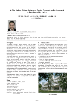
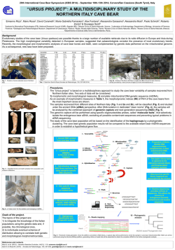
Fig. I Germination of spores in Barbula unguiculata Hedw (1-15)
1-8, early stages in development of bud, X 180
9, Ieafy plant, X50
10-12,
13,
14,
25,
bud of leafy plant (10. 11...XllO, 12...X60)
globose-mass like gametophore. X 60
germination of spores, X 125
filaments showed positive phototrophic character, x 60
r...rhizoid, b...bus. (Yuko Nishida)
57
ment acquired 6 or 7 cells' Iength the branches were formed Lrom the upper part of the
main filament. These branch filaments showed also positive phototrophic character. (Fig
15). In many cases, the branches were produced from the point just anterior to the
cross wall of a filamentous cell. There were two kinds of branch filaments ; the one
crawled on the nutrient media and another grew apart Lrom the media. Several huge
protuberances were found dear the forked points of these filaments. These protuberances
developed new filaments, bearing oblique cross walls, and their width are about 2.5 times
as large as ordinary branch filaments which grew from the main fllaments. These filaments turned into the rhizoid-1ike fllaments. The protonema attained about 2cm. in length
at the end of October, and no were elongation of filaments occurred. Thus, the protonema system was formed. The bud of leafy plant appeared on the main filaments and
on the' branch filaments attached the media on about 45th day after treatment. No buds
were found on the branch filaments which elongated towards the air. The buds appeared as a huge protuberance on the point anterior to the cross walls (Fig. lO). These
protuberances are similar to the Indian-Club in shape and are formed from a terminal
cell and a stalk which holds the cell. The stalk consisted of 3 or 4 cells arranged in a
chain. This was different from that of Bartl a7/lia c7 ispata which is formed from a single
cell. The terminal cell developed on the stalk cell contains abundant chloroplasts and
later forms a globose cell mass by successive segmentation. Several long rhizoids grew
Lrom the basal part of a young shoot. Thus, the leafy plant grew to the size that can
be recognized by the naked eyes at the middle of November (Fig. 9)
Bartralnia crispata Schilnp.
The spores were collected on 19th of April, 1957, and sown on the media on the 14th
of May of the same year. Within 4 days after the treatment, one rarely two germ tubes
appeared from the spore through ruptured exospores. The germ tubes were soon cut by
the cross wall and formed a branch filament by successive cell divisions. There may be
recognized two types in the branch filament ; the one had colorless cell walls and the
cross walls occured at right angle to the longitudinal axis and contained numerous chloro-
plasts, the other had brown cell walls and oblique cross walls and contained few chloro-
plasts. The branch filaments developed in various directions and occasionally some of
them turned into brown rhizoid-like filaments, bearing oblique cross walls. Thus, a pro-
tonema system was formed in about 30 days after the treatment. In the middle of June,
a cell of the main or branch fllaments produced protuberance on the point just anterior
to the cross wall (Fig. 18). The cell derived from the protuberance developed into a
58
leafy plant. The buds of leafy shoot are ovoid and their cells contain rich- cytoplasm
and numerous chloroplasts. At first a diagonal septum was set in the cavity of the initial
cell of a leaLy plant and was followed by two or three additional diagonal ones and thus
divided an apical cell by three divided faces. The rhizoids were produced from the basal
part of a young leafy plant, but it occurred after the formation of two or three leaves
The rhizoids had a numerous spots on the surface of brown cell walls, oblique cross
walls and Lew chloroplasts.
e
,*,*+*********
; :',
+
***"
*
4*'・t*-_,t !'1. 4_
:, ,t・f t*::, _
**:( r; 1
':,,fS
・・:;
? ;ti -
5
4
.
/:': " *
5
,
oo
le
I,,
+ , t¥
'ex *')i t,"!
":'-:
::"':
'¥ ;. ' s;
!; i:;;:
:;; ;_
h , .,. _-: t; i
;;'t}" " *
'
2
A ;
P"'
/
t( ad
a
.' ex
.
t;oees ,=
oaeaLs : ¥._ .
q
:era ai ;:a
,
o
V
v
t'
V
6
s
er
*
,
4Qa
r・
3
3
S S
,
fA
t t
' i
i_
/: i
/
Ll
: ¥
o
od
.
' ;
t{ a
ee
" 's !og
,
,
t,!t
*
l
1
7
¥
L$'
ed'e
e L
8
'$t l i"'/ '.f 'a_
""
<
i* :
i/
7
1
-
_ __ s'"
. 5'
'se
;e
't *
*.'.'... '
:
t"i:4*.';' .'
..
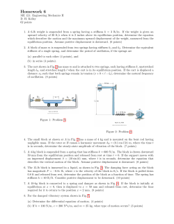
Fig. 2 Germination of spores in Bartramia crispata Schimp (1-9)
l-8, Germination 0L spores (1-4, X 200 5-8, X 150)
9, protonema system, showing bud of leaLy plant X 25
bud of leafy plant, (Yuko Nishida)
ex . . . exospore b
59
a
eS I ;G
S
'OO'
aO O/fOga /P
a(70,¥
v :c:)
aoV
a'a
SS
:t : S
oCQ'
S
: S S
?
0 oo oeos
i b
c o o
oo
oo':20 os
ao
5ao ?oce:
_.o
oc' o!c)co
c)c:s
OOo
Oo : Oo a
n Oe
2a-Z2a
O
O
c20' :Ve7 oooo002
0(::"oo
Oo v gO
v Ooo i aa
1:
uooO
Oaa. ;
10 O O ¥;
(ac i
. V. ,2: O a
! b GS:
¥8l: j o
0q:s
O oo
S ol')
(:t C ?eoo c:)
C)o'p
c)
;!: e o
o(:)1
s'o 1
,
0
S -o o
O ac
o
c'loc)
c)oc:c)
,
・-r _ t
:;'5,::
o
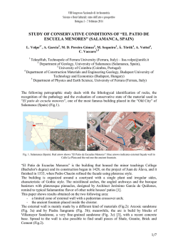
Germination of spores in Bartra77lia c7 ispatr Schilnp. (10-20)
early stages in development of bud, X 200
15-16, a leafy plant with a rhizoid, X 250
17, ditto, X40
18-20, showing a bud of leafy plant on the protonema, (18, 20 X 150, 19 X IIO)
Fig. 3
10-14,
r ... rhizoid, b ... bud. (Yuko Nishida)
Mnium microphyllum Doz. et Molk.
The spores were collected on April 10, 1957 at Sotonakahara-machi, Matsue city and
kept in dry condition until the culture experiments were undertaken. They were sown
on the porous plates and filter papers absorbed Benecke's nutrient solution, bearing a pH
value of 6 on 14th of May of the same year. The spores of this species were large,
measuring about 22p in diameter. They were swollen whithin a few days after the
treatment and enlarged by 35p. The endospores produced one or two germ tubes. The
germ tube was soon divided by a cross wall and developed into a multicellular filament
(Fig. 3). When the filament attained 10-15 cells' Iength several branches were L0rmed
-60
on the apical part of the main filament. In June, the main and branch filaments increased rapidly .their length and showed positive phototrophic character. Two kinks of
branches were recognized ; the one ran on the surface of the substratum or raised the
substratum, and the other crept into the substratum. The former bore colorless, cross
walls at right angle to the longitudinal axis and contained numerous chloroplasts, the
latter was brown and beared oblique wall, and contained chloroplasts. The branches
crept into the agar turned into rhizoidal filaments. The growth of the rhizoids on the
porous plates was better than the filter papers. Thus, a protonema system was formed
i
" ex / :
oo o 'ep::
;eO ,_,
} ..
3
i 'd
a
'O QC'{
6 to a"
7fa
q
4
a o
o
aa 2
o
oo
Qol
5 co
O g
zz2
e
:
iO
li
1
sd
s
i2
8
ex ...'
s
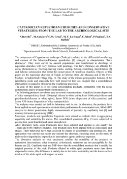
Fig. 4
10-11,
12,
gc'
e
Germination of spores in Mniu/n 7nicrophyllw7:1 D. A/1. (1-12)
germination of spores, (1. 3. >< 300 2. 4. 5. 6. 7. X 150)
8,9,
1-7,
I'
showing a protonema system, X 50
showing a bud of leafy plant on the filament, X 150
early stages in development of a bud, >< 200
showing a leafy plant developed on filament of protonema, X 40
b ... bud . (Yuko Nishida)
ex . . . exospore,
, :61
whithin two months after the treatment. Several huge protuberances were found on the
main and branch fllaments. As is seen in Fig. 9, this protuberance developed as the bud
of a leafy plant. Through the successive segmentation of this cell
Ieafy plant was
established
Pogonatu,n inflexul7:1 Ldb.
The materiales were got on 16th of October, 1957 in Mt. Daisen, Tottori Pref. The
spores were wrapped in the parafline paper and kept in the desicator. They were cultured on the porous plates submerged in the Benecke's nutrient solution bearing a pH
value of 6, on 5th of November of the same year. The spores of this species were about
6-8/ e in diameter. The spores on the porous plates were swollen and became about lOp
in diameter within two weeks after the treatment. The spores germed in the middle of
November. ;rhe feature of the germination was similar to that of the preceding species.
In late November, the germ tube developed into 3-4 cells' Iength but no further develol>
ment of the filament was observed during the winter. In early March of the following
year, they regained activity and became longer and branched as time went on. The
filamentous cells were very slender, measuring about 10p in width and contained rather
few chloroplasts. The branch filaments were extended on the surface of the substratum
for a short distance and turned into the air, showing the positive phototrophic character
(Fig. 6). The branch filaments reached to about 2cm in length. Thus, a protonema
system was formed, but no buds of leafy plant were found both on main and branch
filaments during one year of the culture
..・ ex
2
'
6 "
Fig. 5 Germination of spores in Pogonatanl inflexu7n Ldb. (1-6)
1-5, germination of spore, X 250
6, showing branch filaments, X 200
ex . . . exospore (Yuko Nishida)
62
Discraniw71 japonicum Mitt.
The spores were collected in S ptember 20, 1957, at Rakuzan-park, Matsue city. The
spores were scattered on the porous plates submerged in the Benecke's nutrient solution,
bearing a pH value of 6, on llth of October of the same year. The endospore emerged
as a protuberance through the rupture of exospore after two weeks but the growth of
the filament was stopped during the winter. In early April of the following year, the
filaments regained activity and increased their length and produced several short and
chlorophyllose branch filaments from the upper part of the main filament. There are
two kinds of branch filaments similar to those of the preceding species (Mniwn microphyllmn. Barbular unguiculata). The branch filaments elongated toward the air, showing
the positive phototrophic character, and attained about 1.5cm in length. In late October
the bud of leafy plant appeared at the point just anterior to the transverse walls of
the filamentous cells (Fig. 5, 6). The cells of bud contain rich cytoplasm and numerous
chloroplasts. At the end of October, 1958, a single juvenile plant bearing well formed
leaves and rhizoids formed on a protonema (Fig. 8).
a O
'- ・・- b
O
00 0 . 8
c:'ro ,oloo oo ol:D
o e =oc:' '-q,sosl::ot
l$ ;
(2 5
e ,'v
5
a
2
ooc
6
Fig.
Germination of spores is Dic7-anu/n, japoniculn A/Iitt. (1-9)
1-4,
germination of spore, X 100
5,
a bud of leafy plant developed on protonema, X 125
6,
ditto, X 100
7,
early stage in development of a bud, >< 125
8,
a leafy plant bearing rhizoids and a filament on protonema, X 70
9,
germination of spores, X 60
ex . . . exospore r . . . rhizoid, b . . . bud. (Yuko Nishida)
63
Summary
The spores of Ba7l ula unguiculata, :artrawaa crispata, Mnium microphyllum, Dicranum
Japonicum need one or two weeks for germination. They elongated germ tube from one
or two sides of spore and developed into the filamentous protonema. These filaments
showed the positive phototrophic character. In culture experiment in Spring the protonema
system was formed in about two months respectively. The bud of leafy plant of
;arbula
unb"uiculata, Ba/ tranzia cl ispata, Mnium lnicrophyllw7e:, Dicranm71 japonicum grew on the
main and branch filament which crawled on the substratum but in Pogonatum inflexum,
no buds 0L Ieafy plant were found during one year of the culture. The buds produced
as a protuberance, in which contained rich cytoplasm and chloroplasts
Acknowledgement
This research, subsidized by the Science Department of Ministry of Education, was
directed in full by Mr. Yuko Nishita of the Lower Secondary SchoQl attached to Faculty
of Education, Shimane University. The authors (Yuko Nishida and Shintaro Saito) wish
to express many thanks to Dr. Akira Noguchi, professor at Kumamoto University, for
his suggestions and warmhearted helps afforded to this work.
IJiterature
Samuel L. Meyer (1947) . . . Physioloe"ical studies on Mosses
Gilbert M. Smith (1955) . . . Cryptogamic Botany. Volume II. Bryophytes and Pteridophytes
Noguchi A. & Furuta H. (1956) ... Germination of spores and regeneration of leaves
0L Merceya ligulata and M. gedeana. Hattori Bot. Lab.
Noguchi A. & Miyata I. (1957) . . . Sporeling and regenerants in some mosses. Kumamoto
Jur. Sci.
© Copyright 2026 Paperzz