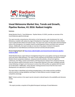
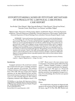
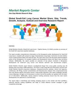
Neurosurgery
68 y/o. Caucasian M
Brain Metastasis from Melanoma
Shintaro Ono
Case: 68 y/o. Caucasian M
HPI
The patient is a 68 y.o. gentleman with h/o metastatic melanoma primarily
on his left neck with spread to adjacent lymph nodes as well as potential
metastases to liver and lung.
On May 21, 2009, He presented to the BI and underwent routine follow up
with brain MRI which showed a large intracranial hemorrhagic lesion in the
right frontal lobe. This was new since his prior scan which was on Nov. 11,
2008.
Past Medical History
- Metastic Melanoma to lung and liver
Allergy NKDA
- DM, HTN, and Af
Family History
- Non-contributory
Past Surgical History
- Left lung lower lobectomy for
metastasis (12/11/08)
- Appendectomy 60 years ago
Medicatoin
- lisinopril, aspirin, warfarin
Social History
- Farmer with extensive sun exposure
- Tobacco; a cigar / week (stopped in 2009)
- EtOH; No,
- illicit drugs; No
Case: 68 y/o. Caucasian M
Physical Examinations
VS: T: 98.5 BP: 146/79 HR: 103 R: 16 O2Sats: 96RA
Gen: WD/WN, comfortable, NAD.
HEENT: Pupils: 3-->1.5, bilat EOMs intact, Neck: Supple.
Lungs: CTA bilaterally.
Cardiac: irregularly irregular. S1/S2.
Abd: Soft, NT, BS+
Extrem: Warm and well-perfused.
Mental status: Awake and alert, cooperative with exam, normal affect.
Orientation: Oriented to person, place, and date.
Recall: 3/3 objects at 5 minutes.
Language: Speech fluent with good comprehension and repetition.
Naming intact. No dysarthria or paraphasic errors.
Case: 68 y/o. Caucasian M
Cranial Nerves:
I: Not tested
II: Pupils equally round and reactive to light. Visual fields are full to confrontation.
III, IV, VI: Extraocular movements intact bilaterally without nystagmus.
V, VII: Very slight left facial nerve droop, otherwise facial nerve intact and muscles
intact, sensation intact to all fields
VIII: Hearing intact to voice.
IX, X: Palatal elevation symmetrical.
XI: Sternocleidomastoid and trapezius normal bilaterally.
XII: Tongue midline without fasciculations.
Motor: Normal bulk and tone bilaterally. Strength full power 5/5 throughout.
Sensation: Intact to light touch, propioception, pinprick and vibration bilaterally.
Coordination: normal on finger-nose-finger, heel to shin
MRI
4mm
4.5 x
3.7cm
May. 21, 2009.
Nov. 11, 2008.
Right frontal craniotomy was done on May 22, 2009.
Post-operative MRI
May. 22, 2009.
May. 21, 2009.
He was discharged on May 26, 2009.
He is taking a course of whole brain radiation therapy.
Brain Metastasis
•The most common intracranial tumors in adults
(>50% in brain tumors).
•In patients with systemic metastasis,
Brain Metastasis occurs 10 to 30 % in adults.
•The incidence of brain metastases is increasing
due to improved imaging tools.
Lung — 16 to 20 %
Renal cell cancer — 7 to 10 %
Melanoma — 7 %
Breast cancer — 5 %
Colorectal cancer — 1 to 2 %
Brain Metastasis from Melanoma
•50% to 75% of malignant melanoma patient end up with
brain metastasis.
•Melanoma is the third most common cause of brain
metastases in US.
•The incidence of malignant melanoma is increasing at rate
greater than any other human cancer.
Treatment
•Whole Brain Radio therapy
WBRT after surgery reduce the rate of recurrence
and possibly prolong survival.
[Wen, PY, Loeffler, JS. Management of brain metastases. Oncology (Huntingt) 1999; 13:941.]
[Skibber, JM, Soong, SJ, Austin, L, et al. Cranial irradiation after surgical excision of brain metastases in
melanoma patients. Ann Surg Oncol 1996; 3:118.]
•Chemotherapy
Brain Metastases from melanoma are generally resistant to chemotherapy.
But, fotemustine and temozolomide have a possibility of treatment.
[Jacquillat, C, Khayat, D, Banzet, P, et al. Final report of the French multicenter phase II study of the
nitrosourea fotemustine in 153 evaluable patients with disseminated malignant melanoma including patients
with cerebral metastases. Cancer 1990; 66:1873.]
[Hwu, WJ, Lis, E, Menell, JH, et al. Temozolomide plus thalidomide in patients with brain metastases from
melanoma. Cancer 2005; 103:2590. ]
Craniotomy vs SRS
There is no confirmed clear advantage of
one treatment over the other.
– No prospective randomized trials have been reported
(M. L. Smith and J. Y. K. Lee 4 Neurosurg. Focus / Volume 22 / March, 2007)
• Craniotomy
– Large, single/dominant, and accessible lesions
– Patients with good performance status
– Patients with herniation or a posterior fossa mass effect
• Stereotactic Radiosurgery (SRS)
– Small(<3cm), multiple(=or<3), and deep lesions
– Patients unlikely to tolerate general anesthesia.
Treatment for the patient
•
•
•
•
•
Resection, Craniotomy?
SRS?
Whole Brain Radiation Therapy (WBRT)?
Chemotherapy?
or Combination?
What the reason for resection surgery
and following WBRT?
Reasons for Surgery
The Patient
•Good performance status (KPS 90 > 70)
•Large (>3cm), dominant Lesion
•Mass Effect
•Resectable
•The lesion cannot be well controlled with external
radiation alone.
Reasons for Surgery
Mayo Clin Proc 2003; 78:1529.
Summary
•Case:
68 y/o. Caucasian M with Brain Metastasis from Melanoma
•The incidence of malignant melanoma is increasing at a rate
greater than any other human cancer.
•Patient with brain metastasis from melanoma still has poor
prognosis.
•New treatment and medical progression is needed for better
prognosis.
Thank you!!
Prognosis
Patient
Age; 68 y.o. (> 65 y.o.)
Karnofsky Performance Score (KPS); 90
Metastasis; Lung, Liver, Brain
Rating
Definition
100 percent
No evidence of disease
90 percent
Normal activity with minor signs of disease
80 percent
Normal activity with effort; signs of disease
70 percent
Cannot do normal activity but cares for self
60 percent
Requires occasional assistance
50 percent
Requires considerable assistance; frequent medical care
40 percent
Disabled, requires special care
30 percent
Severely disabled; hospitalization may be indicated
20 percent
Very sick; hospitalization necessary for supportive treatment
10 percent
Moribund
0 percent
Death
© Copyright 2026 Paperzz