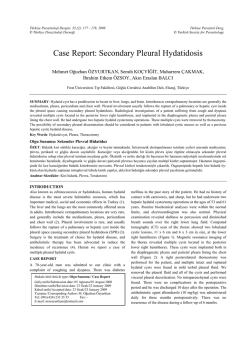
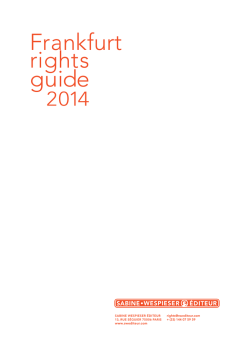
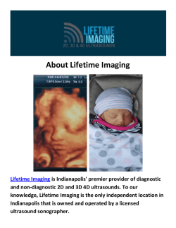
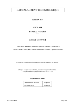
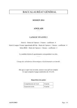
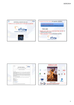
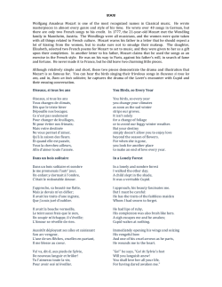
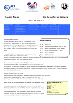
International Journal of Medical Imaging
2014; 2(5): 105-108
Published online September 10, 2014 (http://www.sciencepublishinggroup.com/j/ijmi)
doi: 10.11648/j.ijmi.20140205.12
ISSN: 2330-8303 (Print); ISSN: 2330-832X (Online)
Complicated hydatid cysts of the liver:
A report of two cases
Abdelilah Mouhsine1, *, Ahmed Belkouch2, El Mehdi Atmane1, Redouane Rokhssi1,
Youssef Berrada1, Lahcen Belyamani2, Abdelghani El Fikri1, M’barek Mahfoudi1
1
2
Service de Radiologie Hôpital Militaire Avicenne, faculty of medecine and pharmacy, Marrakech, Maroc
Service des urgences Hôpital Militaire d’Instruction MohamedV, faculty of medecine and pharmacy, Rabat, Maroc
Email address:
[email protected] (A. Mouhsine), [email protected] (A. Belkouch)
To cite this article:
Abdelilah Mouhsine, Ahmed Belkouch, El Mehdi Atmane, Redouane Rokhssi, Youssef Berrada, Lahcen Belyamani, Abdelghani El Fikri,
M’barek Mahfoudi. Complicated Hydatid Cysts of the Liver: A Report of Two Cases. International Journal of Medical Imaging.
Vol. 2, No. 5, 2014, pp. 105-108. doi: 10.11648/j.ijmi.20140205.12
Abstract: The authors report two cases of complicated hydatid cysts of the liver, including one which was spontaneously
broken in the bile ducts and the other fistulized in the right pleura. Patients presented with jaundice and biliary colic in the
first case. Cough basithoracic pain, weakness, and fever in the second. Both patients were investigated by ultrasound and
CT scan followed by a hydatid serology. In both cases the patients improved after surgical treatment. Will be recalled
imaging aspects of these two complications.
Keywords: Liver Hydatid Cyst, Clinical Presentation, Complications, Imaging
1. Introduction
Hydatid cyst of the liver remains a common disease in the
Maghreb countries. Despite the availability of imaging
(ultrasound, CT and MRI), it is still discovered at the stage
of complication. The most common and most serious of
these complications remains the cysto-biliary fistula.
Surgical treatment continues to be improved and should
be as conservative as possible to reduce the morbidity and
mortality of this benign condition.
portal venous phase), objectified two cystic formations; one
was located in segment IV containing peripheral
calcifications, measuring 6 / 4.5cm and fistulized in
intrahepatic bile ducts which were dilated (Figure 1, 2a and b).
The other lesion was located in segment VII, had a fluid
density with membrane peeling in some parts of it, containing
some peripheral calcifications and measuring 6 / 4cm.
2. Case Presentation
2.1. Case 1
A 34 years old man, farmer, with notion of contact with
dogs, complained of hepatalgia since eight months.
One month before his admission to the hospital, the
patient presented a generalized pruritus and a
muco-cutaneous jaundice, without fever.
An abdominal ultrasound showed a cystic formation of
segment VII measuring 6 cm, a calcified formation of
segment IV and a marked dilatation of the intrahepatic bile
ducts and the common bile duct. A CT scan, performed before
and after injection of contrast material (different arterial and
Figure 1. 2a. 2b. Axial CT scans without (Fig1) and with injection of
contrast material (Fig: 2a,b) showing cystic formations including one in
segment IV containing peripheral calcifications and fistulized in the
intrahepatic bile ducts which are dilated.
International Journal of Medical Imaging 2014; 2(5): 105-108
106
Laboratory tests showed inflammatory syndrome
(erythrocyte sedimentation rate (ESR) to 18mm in the first
hour) and a positive hydatid serology.
The patient underwent surgery (unroofing of the hydatid
cyst, sealing of the fistula and drainage).
Postoperative period was unremarkable with no
complications.
2.2. Case 2
A 28 years old woman with no history, with notion of
contact with dogs, complained since 2 months of straight
basithoracic pain when breathing. Ten days before her
admission to the hospital, the patient presented dry cough,
fatigue and fever. Clinical examination revealed right
basithoracic dullness with absence of breath sounds and
reduced tactile fremitus.
The patient had first undergone a chest X-ray (Fig 3),
which showed a right moderate-sized pleural effusion.
Fig 5. Ultrasound section showing the lesion of the dome of the liver
containing a large calcification within it
Biology showed a discrete raised erythrocytes
sedimentation rate (ESR) of 15 mm the first hour with a
positive hydatid serology.
The patient underwent surgery with pleural fluid drainage
and sealing of the fistula. Postoperative period was without
complications.
3. Discussion
Fig 3. chest radiograph showing a right pleural effusion of moderate
abundance without intra parenchymal infection.
A thoraco-abdominal CT scan was performed showing the
presence of a pleural effusion associated with right scissurite,
the effusion communicates through a fistula with a rounded
hypodense well limited hepatic lesion, its dome contained
calcified material and was not enhanced after contrast
material injection (figure 4, 5). Ultrasound complement
confirmed the presence of liver dome lesion with the same
semiology as mentioned above.
Fig 4. CT axial section after injection of contrast material showing pleural
effusion communicating through a fistula with the hepatic lesion.
Hydatid cyst is a parasitic disease caused by the
development of the larval Echinococcus granulosis form.
The usual host is the dog, man being the intermediate host.
All locations are possible.
Hydatid cyst rupture into the biliary tree is the most
common complication (1). These fistulas are found in 30%
according to Moumen (2), 25% according to Abi (3) and
Daali (1) and 31% according to Medarhri (4). It seems
common at the right liver cysts (5, 6), as was the case of our
first patient.
The fistula in the thorax is much uncommon and varies
from 2 to 5% (7, 8) as for our 2nd case. Infection of the
cystic contents, promoted by its fissuring in the biliary tree
varies from 5 to 55% (9, 10).
Clinical symptoms are suggestive when the patient suffers
obstructive jaundice (as for the first patient) or cholangitis
which is a sign of severity (11, 12). But the latent forms are
the most common according to daali (1) and bouzidi (5). The
systematic preoperative search for biliary fistula is
mandatory.
Our second patient’s symptoms were primarily
respiratory; simulating pulmonary tuberculosis and the
diagnosis of hydatid cyst was discovered fortuitously on
ultrasound and was confirmed by hydatid serology.
The radiological aspects of Hydatid cyst depend on the
evolution of the disease. From the young lesion, with cystic
aspect; the alterations are related to the activity of the
germinal membrane, to the thickening and calcification of
the adventitia. Finally the reduction of hypertension
produces intra cavitary membrane peeling and loss of
regular limits.
The duration of each of these events is variable and there
is no strict timeline. Also the classifications reflect
107
Abdelilah Mouhsine et al.:
Complicated Hydatid Cysts of the Liver: A Report of Two Cases
imperfectly the evolution of the disease.
The most widely used due to Gharbi (13) discerns five
types:
Type 1: pure liquid image;
Type 2: total or partial separation membranes;
Type 3: intracavitary native hydatid cysts;
Type 4: solid like mass;
Type 5: calcified walls of a solid mass.
The cross-sectional imaging is the best way to recognize
or diagnose these complications.
Ultrasound examination is often sufficient for simple
uncomplicated hydatid cysts. It can establish the diagnosis
before stage 2 or 3, and strongly suggests it in stage 1 or 5
when epidemiological arguments are present. Serology then
confirms the diagnosis.
Computed tomography scan (CT) is used secondarily.
Thanks to its higher spatial resolution, it overcomes the poor
conditions of examination that usually suffers ultrasound
and it is very useful to show the relationship between the
cyst and the vascular and biliary pedicle (5). A kysto biliary
communication can be recognized when there is a biliary
expansion near a cystic lesion that is not under tension.
Membrane debris can be seen in the common bile duct or in
the pleural effusion associated to a communicating sluice.
The magnetic resonance imaging (MRI) is currently the best
method in the exploration of the bile ducts (14, 15),
especially in case of doubt remaining after CT scan,
especially for lesions of stage 4 (16).
Until now, surgical treatment is the only effective way.
After sterilization of the parasite, the intervention is
designed to eliminate or reduce the residual cavity and treat
the pleural or biliary complication.
The most adopted protocol is unroofing (17, 18, 19),
which remains a simple gesture, but the treatment of biliary
fistula depends mainly on its size. If the fistula is small,
closure and drainage are associated with a simple treatment,
recommended in the first patient, but this method has a great
weak point which is the lack of tightness with risk of
prolonged bile leak and sub phrenic abscess resulting to a
long hospitalization. If the fistula has a large caliber, the
kysto biliary disconnection is the method of choice (20), but
often requires a long hospital stay.
4. Conclusion
The diagnosis of the hydatid cyst is still done at the stage
of complications, despite the current availability of
cross-sectional imaging (ultrasound, CT and MRI).
The treatment is surgical and should be initiated before
the stage of complication to make it conservative with less
morbidity and mortality.
Competing of Interest
The authors don’t have any competing interest.
References
[1]
Daali M, Fakir Y, Hssaida R, Ameur M, Abbar M. Les kystes
hydatiques du foie rompus dans les voies biliaires-à propos
de 64 cas. Ann Chir. 2001;126:242–245.
[2]
MOUMEN. MO., EL ALAOUI M., EL MOKHTARI M., EL
FARES F. Les kystes hydatiques du foie. A propos de 670 cas
dont 552 compliqués. Sem Hôp Paris 69 : 7225,1993.
[3]
Abi F., El Fares F., Bouzidi A. Les kystes hydatiques du foie.
A propos de 181 cas compliqués. Lyon Chir 1988 ; 40 :
418-420
[4]
Medarhri J, EL Ounani M, Echarrab M, Louchi A, Alami F,
Amraoui M, Errogani A, Chkof R, Balafrej S. Les kystes
hydatiques du foie rompus dans les voies biliaires à propos de
88 cas expérience des urgences chirurgicales viscérales Rabat.
Médecine du Maghreb. 2001;86:45–48
[5]
Bouzidi A, Chehab F. Traitement chirurgical des fistules
biliokystiques d’origine hydatique : 83 cas. J Chir 1997 ; 134 :
114-8.
[6]
Serguney S, Tortum O, Haydar A. Les kystes hydatiques
compliqués du foie. Ann Chir 1991 ; 45 : 584-9.
[7]
MOUMEN M., EL FARES F. Les fistules bilio-bronchiques
d’origine hydatique. A propos de 8 cas. J. Chir. 1991,128,
n°4, 188-192.
[8]
MZABI R., BACH HANBA K. Traitement des kystes
hydatiques du foie ouverts dans les bronches. A propos de 24
cas. Mem. Acad Chir. 1978,104,404,412
[9]
CHIGOT J. P., LANGLOIS P., TEBOUL F., GLOT J. P.,
GENTILINI M., MERCADIER M. Le traitement des kystes
hydatiques du foie. Ann. Chir 1986, 40, 177-182.
[10] MOUMEN M., EL ALAOUI M.E., MEHHANE M., JAMIL
D., MOKHTARI M., EL FARES F. La résection du dôme
saillant du kyste hydatique du foie. A propos de 360 cas. J
Chir (Paris). 1990 Feb;127(2):83-6.
[11] Kourias B. Échinococcose péritonéale secondaire. Marseille
chirurgical 1966 ; 18 : 389-99.
[12] Ennabli E, Mouche A, Hachaichi A. Les kystes hydatiques du
foie ouverts dans les voies biliaires. Sem Hôp 1986 ; 62 :
2173-80.
[13] Gharbi HA, Hassine W, Brauner MW, Dupuch K. Ultrasound
examination of the hydatic liver. Radiology. 1981
May;139(2):459–463.
[14] Ernst O, Asselah T, Fakir Y. Cholangio-wirsungographie par
IRM. Ann Chir 1997 ; 51 : 1111-4.
[15] Cervi C, Aube C, Tuech JJ, Pessaux P, Regenet N, Burtin P,
Arnaud JP. La cholangiographie par résonance magnétique
nucléaire en pathologie biliaire. Étude prospective chez 60
patients. Ann Chir 2000 ; 125 : 428-34.
[16] Les kystes hydatiques du foie rompus dans les voies biliaires:
à propos de 120 cas Mountassir Moujahid1,&, Mohamed
Tarik Tajdine1. Pan Afr Med J. 2011;10:43.
[17] Balafrej S, El Alami E, El Madhi T, et al. La rupture du kyste
hydatique dans les voies biliaires. Résultats du traitement
chirurgical et place de la cholédocostomie transhépatico
kystique. Lyon Chir 1995 ; 40 : 177-82.
International Journal of Medical Imaging 2014; 2(5): 105-108
[18] Chehab F, Khail D, Bouzidi A. La surveillance postopératoire
du kyste hydatique du foie opéré. Med Maghreb 1997 ; 86 :
41-2.
[19] Maaouni A. Chirurgie du kyste hydatique du foie. Chirurgie
1989 ; 115 : 61-9.
108
[20] Settaf A, Bargach S, Lamlou M, Oudghiri M. Traitement de
la fistule kystobiliaire du kyste hydatique du foie. J Chir
1991 ; 128 : 133-8.
© Copyright 2026 Paperzz