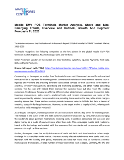
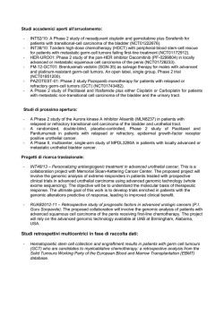
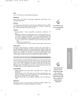
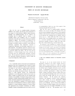
Application Renal Neoplasms: An Update on Immunohistochemical and Histochemical Features Stephen M. Bonsib, MD Ami Bhalodia, MD Albert and Harriet Smith Professor and Chair Louisiana State University Health Science Center Shreveport, LA, USA Renal-Urologic Pathology Fellow Louisiana State University Health Science Center Shreveport, LA, USA Renal Cell Carcinomas The final two decades of the 20th century witnessed a major evolution in our concept of renal cell carcinomas. The extensive histological variability long noted in renal cell carcinoma (RCC) was shown to reflect the existence of a large number of distinct entities having differing prognoses and often harboring unique cytogenetic abnormalities (1-3). Advancement in understanding the molecular misadventures that result from these genetic abnormalities spurned development of promising targeted therapies for a disease in which surgical approaches were previously the sole option (4, 5). The emerging therapeutic possibilities have raised the bar for pathologists mandating accurate classification in an increasingly complex arena. Although, histological assessment remains the mainstay of RCC classification, there are immunohistochemical (IH) and histochemical tools that serve as adjuncts to the histologic analysis. However, cautious interpretation of IH data is recommended since experience in the less common and most recently described tumors may be limited. In this brief review we discuss the use of ancillary immunohistochemistry in RCC classification but admit that space precludes fully addressing the myriad of diagnostic permutations that may arise. Clear Cell Renal Cell Carcinoma Clear cell RCC is overwhelmingly the most common type of renal cancer accounting for 70% of cases (1, 6). The World Health Organization defines CC-RCC as “a malignant neoplasm composed of cells with 178 | Connection 2010 clear or eosinophilic cytoplasm within a delicate vascular network” (6). As this definition implies, cytoplasmic clearing may not be present in every cell, or even in every neoplasm. Other types of RCC may have optically clear cytoplasm and appear indistinguishable. Granular cell, or eosinophilic variant of CC-RCC, a term historically applied to tumors with few or no clear cells, is terminology no longer acceptable since several RCC types are characterized by cells having eosinophilic cytoplasm. Since CC-RCC is so common and histologically diverse, most differential diagnoses are driven by comparative features relative to this tumor, a strategy employed below. Histochemistry of RCCs The cytoplasmic clearing in CC-RCC results from extraction of abundant intracellular glycogen and lipid by organic solvents during tissue processing (Fig. 1). However, sufficient residual glycogen remains after processing that a PAS stain without diastase is usually positive. Demonstration of lipid by an Oil Red O stain is easily accomplished but requires frozen tissue. Although glycogen and lipid are characteristic findings in CC-RCC, these histochemical stains offer little differential diagnostic power in RCC classification since tumors other than CC-RCC may also harbor appreciable lipid and/or glycogen. The single differentiating histochemical stain useful in RCC classification is the Hale’s colloidal iron (CI) stain (7, 8). The CI stain demonstrates the presence of abundant acid mucosubstances in chromophobe cell (Ch) RCC which are absent in other types of RCC (Fig. 2). The acid Figure 1. Electron microscopy of a clear cell renal cell carcinoma showing abundant lipid and glycogen. (L- lipid, G- glycogen). mucosubstances are located within complex cytoplasmic vesicles demonstrable by electron microscopy, a unique feature of this tumor. The CI is often essential to confirm the diagnosis in Ch-RCC because similar to CC-RCC, Ch-RCC shows histological extremes. Some tumors are composed of cells with somewhat clear, so-called transparent cytoplasm, eliciting the differential diagnosis of CC-RCC. Other Ch-RCCs are composed entirely of cells with eosinophilic cytoplasm, the so-called eosinophilic variant, which elicits the differential of a benign oncocytoma or a CC-RCC with eosinophilic cells. Immunohistochemical Differential Diagnosis of RCCs with Cytoplasmic Clearing Although most cases of CC-RCC represent sporadic disease, identical neoplasms may arise in several hereditary and cystic diseases and two tumors are defined by their unique gross or architectural features (Table 1). Family history and presence of extra-renal findings are crucial to establishing the correct diagnosis in these various scenarios because the CC-RCCs that develop may show histological and immunohistochemical identity to sporadic cases. The most common genetic cystic disease with CC-RCC is von HippelLindau disease (9, 10). In this autosomal recessive syndrome CC-RCC is accompanied by clear cell-lined cysts and extrarenal neoplastic disease. CC-RCC also rarely arises in tuberous sclerosis complex (TSC) especially when associated with polycystic kidney disease, known as the contiguous gene syndrome (11, 12). Finally, there is a very rare non-cystic genetic disease known as constitutional chromosome 3 syndrome, an autosomal dominant syndrome defined by the presence of single or multiple, unilateral or bilateral CC-RCC in patients with a balanced chromosome 3 translocation (13, 14). Only seven families have been identified, each with a different translocation. The multilocular cystic renal cell carcinoma, a relatively common (3-5% of RCCs) RCC and defined by its gross, is a tumor that consists entirely of clear cell lined cysts and cyst septae without solid nodules of tumor cells (15). Clear cell papillary RCC, a recently recognized type of RCC originally identified in patients with end stage kidney disease and acquired cystic kidney disease, is now recognized to also develop in native kidneys. This tumor contains clear cells arranged along papillary fronds with basally oriented nuclei (16-18). The papillary architecture Connection 2010 | 179 Figure 2. Colloidal iron stain of a chromophobe cell renal cell carcinoma demonstrating intense cytoplasmic staining. Figure 3. A Xp11.2 translocation carcinoma showing the characteristic TFE3 nuclear staining. 180 | Connection 2010 Clear Cell Carcinomas with Defining Gross, Architectural and Additional Features Clear Cell Tumor Gross or Architectural Features Additional von Hippel-Lindau disease +/- Clear cell lined cysts Extra-renal neoplastic disease Tuberous sclerosis complex +/- Polycystic kidney disease Angiomyolipomas Extrarenal disease Constitutional chromosome 3 syndrome Multiple and/or bilateral CC-RCC Family history of CC-RCC Multilocular cystic RCC Consists entirely of cyst and septae Renal limited pT1 or pT2 Clear cell papillary RCC Papillae formation Often ESKD Table 1. is definitional of this entity; conversely true papillary architecture is an exclusionary criterion for CC-RCC. Many entities with clear and/or eosinophilic cells enter into the differential of RCCs as noted in Tables 2 and 3. The laundry list of antigens provided includes both common and uncommonly employed immunohistochemical analytes. The scope of the differential is smaller with clear cell tumors compared to those with eosinophilic cytoplasm. It includes the two other most common RCCs, papillary RCC, especially when solid, Ch-RCC, translocation carcinomas and urothelial carcinoma which occasionally shows extensive cytoplasmic clearing. Clear Cell RCC expresses vimentin, a variety of low molecular cytokeratins, EMA, CD10 and RCC MA (19, 20). The latter two have some diagnostic utility when a RCC is considered in a metastatic lesion (Table 2). Notable cytokeratin exceptions in CC-RCC are CK7 and high molecular weight cytokeratins. Cytokeratin 7 coupled with AMACR (a-methylacyl-CoA racemase), is useful when solid forms of Pap-RCCs are encountered (21, 22). Several antigens, some uncommonly stocked in immunopathology laboratories, such as parvalbumin, S100A1, caveolin-1, C-kit, PAX2 and E-cadherin have discriminatory efficacy in separating CC-RCC from Ch-RCC (23-30). However, the first line strategy for Ch-RCC should be to stain for CI. For urothelial carcinoma the presence of high molecular weight cytokeratin, CK 7 and p63 permit confirmation of the diagnosis (32-34). Xp11.2 translocation carcinomas are a group of neoplasms characterized by a variety of break points at Xp11.2 with translocation to one of several other chromosomes forming fusion genes (35-37). They occur most frequently in children and young adults. Although histologically variable, the most common tumor cell phenotype are clear cells with voluminous cytoplasm and papillary architecture. Intracellular and stromal calcifications provide additional useful clues to the diagnosis. In general the absence of epithelial markers in translocation carcinomas are powerful clues to the diagnosis. However, a positive reaction for the nuclear transcription factor TFE3 is definitive in the absence of cytogenetic confirmation (Fig. 3). Some cases may also stain for melanocytic markers, but this is more common in TFEB positive t(6:11) translocation carcinomas. Immunohistochemistry in the Differential Diagnosis of RCC with Cytoplasmic Eosinophilia The differential diagnosis for RCCs comprised entirely or predominately of cells with eosinophilic cytoplasm is broad, as reflected in table 3. Although both the mucinous tubular and spindle cell RCC and tubulocystic carcinoma are listed, their distinctive histology drives the diagnosis rather than immunohistochemical phenotype (38-40). The differential diagnosis in tumors with eosinophilic cytoplasm extends beyond carcinomas to include a benign tumor, oncocytoma, and a RCC mimic, the epithelioid angiomyolipoma (41, 42). Epithelioid AML Connection 2010 | 181 Antibody Oncocytoma CC-RCC, Eosin Cytoplasm Chromophobe Carcinoma, Eosinophilic Variant Epithelioid Angiomyolipoma Papillary Renal Cell Carcinoma (Pap RCC), Type 2 Translocation Carcinoma t(6:11) t(6;11) Collecting Duct Carcinoma Medullary Carcinoma Tubulocystic Carcinoma Tubular Mucinous Spindle Cell Carcinoma Immunohistochemical and Histochemical Evaluation of Renal Tumors with Eosinophilic Cytoplasm CAM 5.2 pos pos pos neg pos neg pos pos pos pos AE1/AE3 pos pos pos neg pos neg pos pos pos pos Vimentin neg pos neg neg pos pos pos pos - pos EMA pos pos pos neg pos neg pos pos - pos CD10 pos/neg pos pos/neg neg pos pos pos/neg - pos pos/neg neg pos neg neg pos/neg - - - - neg CK7 “patchy” neg pos neg pos neg pos pos pos pos CK20 neg neg neg neg neg neg neg neg neg neg CK19 neg neg neg neg neg pos/neg pos pos pos/neg pos 34BE12 neg neg neg neg pos/neg neg pos/neg neg pos/neg pos/neg AMACR neg neg neg - pos pos neg - pos pos Parvalbumin pos neg pos - neg - - - pos - S100A1 pos pos neg - pos - - - - - C-kit pos neg pos neg pos/neg - pos/neg - - neg E-cadherin pos neg pos - pos/neg pos pos - - - Kid sp cad pos neg pos - neg neg neg neg neg neg Caveolin-1 neg - pos - - - - - - - Cathepsin K neg neg neg - neg pos - - - - PAX-2 pos pos neg - pos/neg neg neg - pos pos/neg TFE3 neg neg pos - neg pos - - - - TFEB neg neg - - neg neg - - - - UEA-1 neg neg - - pos/neg - pos - pos - HMB 45 neg neg neg pos neg neg neg neg neg neg Melan A neg neg neg pos neg neg neg neg neg neg Coll iron neg neg pos - neg neg neg neg neg neg RCC Ma Table 2. 182 | Connection 2010 Figure 4. An isolated CK 7 positive cell in an oncocytoma. (EpAML), although not a carcinoma, demonstrates malignant behavior in 30% of cases and may lead assist in recognition of the hereditary disease, tuberous sclerosis complex. Most cases of oncocytoma are recognized by their distinctive histology. The presence of mitoses or necrosis should prompt great hesitation in rendering an oncocytoma diagnosis regardless of an otherwise typical histology. Although there are several antigens that can be employed to separate oncocytoma from CC-RCC with eosinophilic cells and the eosinophilic variant of Ch-RCC, because of histogenetic similarity between oncocytoma and Ch-RCC, immunohistochemical stains in these two tumors show substantial overlap. Thus parvalbumin, S100A1, C-kit, caveolin-1, E-cadherin and kidney specific cadherin are usually positive in both (23-30). The CK 7, however, can be very helpful in oncocytoma where most cells are completely negative but a small population of strongly positive cells is invariably present imparting a distinctive staining pattern (Fig. 4). Recently PAX2 has been reported to discriminate between oncocytoma and Ch-RCC (25, 26). When combined with C-kit and CK this profile of three antigens is useful in the separation of the four most common RCCs with eosinophilic cytoplasm as noted in Table 4. Identification of a translocation carcinoma as previously described is facilitated by negative cytokeratin stains and EMA stain. The presence of a positive melanocytic marker is helpful, but a positive stain for the nuclear transcription factor TFEB or TFE3 is required for confirmation. Although, EpAML like translocation carcinomas is negative for cytokeratin and EMA stains and positive for melanocytic markers, its distinctive histology with large ganglion-like cells should permit recognition (40, 41). In uncertain case TFEB and TFE3 stains should be employed. When collecting duct carcinoma (CDC) is considered the histologic features of a high grade tubulopapillary tumor with desmoplastic stroma and infiltrative growth among native nephron elements is characteristic and markedly distinct from all other renal cancers except for urothelial carcinoma (42, 43). The presence of high molecular weight cytokeratin and positive reaction for UEA-1 stain distinguish CDC from other RCCs. A related carcinoma, renal medullary carcinoma (RMC), is recognized by its mucinous or mucoid stroma and infiltrating inflammatory cells. Since development of RMC to date has been limited to patients with sickle cell trait or SC disease, this laboratory feature must be present to support the diagnosis. Immunohistochemistry rarely plays a role. Connection 2010 | 183 Immunohistochemical and Histochemical Evaluation of Renal Tumors with Clear Cytoplasm Antibody Clear Cell RCC Papillary RCC, Type 1 Chromophobe RCC Translocation Carcinoma Urothelial Carcinoma CAM 5.2 pos pos pos neg pos AE1/AE3 pos pos pos neg pos Vimentin pos pos neg pos pos EMA pos pos pos neg pos CD10 pos pos pos/neg pos pos RCC Ma pos pos neg pos pos CK7 neg pos pos neg pos CK20 neg neg neg neg neg CK19 neg neg - neg neg 34BE12 neg neg neg neg pos Parvalbumin neg neg pos - - S100A1 pos pos neg - - C-kit neg neg pos neg neg AMACR neg pos neg pos neg E-cadherin neg - pos pos/neg neg Kid sp cad neg neg pos neg - Cathepsin K neg neg neg pos - PAX-2 pos pos neg neg neg TFE3 neg neg neg pos neg TFEB neg neg neg neg neg HMB 45 neg neg neg neg neg Melan A neg neg neg neg neg Coll iron neg neg pos neg neg P63 neg neg neg - pos Table 3. 184 | Connection 2010 of lipid by an Oil Red O stain is “Demonstration easily accomplished but requires frozen tissue. ” Discussion The classification of RCC has blossomed in recent years with the description of many new entities. The 20th century acceptance of an astounding degree of morphologic diversity within a single nosologic entity RCC has been replaced by a broad menu of diagnostic choices that the pathologist must consider. One member, CC-RCC, remains the most common of RCC. Although CC-RCC is usually sporadic, similar appearing tumors can arise in patients with several genetic cystic diseases. Furthermore, CC-RCC is histologically diverse and may resemble several other cytogenetic and prognostically different tumors. Therefore, the pathologist, must have pertinent history and be well versed in histological nuances of renal tumor classification. Histology remains the cornerstone for diagnosis; immunohistochemistry serves as a useful adjunct to the diagnosis in selected circumstances. When considering immunohistochemistry in differential diagnosis, profiles of antigens are required since any type of RCC may not fully express the IH profiles listed in the tables provided where positive entries do not indicate 100% incidence, nor do negative entries indicate 0% incidence of reaction. Selected Immunohistochemical Profile For The Four Most Common Renal Neoplasms Antibody CK 7 C-Kit PAX2 Clear cell RCC neg neg pos Chromophobe cell RCC pos pos neg Papillary RCC pos neg pos Oncocytoma neg pos pos Table 4. The IH profiles although useful are not a substitute for careful histologic assessment. In a practical sense, more blocks of tissue are often more cost effective than immediate initiation of IH. Lastly, do not feel obligated to place every tumor within a currently defined category. Renal cell carcinoma, unclassified is a valid designation and should account for several percent of RCC diagnoses rendered. It is from this category that new entities may emerge of academic interest that may demonstrate differing biologies with important prognostic or therapeutic implications. Connection 2010 | 185 References 1. Eble JN, Togashi K, Pisani P. Renal cell carcinoma. In: The WHO Classification of Tumours of the Urinary System and Male Genital Organs, edited by Eble JN, Sauter G, Epstein JI, Sesterhenn IA, Lyon, IARC, 2004, pp. 12-14. 16. Tickoo SK, dePeralta-Venturina MN, Harik LR, et. al. Spectrum of epithelial neoplasms in end-stage renal disease. an experience for 66 tumor-bearing kidneys with emphasis on histologic patterns distinct form those in sporadic adult renal Neoplasia. Am J Surg Pathol. 2006; 30:141-153. 30. Garcia E, Li M. Caveolin-1 immunohistochemical analysis in differentiating chromphobe renal cell carcinoma from renal oncocytoma. Am, J Clin Pathol 2006; 125:392-398. 31. Parker DC, Folpe AL, Bell J, et. al. Potential utility of uroplakin III, thrombomodulin, high molecular weight cytokeratin and cytokeratin 20 in noninvasive, invasive and metastatic urothelial (transitional cell) carcinomas. Am J Surg Pathol 2003; 27:1-10. 2. Lohse CM, Cheville JC: A review of prognostic pathologic features and algorithms for patients treated surgically for renal cell carcinoma. Clin Lab Med 2005; 25:433-464. 17. Gobbo S, Eble JN, Grignon DJ, et. al. Clear cell papillary renal cell carcinoma. A distinct histopathologic and molecular genetic entity. Am J Surg Pathol 2008; 32:1239-1245. 3. Moch H, Gasser T, Amin M B, et. al. Prognostic utility of the recently recommended histologic classification and revised TNM staging system of renal cell carcinoma: a Swiss experience with 588 tumors. Cancer 2000; 89:604-614. 18. Gobbo S, Eble JN, MacLennan GT, et. al. Renal cell carcinomas with papillary architecture and clear cell component. The utility of immunohistochemical and cytogenetical analyses in differential diagnosis. Am J Surg Pathol 2008; 32:1780-1786. 4. Cheng L, Zhang S, MacLennan et. al. Molecular and cytogenetic insights into the pathogenesis, classification, differential diagnosis, and prognosis of renal epithelial neoplasms. Hum Pathol 2009; 40:10-29. 19. Skinnider BF, Folpe AL, Hennigar RA, et. al. Distribution of cytokeratins and vimentin in adult renal neoplasms and normal renal tissue. Am J Surg Pathol 2005; 29:747-754. 34. Argani P, Olgac S, Tickoo SK, et. al. Xpll translocation renal cell carcinoma in adults: expanded clinical, pathologic, and genetic spectrum. Am J Surg Pathol 2007; 31:1149-1160. 5. Clark PE. The role of VHL in clear-cell renal cell carcinoma and its relation to argeted therapy. Kidney Int 2009; 76:939-945. 6. Grignon DJ, Eble JN, Bonsib SM, Moch H. Clear cell renal cell carcinoma. In: The WHO Classification of Tumours of the Urinary System and Male Genital Organs, edited by Eble JN, Sauter G, Epstein JI, Sesterhenn IA, Lyon, IARC, 2004, pp. 23-25. 20. Avery AK, Beckstead J, Renshaw AA, Corless CL. Use of antibodies to RCC and CD10 in the differential diagnosis of renal neoplsms. Am J Surg Pathol 2000; 24:203-210. 35. Wu A, Kunju LP, Cheng L, Shah RB. Renal cell carcinoma in children and young adults: analysis of clinicopathologic, immunohistochemical and molecular characteristics with an emphasis on the spectrum of Xpll.2 translocation-associated and unusual clear cell subtypes. Histopathology 2008; 53:533-544. 7. Tickoo SK, Amin MB, Zarbo RJ. Colloidal iron staining in renal epithelial neoplasms, including chromophobe renal cell carcinoma. Am J Surg Pathol 1998; 22:419-424. 8. Abrahams NA, MacLennan GT, Khoury JD, et. al. Chromophobe renal cell carcinoma: a comparative study of histological, immunohistochemical and ultrastructural features using high throughput tissue microarray. Histopathol 2004; 45:593-602. 9. Neumann HPH, Zbar B: Renal cysts, renal cancer and von Hippel-Lindau disease. Kidney Int 1997; 51:16-26. 10. Shehata BM, Stockwell CA, Castellano-Sanchez AA, et. al. von Hippel-Lindau (VHL) disease: an update on the clinico-pathologic and genetic aspects. Adv Anat Pathol 2008; 15:165-171. 11. Rakowski SK, Winterkorn EB, Paul E, et. al. Renal manifestations of tuberous sclerosis complex incidence, prognosis and predictive factors. Kidney Int. 70:17771782, 2006. 12. Martignoni G, Bonetti F, Pea M, et. al. Renal disease in adults with the TSC2/PKD1 contiguous gene syndrome. Am J Surg Pathol 2002; 26:198-205. 13. van Kessel AG, Wijnhoven H, Bodmer D, et. al. Renal cell cancer: chromosome 3 translocations as risk factors. J Natl Cancer Inst 1999; 91:1159-1160. 14. Kanayama H, Lui WO, Takahashi M, et. al. Association of a novel constitutional translocation t(1q;3q) with familial renal cell carcinoma. J Med Genet 2001; 38:165-170. 15. Suzigan S, López-Beltrán A, Mantironi R, et. Al. Multilocular cystic renal cell carcinoma. A report of 45 cases of a kidney tumor of low malignant potential. Am J Clin Pathol 2006; 125:217-222. 186 | Connection 2010 21. Delahunt B, Eble JN. Papillary renal cell carcinoma: a clinicopathologic and immunohistochemical study of 105 tumors. Mod Pathol 1997; 10:537-544. 22. Molinié V, Balaton A, Rotman S, et. al. Alpha-methyl CoA racemace expression in renal cell carcinomas. Hum Pathol 2006; 37:698-703. 32. McKenney JK, Amin MB. The role of immunohistochemistry in the diagnosis of urinary bladder neoplasms. Sem Diagn Pathol 2005; 22:69-87. 33. Coleman JF, Hansel DE. Utility of diagnostic and prognostic markers in urothelial carcinoma of the bladder. Adv Anat Pathol 2009; 16:67-78. 36. Argany P, Aulmann S, Kuranjawala Z, et. al. Melanotic Xp11 translocation renal cancers. A distinctive neoplasm with overlapping features of PEComa, carcinoma and melanoma. Am J Surg Pathol 2009; 33:609-619. 23. Martignoni G, Pea M, Chilosi M, et. al. Parvalbumin is constantly expressed in chromophobe renal carcinoma Mod Pathol 2001; 14:760-767. 37. Amin MB, MacLennan GT, Gupta R, et. al. Tubulocystic carcinoma of the kidney. Clinicopathologic analysis of 31 cases of a distinctive rare subtype of renal cell carcinoma. Am J Surg Pthoil 2009; 33:384-392. 24. Rocca PC, Brunelli M, Gobbo S, et. al. Diagnostic tility of S100A1 expression in renal cell neoplasms: an immunohistochemical and quantitative RT-PCR study. Mod Pathol 2007; 20:722-728. 38. MacLennan GT, Bostwick DG. Tubulocystic carcinoma, mucinous tubular and spindle cell carcinoma, and other recently described rare renal tumors. Clin Lab Med 2005; 25:393-316. 25. Memeo L, Jhang J, Assaad AM, et. al. Immunohistochemical analysis for cytokeratin 7, Kit and PAX2. Am J Clin Pathol 2007; 127:225-229. 39. Paner GP, Srigley JR, Radhakrishnan A, et. al. Immunohistochemical analysis of mucinous tubular and spindle cell carcinoma of the dkideny and papillary renal cell carcinoma of the kidney. Am J Surg Pathol 30; 13-19. 26. Gupta R, Balzer B, Picken M, et. al. Diagnostic implications of transcription factor Pax 2 protein and transmembrane enzyme complex carbonic anbhydrase IX immunoreactivity in adult renal epithelial neoplasms. Am J Surg Pathol 2009:33:241-247. 40. Martignoni G, Pea M, Bonetti F, et. al. Carcinomalike monotypic epithelioid angiomyolipoma in patients without evidence of tuberous sclerosis. Am J Surg Pathol 1998; 22:663-672. 27. Huo L, Sugimura J, Tretiakova MS. C-Kit expression in renal oncocytomas and chromophobe renal cell carcinoma. Hum Pathol 2005; 36:262-268. 41. Graziano A, Santangelo M, Umana DS. Clinical evaluation of epithelioid angiomyolipoma. Ann Ital Chir 2008; 79::135-138. 28. Wang H-Y, Mills SE. Kit and RCC are useful in distinguishing chromophobe renal cell carcinoma from the granular cell variant of clear cell renal cell carcinoma. Am J Surg Pathol 2005; 29:640-646. 42. Kobayashi N, Matsuzaki A, Shirai S, et. al. Collecting duct carcinoma of the kidney: an immunohistochemical evaluation of the use of antibodies for differential diagnosis. Hum Pathol 2008; 39:1350-1359. 29. Kuehn A, Paner GP, Skinnider BF, et. al. Expression of kidney-specific cadherin in a wide spectrum of traditional and newly recognized renal epithelial neoplasms: diagnostic and histogenetic implications. Am J Surg Pathol 2007; 31:1528-1533. 43. Swartz MA, Karth J, Schneider DT, et. al. Renal medullary carcinoma: clinical pathologic, immunohistochemical and genetic analysis with pathogenetic implications. Urology 2002; 60:1083-1089.
© Copyright 2026 Paperzz