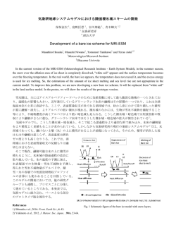
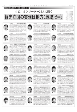
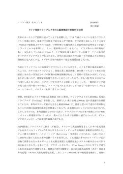
後方散乱電子回折(EBSD)による氷結晶中の微小方位差測定
繁山
航 1,永塚尚子 2,本間智之 3,高田守昌 3,東 久美子 2, 4,
Ilka Weikusat5,Martyn R. Drury6,Ernst-Jan Kuiper6,Gill Pennock6,東 信彦 3
1
長岡技術科学大学大学院
2
国立極地研究所
3
長岡技術科学大学
4
総合研究大学院大学
5
アルフレッド・ウェゲナー極地海洋研究所
6
ユトレヒト大学
Measurement of misorientations in ice crystals by using electron backscatter diffraction (EBSD)
Wataru Shigeyama1, Naoko Nagatsuka2, Tomoyuki Homma3, Morimasa Takata3, Kumiko Goto-Azuma2, 4,
Ilka Weikusat5, Martyn R. Drury6, Ernst-Jan Kuiper6, Gill Pennock6, Nobuhiko Azuma3
1
Graduate School of Engineering, Nagaoka University of Technology
2
National Institute of Polar Research
3
Nagaoka University of Technology
4
The Graduate University for Advanced Studies
5
Alfred-Wegener-Institut Helmholtz-Zentrum für Polar- und Meeresforschung
6
Faculty of Earth Science, Utrecht University
In order to understand dynamics of ice sheet flow, clarifying flow law is demanded. Conventional flow laws are based on
Glen’s flow law (Glen, 1955), which describes that ice deforms due to movements of dislocations on crystallographic basal
planes. However, strong contributions of grain boundary sliding and diffusion have been suggested at low strain rate in ice sheets
(Goldsby and Kohlstedt, 1997; Faria et al., 2014). Especially in high-impurity layers, where grains are small, it could be possible
that grain boundary sliding/diffusion activate deformation. If the mechanism of formation of those fine grains is discriminated
either as impeding grain growth by impurities or as recrystallization caused by coalescence of dislocations, this knowledge can
contribute to clarifying the flow law. To reveal such a mechanism of fine-grain formation, investigation of distribution of submicrometer impurities (solid particles) and misorientations around grain/subgrain boundaries are important. We therefore use
scanning electron microscope (SEM) and electron backscatter diffraction (EBSD) for high-resolution observations and
measurements of crystal orientation. This report shows our recent progresses for EBSD on ice, focusing on evaluation of accuracy
of measured crystal orientations for analyses of misorientations, rotation axes and their distribution. Examples of such
microstructural features found in deformed laboratory-made ice will be shown.
1.はじめに
海面上昇や気候変動の予測において重要な氷床流動の動態を理解するため,氷床氷の流動則を解明する必要が
ある.これまでの氷の力学試験やフィールド観測から,氷床の流動は主に氷結晶の基底面上の転位の運動による
塑性変形で生じると考えられ,Glen の流動則(Glen, 1955)が用いられているが,氷床中のような低ひずみ速度で
は粒界すべりや拡散の寄与が大きいことが示唆されている(Goldsby and Kohlstedt, 1997; Faria et al., 2014).特に不
純物を多く含む層では結晶粒が小さく,粒界すべり・拡散が変形を活性化している可能性がある.この高濃度不
純物層での細結晶粒化が不純物(固体微粒子)の粒成長抑制によるものか,或いは転位の蓄積による亜粒界・粒
界形成によるものかが明らかになれば流動則の構築に大きく貢献することになる.これには,サブミクロンサイ
ズの固体微粒子の存在状況や亜粒界・粒界近傍の結晶方位差を詳細に調べる必要があり,走査型電子顕微鏡
(SEM)および後方散乱電子回折(EBSD)が有効である.我々はこれら方法を氷に適用するため,観察・測定法
の改良を行ってきた.
本研究では,EBSD を用いてひずみを受けた氷結晶方位の詳細な測定から結晶粒内や粒界近傍の微小な結晶方位
差を調べ,転位の蓄積よる亜粒界の形成ついて考察を行う.また,EBSD による結晶方位の測定値に含まれる誤差
の影響を明らかにするため,本報では,結晶方位測定値の精度評価と変形氷の結晶組織に見られる結晶方位差と
その回転軸方位,分布について報告する.
2.後方散乱電子回折(EBSD)
EBSD は,試料に照射した電子線の菊池パターンから結晶方位を決定するものである.角度分解能は 0.5°から
1.5°であり,電子線の照射位置を移動させることで,最高 10nm オーダーの空間分解能で連続的な測定を行うこと
ができる(Humphreys, 2001).六方晶の氷の場合,結晶主軸(c 軸)・副軸(a 軸)方位の両者を同時に測定でき,
結晶内部の微小な結晶方位差とその回転軸方位,分布の分析が可能である.従来,氷の結晶方位測定には,ファ
ブリック・アナライザー,エッチピット法,X 線回折等が用いられてきたが,これらの方法はこのような氷の微小
な結晶方位差とその分布の分析に十分な分解能を持たない.本研究では,高い角度・空間分解能を持つ EBSD によ
って,結晶粒内や粒界近傍の微小な結晶方位の詳細な測定を行う.
装置
使用する EBSD 分析装置(Oxford Instruments HKL NordlysNano)は SEM(FEI Quanta 450 FEG)に搭載されてい
る.また,氷を SEM の真空チャンバーにおいて維持するため,クライオシステム(Quorum Technologies PP3010)
が搭載されており,試料ステージは液体窒素を用いて-190℃までの任意の温度に設定することができる.Fig. 1 に
装置の概略図を示す.菊池パターンの取得には,試料表面を平滑に処理し,SEM チャンバーにおいて 70°傾斜さ
せる必要がある.しかし,チャンバー内の電子銃や EBSD 分析装置,冷却装置等の位置関係から試料ステージを
70°傾斜させることが困難であるため,35°の予備傾斜を持つ試料台を製作し,試料ステージをさらに 35°傾斜
させることで計 70°の試料傾斜を実現した.試料表面処理には,マイクロトームを用いた.35°傾斜した試料の
表面処理を行うため,氷を固定した状態でマイクロトームの使用を可能とする治具を製作し,より正確な傾斜角
を実現している(Fig. 2).
3.
EDS analyzer
Dry N2
Emitter
Cold trap
SEM chamber
Sample and holder
SE detector
Preparation chamber
35°pre-tilt
holder
Cold
stage
LN2
Heat
exchanger
Fig. 1
EBSD analyzer
Cold
stage
Schematic illustration of the instrumentation
Microtoming jig
10mm
Fig. 2
35°pre-tilt holder and microtoming jig
4.結果
製作した試料台を使用した場合に測定される結晶方位の誤差について理解するため,内部ひずみが少ない焼鈍
人工氷と内部ひずみを多く含む変形人工氷を用いて結晶内部の方位測定値の精度評価を行った.発表において
は,この結果に併せて EBSD によって明らかとなった変形氷の結晶組織に見られる結晶方位差とその回転軸方
位,分布を示す.
参考文献
Faria, S.H., Weikusat, I. and Azuma, N., 2014, The microstructure of polar ice. Part II: State of the art, J. Struct. Geol. 61,
21-49
Glen, J.W., The creep of polycrystalline ice, 1955, Proc. Roy. Soc. London, A228, 519-538
Goldsby, D.L. and Kohlstedt, D.L., 1977, Grain boundary sliding in fine-grained ice I, Scr. Mater., 37, 1399-1406
Humphreys, F.J., 2001, Grain and subgrain characterization by electron backscatter diffraction, J. Mat. Sci. 36, 3833-3854
© Copyright 2026 Paperzz