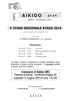
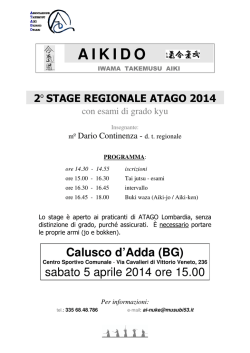
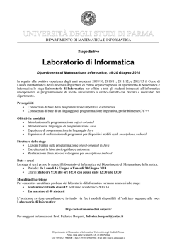
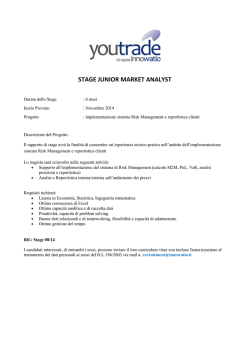
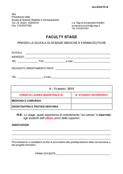
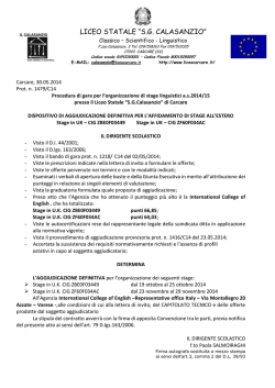
THE INTERNATIONAL JOURNAL OF
Int. J. Dev. Biol. 53: 129-134 (2009)
DEVELOPMENTAL
BIOLOGY
doi: 10.1387/ijdb.082617mz
www.intjdevbiol.com
The 2-cell block occurring during development of outbred
mouse embryos is rescued by cytoplasmic factors
present in inbred metaphase II oocytes
MARIO ZANONI1, SILVIA GARAGNA1, CARLO A. REDI2 and MAURIZIO ZUCCOTTI*,3
1Laboratorio
di Biologia dello Sviluppo, Dipartimento di Biologia Animale and Centro di Ingegneria Tissutale,
Universita' degli Studi di Pavia, Pavia, Italy, 2Fondazione I.R.C.C.S. Policlinico San Matteo, Pavia, Italy and
3Sezione di Istologia ed Embriologia, Dipartimento di Medicina Sperimentale, Universita’ degli Studi di Parma, Parma, Italy.
ABSTRACT In mice, completion of preimplantation development in vitro is restricted to certain
crosses between inbred strains. Most of the outbred and inbred strains cease development at the
2-cell stage, a phenomenon known as the "2-cell block". Reciprocal mating between blocking and
non-blocking strains has shown that the 2-cell block is dependent upon female, but not male,
developmental information. One question that still remains unanswered is whether the genome
of the metaphase II (MII) oocyte is genetically programmed to express, during the very early stages
of development, some factor(s) required to determine developmental competence beyond the 2cell stage. In the present study, we have addressed this question by performing reciprocal MIIchromosome plate transfer between MII oocytes of a non-blocking inbred strain and MII oocytes
of a blocking outbred strain. Here, we report that development beyond the 2-cell stage does not
depend on the MII genome, but instead it relies on a cytoplasmic factor(s) already present in
ovulated non-blocking oocytes, but absent, inactive or quantitatively insufficient in blocking
oocytes. Further evidence of the ooplasmic origin of this component(s) was obtained by transferring a small quantity of ooplasm from non-blocking MII oocytes to blocking MII oocytes or 2-cell
embryos. Following the transfer, a high percentage of blocking oocytes/embryos acquired
developmental competence beyond the 2-cell stage and reached the blastocyst stage. This study
shows that development beyond the 2-cell stage relies also on a factor(s) already present in the
ovulated oocyte.
KEY WORDS: preimplantation, 2-cell block, nuclear transfer, cytoplasm transfer
Introduction
In mice, completion of preimplantation development in vitro is
restricted to certain crosses between inbred strains. Most of the
outbred and inbred strains cease development at the 2-cell stage,
a phenomenon known as the ‘2-cell block’. Although it occurs in
particular in vitro culture conditions (for a review see Biggers,
1998), the 2-cell block may represent a model study that provides
important insights on a critical stage of development, when zygotic
genome activation (ZGA) occurs (Schultz, 2002). ZGA marks the
passage from maternal to embryonic control of development, and
represents the first crucial hurdle in the life of the new individual as
inhibiting the activation of embryonic genes arrests development
(Schultz and Worrad, 1995). Blocks to development at the time
when ZGA occurs have been described in other mammals (Schultz,
2002), including humans at the 4-cell stage, when a high percentage of IVF embryos is lost.
The 2-cell block is rescued when cytoplasm is transferred from
2-cell non-blocking embryos to 2-cell blocking embryos, allowing
completion of preimplantation development (Muggleton-Harris et
al., 1982). Rescue does not occur when cytoplasm is injected from
a 1-cell G1 stage non-blocking embryo to a blocking 2-cell embryo
(Muggleton-Harris et al., 1982), suggesting that the component(s)
that allows to overcome the 2-cell block is yet not present, it has not
been translated or it is not functioning. Instead, rescue occurs
Abbreviations used in this paper: MII, metaphase II
*Address correspondence to: Maurizio Zuccotti. Sezione di Istologia ed Embriologia, Dipartimento di Medicina Sperimentale, Univesita’ degli Studi di Parma
43100, Parma, Italy. e-mail: [email protected]
Accepted: 6 June 2008. Published online: 18 December 2008.
ISSN: Online 1696-3547, Print 0214-6282
© 2008 UBC Press
Printed in Spain
130
M. Zanoni et al.
consistently when injection is performed from a 2-cell G1 or G2
stage, but not S phase, non-blocking embryo to a 2-cell blocking
embryo at any phase of the cell cycle (Pratt and Muggleton-Harris,
1988). The blocking activity is not due to a blocking factor, since the
transfer of cytoplasm from blocking to non-blocking 2-cell embryos
did not impair their ability to complete preimplantation development (Pratt and Muggleton-Harris, 1988).
The cyclic nature of these molecules indicates either a transcriptional control of their expression by the embryonic genome or a
translational/post-translational regulation of transcripts/proteins.
Reciprocal mating between blocking and non-blocking strains
has shown that the 2-cell block is dependent upon a female, but not
male, developmental information (Goddard and Pratt, 1983). One
question that still remains unanswered is whether the genome of
the MII oocyte is genetically programmed to express, during the
very early stages of development, some factor(s) required to
determine the developmental competence beyond the 2-cell stage.
In the present study we have addressed this question by
performing reciprocal MII-chromosome plates (MII-plates) transfer
between MII oocytes of a non-blocking inbred strain and MII
oocytes of a blocking outbred strain. We report that development
beyond the 2-cell stage does not depend on the MII genome, but
instead it relies on a cytoplasmic factor(s) already present in
ovulated non-blocking oocytes, but absent, inactive or quantitatively insufficient in blocking oocytes. Further evidences of the
ooplasmic origin of this component(s) were obtained by transferring a small quantity of ooplasm from non-blocking MII oocytes to
blocking MII oocytes or 2-cell embryos. Following the transfer, a
high percentage of blocking oocytes/embryos acquired develop-
A
B
C
D
Fig. 1. An MII-plate was removed from an F1 MII oocyte (A,B) and
transferred to another, previously enucleated, F1 MII oocyte (C,D).
All the other MII-plate transfers were obtained following the same
procedure. Arrows: an MII-plate (A); an MII-plate that is being removed
from the oocyte (B); an MII-plate enclosed into a small portion of
cytoplasm and oolemma, removed using a micropipette (C); an MII-plate
few minutes after its transfer into a previously enucleated oocyte (D).
mental competence beyond the 2-cell stage and reached the
blastocyst stage.
Results
The presence in non-blocking embryos of a functional rescuingfactor(s) beginning with the 2-cell stage (Pratt and MuggletonHarris, 1988) suggests either a developmentally regulated transcription or a translational/post-translational control of transcripts/
proteins that could be present already in the ovulated MII oocyte.
Since the male genome is unimportant in determining/overcoming
the 2-cell block (Goddard and Pratt, 1983), to understand whether
the female genome is programmed to express the rescuing activity
or whether the rescuing factor(s) is already present in the MII
ooplasm, in the present study we have performed reciprocal MIItransfer between MII oocytes of a non-blocking inbred strain
(B6C3F1, F1) and MII oocytes of a blocking outbred strain (CD1).
We argued that if the MII-plate of a non-blocking strain transferred
into an enucleated oocyte of a blocking strain was supporting
development beyond the 2-cell stage, whereas the reciprocal
transfer was not, then we could assume that the female F1 genome
was programmed to control the expression of this factor(s) in a
developmentally regulated fashion. On the contrary, we could infer
that the factor(s) was already present in the ooplasm of nonblocking MII oocytes.
Reciprocal MII-plates transfer between MII oocytes of a nonblocking inbred strain and MII oocytes of a blocking outbred
strain
To this end, four different types of reconstructed MII oocytes
were produced: F1F1MII (F1MII-plates transferred into F1 enucleated oocytes), F1CD1MII (CD1MII-plates transferred into F1 enucleated oocytes), CD1CD1MII (CD1MII-plates transferred into CD1
enucleated oocytes) and CD1F1MII (F1MII-plates transferred into
CD1 enucleated oocytes). Following MII-transfer (Figure 1), oocytes were inseminated in vitro with capacitated spermatozoa
isolated from the epidydimes of F1 males; 6 hr later, those oocytes
that presented two polar bodies (PBs) were transferred to M16
medium for further embryonic development. Control in vitro
fertilisation (IVF) experiments were conducted in parallel. For each
type of experiment, about 10 of the inseminated oocytes with two
PBs were fixed, stained with acetic orcein and observed under
phase microscopy for the presence of a male and a female
pronucleus. The great majority (98%) of all the reconstructed
oocytes presented two polar bodies and were correctly fertilised by
one single sperm (Fig. 2A). Then, 1-cell embryos were cultured for
up to 96 hrs to observe preimplantation development.
The MII-transfer procedure was tested, from fertilisation to birth,
on F1F1MII reconstructed oocytes. These experiments showed that
the whole procedure did not affect the preimplantation developmental potential of the reconstructed oocytes. The frequency of
F1F1MII 2-cell embryos that developed to the blastocyst stage
(63.3%; Table 1) was not significantly different compared to that
obtained after IVF of normal F1 MII oocytes (71.4%; p= 0.218,
statistically not significant). When eight blastocysts obtained from
F1F1MII oocytes where transferred to a pseudopregnant female
recipient, three of them reached full-term development giving birth
to healthy mice (Fig. 2B). The majority of the zygotes obtained from
CD1CD1MII or CD1F1MII oocytes stopped development at the 2-cell
Ooplasmic factors rescue the 2-cell block 131
A
D
B
E
C
F
Fig. 2. Development of embryos obtained by reciprocal MII-plate
transfer. (A) About ten of the oocytes showing two polar bodies were
fixed and stained with acetic orcein and observed under phase microscopy. The micrograph shows the male (black harrowhead) and female
(white arrow) pronuclei. The male pronucleus is recognisable for the
presence of the sperm tail. (B) Three F2 mice born following IVF
fertilisation and embryo transfer of blastocysts obtained after insemination of F1F1MII reconstructed oocytes with F1 sperm. The mouse with the
white coat colour is the recipient female. (C) Most of the CD1F1MII
stopped development at the 2-cell stage. (D) The majority of F1CD1MII 2cell embryos developed to the blastocyst stage. (E) Summary of the
percentage of development of the 2-cell embryos obtained from the four
different types of reconstructed oocytes. (F) Blastomeres of a single
blastocyst developed from an F1CD1MII reconstructed oocyte.
stage (73.5% and 81.2%, respectively) (Table 1, Fig. 2C), a
number not significantly different (p= 0.351, statistically not significant) to that obtained after IVF of CD1 oocytes with F1 sperm
(75.6%). As shown in Table 1, 5.9% of CD1CD1MII, none CD1F1MII
and 42.2% F1CD1MII 2-cell embryos developed to the blastocyst
stage (Fig. 2D).
As reported before (Whittingham, 1974; Pratt and MuggletonHarris, 1988), the 2-cell block is not an ‘all or none’ phenomenon,
but some of the embryos from blocking strains may develop
beyond the 2-cell stage and reach blastocyst. In the reconstructed
oocytes that we produced with a CD1 ooplasm, the number of
blastocysts varied from none (CD1F1MII) to 5.9% (CD1CD1MII). The
reconstructed embryos that best developed beyond the 2-cell
stage were those with an F1 ooplasm, namely F1F1MII and F1CD1MII.
The percentage of F1CD1MII 2-cell embryos that reached the 4-cell
stage (78.9%) was not significantly different (p= 0.085, statistically
not significant) to that of F1F1MII 2-cell embryos (89.4%) (Table 1,
Fig. 2E); however, although the majority of F1CD1MII embryos
developed beyond the 2-cell stage, further preimplantation development was significantly different comparing the two types of
reconstructed embryos as 75.7% F1F1MII and 54.9% F1CD1MII 2-cell
embryos (p<0.05) reached the morula stage and 63.3% F1F1MII and
42.2% F1CD1MII embryos (p<0.05) reached the blastocyst stage
(Table 1). In those F1CD1MII embryos that reached the blastocyst
stage, the blastomere number was attested at 35+8 (Fig. 2F), a
number of cells not significantly different (p= 1, statistically not
significant) to that found in control IVF F1 embryos (35+5).
Cytoplasm transfer from non-blocking to blocking oocytes
Since this rescuing factor(s) is present in the ooplasm of
developmentally competent gametes, we then argued whether the
transfer of cytoplasm from F1 MII oocytes to CD1 MII oocytes could
result in the acquisition of developmental competence by this latter
blocking strain. When we injected approximately 8-10 pl of MII-F1
ooplasm into MII-CD1 oocytes, the majority of CD1 2-cell embryos
developed to the 4-cell stage (34 4-cell out of 49 2-cell embryos,
69.4%), and 38.7% of them developed to blastocyst (Table 2).
Cytoplasm transfer from MII-CD1 to MII-CD1 oocytes did not
rescue these oocytes from the 2-cell block (Table 2). This result
shows that a small quantity of F1 ooplasmic factor(s) is capable of
rescuing the majority of blocking CD1 oocytes to develop beyond
the 2-cell stage and also that the CD1 ooplasm does not represent
an inhibitory environment since it did not inactivate the functionality
of the transferred F1 factor(s). While the transfer of a small quantity
of ooplasm from F1 oocytes to CD1 oocytes rescues the latter from
the 2-cell block, the transfer of the whole F1 MII-plate to an
enucleated CD1 oocyte did not give any developmental improvement, suggesting that the rescuing factor is not present on or
around the MII-plate.
Cytoplasm transfer from non-blocking MII oocytes to blocking 2-cell embryos
In a third type of experiment we addressed the question of
whether the ooplasm factor(s) activates a series of events, comprised between the oocyte and the 2-cell embryo, that lead to
completion of the second mitotic division or whether itself can
TABLE 1
RECIPROCAL MII-PLATE TRANSFER
N. of
Enucleated Transferred reconstructed
MII oocyte MII-plate
MII oocytes
% ± S.D. (N.) of preimplantation embryos
2-cell
4-cell*
Morula
Blastocyst
F1
F1
89
100 (66)
89.4 ± 10.1
(59)
75.7 ± 9.1
(50)
63.3 ± 13.8
(42)
CD1
CD1
54
100 (34)
26.5 ± 13.2
(9)
20.6 ± 11.2
(7)
5.9 ± 7.3
(2)
CD1
F1
87
100 (69)
18.8 ± 6.3
(13)
7.2 ± 3.6
(5)
0
F1
CD1
89
100 (71)
78.9 ± 10.1
(56)
54.9 ± 9.1
(39)
42.2 ± 10.1
(30)
Reciprocal MII-plate transfer between enucleated MII oocytes of a non-blocking inbred strain (F1)
and enucleated MII oocytes of a blocking outbred strain (CD1). Reconstructed oocytes were
inseminated with F1 spermatozoa and preimplantation development beyond the 2-cell stage was
analysed.
*: the percentage was calculated considering the 2-cell stage as 100%.
132
M. Zanoni et al.
TABLE 2
TRANSFER OF OOPLASM FROM F1-MII TO CD1-MII RESCUES
THE DERIVED EMBRYOS FROM 2-CELL BLOCK
N. (%) of preimplantation embryos
MII oocyte
recipient
MII ooplasm
transferred
N. of oocytes
2-cell
CD1
F1
86
49
CD1
CD1
82
32
4-cell
Morula Blastocyst
34 (69.4) 26 (53.1)
8 (25)
5 (15.6)
19 (38.7)
1 (3.1)
participate to unblock the second mitotic division. We have injected
approximately 10 pl of cytoplasm taken from F1 oocytes at the
opposite side of the MII-plate, into one single blastomere of early
(21 hr post insemination) 2-cell CD1 embryos obtained following
IVF with F1 spermatozoa. Out of a total of 58 CD1 2-cell embryos
injected, 36 (62%) developed to the 4-cell stage, 26 (44.8%) to the
morula and 23 (39.6%) to the blastocyst stage. Development
beyond the 2-cell stage in these experiments was significantly
(p<0.05) improved compared to that of embryos obtained after IVF
of CD1 MII oocytes. These results demonstrate that the same
factor(s) present in the F1 ooplasm is capable of rescuing CD1 2cell embryos and to induce further development, with a percentage
of embryos that reached the blastocyst stage similar (39.6%) to
that of reconstituted F1CD1MII embryos (42.2.%, p = 0.668, statistically not significant; Table 1). Also, the small quantity of cytoplasm
that is transferred in one blastomere is sufficient to induce the
second mitotic division in both blastomeres, as already shown
before (Pratt and Muggleton-Harris, 1988) when cytoplasm was
transferred between two 2-cell embryos. We think that a passage
of molecules from the injected blastomere to the other may occur
and induce cell division in the uninjected blastomere. This migration has been demonstrated to occur through gap junctions between blastomeres of cleavage embryos and fibroblasts, these
latter shown to express Oct-4, an embryonic-specific transcription
factor (Burnside and Collas, 2002).
Discussion
The results of our study show that development beyond the 2cell stage does not depend on the MII genome, but instead it relies
on a cytoplasmic factor(s) already present in ovulated non-blocking oocytes, but absent, inactive or quantitatively insufficient in
blocking oocytes. These results also indicate that the CD1 MIIplate, within an F1 cytoplasm, does not gain full developmental
competence, suggesting a strain-specificity of this genome. At
these very early stages of development, epigenetic modifications
from a gametic to an embryonic type of chromatin organisation
mold an expression profile required for development. Methylation
of cytosines in CpG sites is crucial for regulating the temporal,
spatial and parent-specific gene expression patterns (Haaf, 2006).
The paternal genome is actively demethylated within a few hours
after fertilisation, whereas the maternal genome is passively
demethylated after the 2-cell stage (Mayer et al., 2000; Barton et
al., 2001). Recent studies have demonstrated a strain-specific
difference in the efficiency of methylation reprogramming during
preimplantation development (Shi and Haaf, 2002; Haaf, 2006). In
vitro development of NMRI (an outbred strain that shows the 2-cell
block) 2-cell embryos show a higher frequency of abnormal methylation patterns than B6C3F1, suggesting a functional link between
methylation reprogramming defects and lower developmental com-
petence (Shi and Haaf, 2002). The transfer of the CD1 genome
within the F1 ooplasm clearly improves its developmental competence, but the reprogramming activity is not completely efficient.
We have given a clear indication on the presence within the
mouse F1 ooplasm of a component(s) involved in conferring
developmental competence beyond the 2-cell stage. However, its
molecular feature remains unknown. The data of earlier studies
and a number of considerations that we may draw out from our
results may help its search.
One marked evidence is that this factor(s), or its functional form,
has a cyclic nature: it is present in MII oocytes (our data), it
disappears in the early 1-cell embryo, reappears in the early 2-cell
embryo, disappears in S-phase of the second cell cycle and
reappears again in the following G2-phase (Pratt and MuggletonHarris, 1988). This cyclic appearance speaks for a possible role of
this component(s) in the regulation of the embryo’s cell cycle and
since we have shown that its injection into a blastomere rescues 2cell blocking embryos from the block, the rescuing molecule is
probably the same during the first two cell cycles. At this regard, the
maturation promoting factor (MPF) has been considered a possible candidate. MPF is a cell cycle regulator in all eukaryotes and
failure of MPF activation leads to arrest of the cell cycle at the G2/
M phase (Norbury and Nurse, 1992). MPF is a complex of cyclindependent kinase p34cdc2 and cyclin B; p34cdc2 kinase is
dephosphorilated and activated by cdc25 phosphatase (King et al.,
1994; Lew e Kornbluth, 1996) and cyclin B is phosphorylated by
Polo-like kinase I and translocated into the nucleus (Pines and
Hunter, 1991; Ookata et al., 1992). Microinjection of MPF purified
from Xenopus laevis eggs into 2-cell mouse embryos, rescued
these embryos from the block (Nakano et al., 2001). An earlier work
has proposed that 2-cell blocked embryos contain enough p34cdc2
to induce mitosis, but the mechanisms for its dephosphorilation
and activation is inefficient (Aoki et al., 1992). Absence of MPF
activation in G2/M-arrested 2-cell embryos occurs in the presence
of phosphate in the culture medium, it affects the dephosphorilation
of phosphorylated p34cdc2 (Aoki et al., 1992; Haraguchi et al.,
1996) and is correlated to a decrease in the levels of cyclin B and
cdc25B mRNAs (Haraguchi et al., 1999). When phosphate is
absent from the medium, the activation pattern of p34cdc2 kinase
during the second cell cycle is similar to that in the first cell cycle,
allowing completion of preimplantation development (Haraguchi et
al., 1996).
Although a possible involvement of MPF as a crucial molecule
in rescuing embryos from the 2-cell block is intriguing and supported by a number of observations, our results suggest that it
might not be the sole factor. In mouse MII oocytes MPF is
compartmentalised, with the majority of this kinase around the MIIplate (Fulka et al., 1995). The MII-transfer technique that we used
removes the whole MII-plate together with some cytoplasm around
the spindle (Figure 1), thus MPF from an F1 oocyte should be
transferred altogether into the recipient enucleated CD1 oocyte.
Since our results clearly show that the reconstituted CD1F1MII
oocytes maintained the 2-cell block (Table 1), MPF present in MII
oocytes is likely not enough to release 2-cell embryos from the
block. MPF is involved in the first mitotic division which occurs also
in blocking embryos, but some other molecule(s), perhaps other
cell cycle molecules, already present in the F1 ooplasm in an active
form (as shown by the transfer of small quantities of F1 cytoplasm
to MII oocytes or 2-cell CD1 embryos), is needed to complete the
Ooplasmic factors rescue the 2-cell block 133
second cell cycle.
Other candidates might be found focusing on the events that
most characterise these early developmental stages. For example,
the female gamete accumulates during growth a group of gene
transcripts and proteins that become necessary, after fertilisation,
for successful embryogenesis. These maternal-effect genes are
crucial during the oocyte-to-embryo transition (Zheng and Dean,
2007), since their misregulation determines a developmental block,
mainly at the 2-cell stage. Our next experiments will try to compare
the pattern of expression of these genes and of their proteins in
preimplantation embryos derived from oocytes of blocking and
non-blocking strains.
Materials and Methods
Animals
Mice of the strains B6C3F1 (3-4 month-old males and 4-5 week-old
females) and CD1 (4-5 week-old females) were purchased from Charles
River (Como, Italy). Animals were maintained under controlled room
conditions (22°C, with 60% air moisture and 14L:10D photoperiod) and
investigations were conducted in accordance with the guiding principles of
European (n. 86/609/CEE) and Italian (n. 116/92, 8/94) laws protecting
animals used for scientific research.
Reagents
All reagents were purchased from Sigma Chemical Co. (Milano, Italy),
unless otherwise stated.
Micromanipulation of metaphase II oocytes
Ovulated MII oocytes were collected from the oviducts of females
injected with 3.5 I.U. Folligon (Intervet International, Netherlands), followed
48 hr later by an injection of 3.5 I.U. Corulon (Intervet International,
Netherlands). MII oocytes were placed in M2 medium containing hyaluronidase (Type II, 500 IU/ml in BSA-free M2—medium) for 4-5 min., then
cumulus-free oocytes were thoroughly washed in Whittingham medium
(Whittingham, 1971) and transferred to a drop of fresh Whittingham
medium in a 35x10 mm plastic petri dish (Corning, Bibby Sterilin, UK) under
mineral oil and placed in incubator at 37°C and 5% CO2, before further
treatment. Groups of 20-30 MII oocytes were then transferred to HEPESbuffered CZB (H-CZB) medium (Chatot et al., 1989) containing 0.1% PVP
(Polyvinylpyrrolidone), 5 µg/ml cytochalasin B and 0.1% DMSO, for enucleation. Enucleation and MII transplantation were performed using piezodriven (PMM 150 FU, Prime Tech, Ibaraki, Japan) borosilicate micropipettes (Clark Instruments, Edenbridge, UK) under an inverted microscope
equipped with DIC optics (Olympus, Ibaraki, Japan). Following enucleation, oocytes were transferred in M16 medium (Biggers, 1998) and
incubated at 37° C in 5% CO2 in air for 1 1/2 hr. Then they were transferred
in H-CZB containing 1% PVP and 5 µg/ml cytochalasin B and following MII
transplantation, reconstructed oocytes were incubated in M16 for 1 1/2 hr,
washed in Whittingham medium and finally transferred in fresh Whittingham
medium for insemination with capacitated spermatozoa.
In vitro fertilisation and embryo culture
Sperm were isolated as previously described (Zuccotti et al., 1998) and
incubated for 60 min in 100 µl drops of Whittingham medium at a final
concentration of 1.8x10⁶ sperm/ml. MII oocytes were transferred into the
insemination drop and incubated at 37°C under a 5% CO2 in air for 2 hr.
Based on the presence of a second polar body, as a sign that fertilisation
had occurred, presumptive 1-cell stage embryos were pooled, transferred
from Whittingham medium to a 30 µl drop of M16 medium (Biggers, 1998),
for further development.
For each experiment type, about 10 oocytes with two polar bodies were
used to check that fertilisation was occurred. Oocytes were placed under
a coverslip suspended with drops of beeswax at each corner; the coverslip
was slightly pushed down until the oocytes were held in place. Oocytes
were then treated with a series of fixatives and washing. Glutaraldehyde
(2.5%) was drawn under the coverslip using a filter paper placed at one side
of the coverslip; this fixative was followed by 10% formalin, a wash with
distilled water, 95% ethanol and finally 1% acetic orcein.
Statistical analysis
Statistical analysis was done with the SigmaStat 3.0 software, introducing the raw data of each single experiment performed and using the
Student’s t test. A value of p<0.05 was considered statistically significant.
Acknowledgements
This work was supported by grants from: FIRB 2005 (Project N.
RBIP06FH7J), Regione Lombardia (Project REGLOM06), Millipore, Olympus Foundation Science for Life, Fondazione CARIPLO.
References
AOKI, F., CHOI, T., MORI M., YAMASHITA, M., NAGAHAMA, Y. and KOHMOTO, K.
(1992). A deficiency in the mechanism for p34cdc2 protein kinase activation in
mouse embryos arrested at 2-cell stage. Dev Biol 154: 66-72.
BARTON, S.C., ARNEY, K.L., SHI, W., NIVELEAU, A., FUNDELE, R., SURANI, M.A.
and HAAF T. (2001). Genome-wide methylation patterns in normal and uniparental early mouse embryos. Hum Mol Genet 10: 2983-2987.
BIGGERS, J.D. (1998). Reflections on the culture of the preimplantation embryo. Int
J Dev Biol 42: 879-884.
BURNSIDE, A.S. and COLLAS, P. (2002). Induction of Oct-3/4 expression in somatic
cells by gap junction-mediated cAMP signaling from blastomeres. Eur J Cell Biol
81: 585-591.
CHATOT, C.L., ZIOMEK, C.A., BAVISTER, B.D., LEWIS, J.L. and TORRES, I. (1989).
An improved culture medium supports development of random-bred 1-cell mouse
embryos in vitro. J Reprod Fertil 86: 679-688.
FULKA, J. JR, OUHIBI, N., FULKA, J., KANKA, J. and MOOR, R.M. (1995).
Chromosome condensation activity (CCA) in bisected C57BL/6JxCBA mouse
oocytes. Reprod Fertil Dev 7: 1123-1127.
GODDARD, M.J. and PRATT, H.P. (1983). Control of events during early cleavage of
the mouse embryo: an analysis of the ‘2-cell block’. J Embryol Exp Morphol 73:
111-133.
HAAF, T. (2006). Methylation dynamics in the early mammalian embryo: implications
of genome reprogramming defects for development. Curr Top Microbiol Immunol
310: 13-22.
HARAGUCHI, S., NAITO, K., AZUMA, S., SATO, E., NAGAHAMA, Y., YAMASHITA,
M. and TOYODA, Y. (1996). Effects of phosphate on in vitro 2-cell block of AKR/
N mouse embryos based on changes in cdc2 kinase activity and phosphorylation
states. Biol Reprod 55: 598-603.
HARAGUCHI, S., NAITO, K. and SATO, E. (1999). Phosphate exposure during the
late 1-cell and early 2-cell stages induces a time-specific decrease in cyclin B and
cdc25B mRNAs in AKR/N mouse embryos in vitro. Zygote 7: 87-93.
KING, R.W., JACKSON, P.K. and KIRSCHNER, M.W. (1994). Mitosis in transition.
Cell 79: 563-571.
LEW, D.J. and KORNBLUTH, S. (1996). Regulatory roles of cyclin dependent kinase
phosphorylation in cell cycle control. Curr Opin Cell Biol 8: 795-804.
MAYER, W., NIVELEAU, A., WALTER, J., FUNDELE, R. and HAAF T. (2000).
Demethylation of the zygotic paternal genome. Nature 403: 501-502.
MUGGLETON-HARRIS, A., WHITTINGHAM, D.G. and WILSON, L. (1982). Cytoplasmic control of preimplantation development in vitro in the mouse. Nature 299:
460-462.
NAKANO, H. and KUBO, H. (2001). Rescue of mouse embryos from 2-cell blocks by
microinjection of maturation promoting factor. Fertil Steril 75: 1194-1197.
NORBURY, C. and NURSE, P. (1992). Animal cell cycles and their control. Annu Rev
Biochem 61: 441-470.
OOKATA, K., HISANAGA, S., OKANO, T., TACHIBANA, K. and KISHIMOTO, T.
(1992). Relocation and distinct subcellular localization of p34cdc2-cyclin B com-
134
M. Zanoni et al.
plex at meiosis reinitiation in starfish oocytes. EMBO J 11: 1763-1772.
PINES, J. and HUNTER, T. (1991). Cyclin-dependent kinases: a new cell cycle motif?
Trends Cell Biol 1: 117-121.
an indicator of early developmental failure. Mol Reprod Dev 63: 329-334.
WHITTINGHAM, D.G. (1971). Culture of mouse ova. J Reprod Fertil Suppl 14: 7-21.
PRATT, H.P. and MUGGLETON-HARRIS, A.L. (1988). Cycling cytoplasmic factors
that promote mitosis in the cultured 2-cell mouse embryo. Development 104: 115120.
WHITTINGHAM, D.G. (1974). Fertilisation, early development and storage of mammalian ova in vitro. In The Early development of mammals. Brittish Society for Devl
Biol. Symposium 2 (Eds. Balis M. and Wild A.E.) Cambridge University Press pp.
1-24.
SCHULTZ, R.M. and WORRAD, D.M. (1995). Role of chromatin structure in zygotic
gene activation in the mammalian embryo. Semin Cell Biol 6: 201-208.
ZHENG, P. and DEAN, J. (2007). Oocyte-specific genes affect folliculogenesis,
fertilization, and early development. Semin Reprod Med 25: 243-251.
SCHULTZ, R.M. (2002). The molecular foundations of the maternal to zygotic
transition in the preimplantation embryo. Hum Reprod Update 8: 323-331.
ZUCCOTTI, M., PICCINELLI, A., GIORGI ROSSI, P., GARAGNA, S. and REDI, C.A.
(1995). Chromatin organisation during mouse oocyte growth. Mol Reprod Dev 41:
479-485.
SHI, W. and HAAF, T. (2002). Aberrant methylation patterns at the two-cell stage as
Further Related Reading, published previously in the Int. J. Dev. Biol.
See our recent Special Issue Fertilization, in honor of David L. Garbers and edited by Paul M. Wassarman and Victor D. Vacquier at:
http://www.ijdb.ehu.es/web/contents.php?vol=52&issue=5-6
See our Special Issue Mammalian Reproduction and Development in honor of Anne McLaren and edited by Brigid Hogan at:
http://www.ijdb.ehu.es/web/contents.php?vol=45&issue=3
A reversible block at the G1/S border during cell cycle progression of mouse embryos.
N Ouhibi, J Fulka, J Kanka and R M Moor
Int. J. Dev. Biol. (1994) 38: 731-736
2006 ISI **Impact Factor = 3.577**
© Copyright 2026 Paperzz