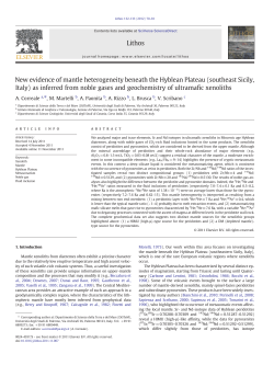
This article appeared in a journal published by Elsevier. The attached copy is furnished to the author for internal non-commercial research and education use, including for instruction at the authors institution and sharing with colleagues. Other uses, including reproduction and distribution, or selling or licensing copies, or posting to personal, institutional or third party websites are prohibited. In most cases authors are permitted to post their version of the article (e.g. in Word or Tex form) to their personal website or institutional repository. Authors requiring further information regarding Elsevier’s archiving and manuscript policies are encouraged to visit: http://www.elsevier.com/copyright Author's personal copy Immunology Letters 131 (2010) 67–72 Contents lists available at ScienceDirect Immunology Letters journal homepage: www.elsevier.com/locate/ Seroma fluid subsequent to axillary lymph node dissection for breast cancer derives from an accumulation of afferent lymph Erika Montalto a , Salvatore Mangraviti b , Gregorio Costa a , Paolo Carrega c , Barbara Morandi d , Gaetana Pezzino a , Irene Bonaccorsi a , Antonino Cancellieri e , Maria Cristina Mingari d,f , Mario Mesiti g , Guido Ferlazzo a,∗ , Giovanni Melioli b a Laboratorio di Immunologia e Biotecnologie Terapeutiche, Dipartimento di Patologia Umana, Università di Messina, Via Consolare Valeria 1, Messina - 98125, Italy Laboratorio Centrale di Analisi, Istituto Giannina Gaslini, Genova, Largo G. Gaslini 5, Genova - 16148, Italy c Laboratorio di Immunologia Clinica e Sperimentale, Istituto Giannina Gaslini, Largo G. Gaslini 5, Genova - 16148, Italy d Dipartimento di Medicina Sperimentale, Università degli Studi di Genova, Via Leon Battista Alberti 2, Genova - 16132, Italy e Unità di Chirurgia Generale ad Indirizzo Oncologico, Università di Messina, Via Consolare Valeria 1, Messina - 98125, Italy f Istituto Nazionale per la Ricerca sul Cancro, Largo Rosanna Benzi 10, Genova - 16132, Italy g Dipartimento di Protezionistica Ambientale, Sanità Sociale ed Industriale, Università di Messina, Via Consolare Valeria 1, Messina - 98125, Italy b a r t i c l e i n f o Article history: Received 12 January 2010 Received in revised form 1 March 2010 Accepted 10 March 2010 Available online 16 March 2010 Keywords: Seroma Breast neoplasms Lymph Lymphadenectomy Interleukin-6 a b s t r a c t Seroma is a frequent complication of breast cancer surgery, the etiology of which remains indefinite. It represents a subcutaneous accumulation of fluid frequently reported after surgical procedures such as axillary lymph node dissection. Despite previous studies have associated seroma fluid to an inflammatory exudate, the surgical removal of draining lymph nodes may indicate that seroma might not represent a mere exudate but rather an accrual of lymph drained from tributary tissues. To verify this hypothesis, seromas were collected at different intervals of time in patients operated upon for axillary lymph node removal. Fluids were analyzed in details by flow cytometry and biochemical assays for their cellular content and for their molecular features and relevant cytokine content. Lymphocytes and other peculiar blood mononuclear cells were present, while erythrocytes, platelets and granulocytes were absent or extremely rare. The protein concentration resulted lower (median 64%) than in peripheral blood. However, specific proteins related to locoregional tissues resulted highly concentrated (e.g. up to 500% for ferritin and 300% for lactate deydrogenase and exclusive presence of interleukin-6) whereas all enzymes and proteins synthesized in the liver or other organs (e.g. alkaline phosphatase, ALT, ␥GT, prealbumin, transferrin, ceruloplasmin, C3 and C4, ␣2 macroglobulin from liver; apolipoproteins from liver and gut; amylase and lipase from pancreas) were represented in reduced concentrations, thus ruling out that seroma proteins derive directly from blood serum. As a whole, this comprehensive cytological and molecular analysis provided evidences that seroma is constituted by serum ultrafiltrated-derived extracellular fluid of regions located upstream of removed lymph nodes. This fluid is then enriched by proteins and cells collected in the drained regions. Remarkably, seroma fluids collected in the same patient at different time points (up to 50 days following surgery) displayed similar biochemical features, clearly indicating that fluid composition was not significantly affected by postsurgical locoregional flogosis. Finally, the period of seroma formation indicates that lymph accumulates in the axillary region during the interval of time needed for afferent lymphatic vessels to re-anastomose with the efferent ducts. Therefore, seroma fluid represents a font of biological material suitable for investigating the biology of breast cancer, healing tissues and lymph. © 2010 Elsevier B.V. All rights reserved. 1. Introduction ∗ Corresponding author at: Laboratory of Immunology and Biotherapy, University of Messina, Italy. Tel.: +39 0 90 221 2040; fax: +39 0 90 221 2043. E-mail address: [email protected] (G. Ferlazzo). 0165-2478/$ – see front matter © 2010 Elsevier B.V. All rights reserved. doi:10.1016/j.imlet.2010.03.002 Afferent lymph stream moves from peripheral interstitial spaces to the draining lymph node stations through lymphatic vessels [1]. Following lymph nodes, the efferent lymph moves trough lymphatic vessels to thoracic duct and finally to the venous circulation. It is common notion that lymph from thoracic duct is character- Author's personal copy 68 E. Montalto et al. / Immunology Letters 131 (2010) 67–72 ized by a high concentration of lipids [2] derived from food intake and absorbed by the gut, thus suggesting that lymph may be considered a continuous sampling of peripheral anatomic districts [3,4]. Seroma is a subcutaneous accumulation of noninfected fluid, frequently observed in certain medical conditions after abdominal surgery [5] and axillary lymph node dissection for breast cancer [6]. The pathophysiology of seroma is largely unknown [as reviewed in 7]. Although seroma was originally considered the results of a pure lymphatic obstruction, thus far most of the studies have suggested that seroma is an essudate, with specific characteristics largely superimposable to those defined by Light rules [8–13]. Indeed, most of these studies analyzed seroma fluid content a few days following surgery, when a strong inflammatory component in the axillary region is conceivable. Nevertheless, the surgical removal of axillary lymph nodes corresponds to a physical interruption of lymphatic vessels draining lymph from interstitial spaces: in this context, subcutaneous accumulation of sterile seroma fluid could also be considered as an accumulation of afferent lymph in the absence of a guided lymph flow to locoregional lymph nodes. In the case of seroma consequent to axillary lymph node dissections, the district drained by sliced lymphatic vessel was hosting a breast cancer, with all the possible variation of protein composition related to different cancer stages, different inflammation (including postsurgical flogosis), different surgical procedures and, in some cases, neo-adjuvant therapies. Very few papers have described the characteristics of afferent lymph: in an animal model [14], it has been shown that lymph of pre-mammary gland contained 7, 6, and 10 times less protein, albumin, and globulin, respectively, than plasma. Glucose concentrations were equivalent and in lymph, only 6,9% of serum cholesterol and 50% of triglyceride and calcium were found. Gamma-glutamyl-transaminase and aspartate transaminase were substantially higher in plasma than in lymph. Thus, substantial differences are evident when plasma is compared to lymph. In another studies in humans [15], it has been described that afferent lymph had selective features, in particular the concentration of protein was significantly lower than in plasma (<25%) and specific molecules (i.e. interleukin (IL)-6 and IL-8) were extremely well represented in lymph and virtually absent in human plasma. In this report, we describe a series of biochemical and cellular characteristics of seroma fluid collected in women operated upon breast cancer and provide evidences strongly favouring the hypothesis that this fluid represents afferent lymph, a biological fluid otherwise extremely difficult to investigate in humans. 2. Materials and methods 2.1. Patients Eleven seroma samples were collected in seven different patients operated upon breast cancer and removal of axillary lymph nodes. Seroma was collected using needle aspiration between days 15 and 50 after axillary dissection. In three patients more than 1 sample was obtained at 1–2-week interval, while in other patients only one sample was obtained. Aliquots of seroma were used to identify cell subsets present in the fluid. The remaining fluid was immediately centrifuged, cell pellet was removed and aliquots of supernatants were stored frozen at −80 ◦ C until analysed. In all patients, a sample of serum was obtained at the first collection of seroma fluid. An informed consent to the study of the biological characteristics of the fluid was given by all patients while no ethical committee permission was required because the draining procedure is routinely performed in the treatment of seroma. 2.2. Cytologic analysis The population of cells present in the seroma fluid was studied using flow cytometry. Briefly, samples were analysed on a FACScanto II (BD, Montaing View, CA) and the percentage of granulocytes, lymphocytes and monocytes were calculated using a linear forward scatter and side scatter dotplot after electronic removal of debris. Anti-CD3 (T lymphocytes), -CD14 (monocytes), -CD15 (granulocytes) monoclonal antibodies were also employed for the analyses. 2.3. Cytokine assay Cytokines were evaluated in both seromas and autologous sera by flow cytometry employing a fluorescent bead immunoassay (FlowCytomix, Bender MedSystems, Vienna, Austria) according to manufacturer instructions. 2.4. Biochemical analysis The following laboratory tests were performed in all seroma samples. Total protein, albumin, total immunoglobulin A (IgA), total immunoglobulin G (IgG), total immunoglobulin M (IgM), Alpha1 antitrypsin (AAT), aptoglobin (APG), apo-liproprotein A1 (APOA1), Apolipoprotein B (APOB), ferritin, transferrin, alpha1 acid glycoprotein (A1AG), prealbumin, ceruloplasmin, Complement-C3 (C3), Complement-C4 (C4), were performed using a turbidimetric assay. Alkaline phosphatase (ALP), aspartate-leucine transferase (ALT), aspartate amino transferase (AST), lactate dehydrogenase (LDH), gamma-glutamyl transpeptidase (GGT), creatine kinase (CK), lipase and amylase were assayed on the same instruments using optimized enzyme kinetics. Ions chloride (Cl), potassium (K), sodium (Na), bicarbonate, phosphor (P) and calcium (Ca) were assayed using a potenziometric approach. Nitrogen, glucose, total bilirubin, triglycerides and cholesterol were assayed using enzymatic kinetics. All these tests were performed on Cobas 800 Roche analyser using the protocol for serum and accordingly to the producer’s recommendations. Capillary electrophoresis for the identification of serum fractions was performed on a Sebia Hydrasis. Fibronectin and alpha2 macroglobulin (␣2M) were assayed on a Siemens BNII nephelometer. Finally beta2 microglobulin (2M), was assayed on a Siemens Immulite 2000 using chemiluminescence. The same protocols were also used to detect the presence of all analytes on autologous serum samples. 3. Results In this study, we observed that seroma fluids were, in some cases, still accumulating 45 days after axillary lymph node dissection, i.e. when surgical wound healing had been fully reached and no sign of local inflammation was present. These evidences support the notion that seroma fluids is not necessarily associated to an inflammatory exudate secondary to flogosis of the axillary region caused by the surgical procedure. The cellular component of seroma fluid was studied using flow cytometry and immunophenotyping. Fig. 1 shows the results of a representative analysis. The percentage of lymphocytes ranged between 58.4 and 93.8% (median value 84.7%), the percentage of CD14+ monocytes and other CD3− CD19− large mononuclear cells ranged between 3.5 and 20.5% (median value 7.5%) and the percentage of granulocytes ranged between 0.1 and 44.0% (median value 0.9%). These results were different from results obtained analyzing the same populations in peripheral blood, which were, as expected, 20–30% for lymphocytes, 2–8% for CD14+ monocytes and 55–70% for granulocytes (Fig. 1). Remarkably, CD14+ monocytes of peripheral blood are substituted in Author's personal copy E. Montalto et al. / Immunology Letters 131 (2010) 67–72 69 Fig. 1. Cytofluorimetric analysis of cells derived from a seroma and peripheral blood of a representative patient. Gra90ent in seroma and well represented in blood. On the contrary, lymphocytes (L) are well represented in both samples. CD14 positive monocytes (M) of peripheral blood are substituted, in seromas, by cells with heterogenous physical characteristics (forward scatter and side scatter) only partially expressing CD14. seroma by cells with more heterogenous physical characteristics (flow cytometric forward scatter and side scatter) and only partially expressing CD14. Lymphocyte subsets in seroma displayed a peculiar composition as well, being T lymphocytes ranging between 79 and 95% (median value 87.1), B cells from 1.5 to 12.8% (median value 4.4) and NK cells from 1.4 to 6.6% (median value 2.3). Tables 1 and 2 show the results of the biochemical analysis of 11 seroma fluids analyzed in this study. Of note, the concentrations of ions was largely superimposable to those observed in the peripheral blood. The presence of a physiologic osmolarity is in agreement with the cytometric evidence of living cells with intact physical properties in the seromas. The average protein concentration in seromas were 64% (range 52–76%) of serum proteins. When these proteins were fractionated and studied using capillary electrophoresis, it was evident that the proteins belonging to the seroma had a highly similar composition of serum proteins, even if more diluted. Indeed, while the percentages were very similar, the concentration of different fractions was lower than that observed in patients’ sera. As far as immunoglobulins concerns, electrophoretic data were supported also by immunometric quantization of these proteins: indeed, IgG and IgA were more diluted in seroma (55 and 28% of serum counterpart) than expected on the basis of serum proteins (64%, see before), while IgM were much more diluted (18% of serum concentration). Along this line, small molecules (such as nitrogen, glucose, bilirubin, triglycerides, cholesterol) were only slightly modified respect to serum level, while large proteins (represented by AAT, APG, APOA1, APOB, transferrin, A1AG, prealbumin, ceruloplasmin, C3, C4, ␣2M and fibronectin) resulted as diluted as total serum proteins. Of note, 2M (272% of serum counterpart), ferritin (523%), and the enzymes CK (149%), LDH (302%) and AST (92%), all belonging to the proteins strictly related to the “damaged” or inflamed breast tissue following a surgical operation, were significantly increased, especially if considered the total protein dilution consistently observed in seroma fluids. These data are particularly relevant because other biomarkers, such as lipase, spe- cific of pancreas (47%) and ALT, mainly derived from liver (28%), were as diluted as expected on the basis of total protein dilution in seroma. Remarkably, as shown in Fig. 2, there was a significant inverse correlation between the molecular weight of proteins and the concentration of these proteins in the seroma fluid, thus suggesting that a specific mechanism of “physical” filtration was operative throughout seroma fluid accumulation. Nevertheless, proteins and enzymes derived from peripheral tissues (2M, ferritin, CK, LDH and AST) resulted highly concentrated in seroma fluids. In addition, in 3 patients, seroma samples were collected at least twice at interval of time of 10–20 days (3 samples collected in pt5 and 2 samples in pt6 and pt7). Remarkably, fluid compositions were highly comparable when collected from the same patient either 1 week after surgery or 40 days after surgery (see Tables 1 and 2). This evidence further supports Fig. 2. Correlation between seroma protein concentrations and their molecular weight. On horizontal axis, the log of molecular weight. On the vertical axis, the percent variation of protein concentration. The line represents the observed serum values. Five proteins resulted more concentrated than predicted on the basis of the exponential correlation line: (A) ferritin, (B) 2 microglobulin, (C) lactate dehydrogenase, (d) creatine kinase, and (e) aspartate amino transferase. Author's personal copy 70 E. Montalto et al. / Immunology Letters 131 (2010) 67–72 Table 1 Ions and low molecular weight molecules contained in seromas and comparison with related sera (% of serum concentration). PT: patient. Chlorine Potassium Sodium Calcium Phosphor Bicarbonate Nitrogen Glucose Bilirubin Triglycerides Cholesterol PT1 PT2 PT3 PT4 PT5.1 PT5.2 108 4.61 142 4.0 3.72 30.9 56 91 0.59 32 101 107 3.64 143 3.5 3.66 35.7 40 99 0.36 24 65 96.9 3.71 125 3.6 3.59 30.1 37 78 0.51 19 59 106 3.6 139 3.7 3.6 27.9 40 115 0.36 28 83 101 3.57 134 3.8 3.53 32.7 38 74 0.82 35 97 104 3.54 140 3.9 3.7 36.3 47 78 0.5 25 105 PT5.3 99.9 3.51 133 4.0 3.34 23.8 41 84 0.47 20 95 Fig. 3. Levels of IL-6 detectable in seromas but not in the sera of the same patients. Data shown represent median values of results obtained in nine patients analyzed. the notion that seroma content is not strictly related to postsurgical inflammation in the axillary region, which is expected in the early days following surgery. Finally, we evaluated the content of IL-6 in the seroma since this cytokine has been reported to be highly concentrated in human lymph but not in the serum of the same donors [15]. As shown in Fig. 3, we found high level of IL-6 (median value 825 pg/mL, range 5800–514 pg/mL) but not of IL1 and IL-12 (the latter not shown), two other prototypical inflammatory lymphokines released by activated monocytes/macrophages. As previously reported [15], these cytokines were present in very limited amount, if any, in the sera of the same patients. 4. Discussion The composition of seroma fluid has been rarely described in published reports and considered as an adverse effect to be avoided by therapeutic strategies [16]. Nevertheless, the importance of defining accurately the origin of seroma fluid in a pathophysiology context is also paralleled by the major interest that such a fluid has in clinical pathology. Indeed, in breast cancer, the origin of the fluid itself (anatomical regions including tissues that gave shelter to neoplastic cells) is particularly exciting because both specific biomarkers and immuno-competent cells possibly primed by the autologous tumor could be detected: already in 1990, interest in this fluid has been raised and the “potential benefit of an immunotherapeutic approach” of cells derived from seroma [17] was suggested. In a recent paper, the composition of seroma fluid as well as the composition of lymph have been reported, suggesting that the serous fluid formed under the flap in the early post-operative period after an abdominoplasty is reminiscent of an inflamma- PT6.1 PT6.2 PT7.1 PT7.2 Average % of serum concentration Units 110 3.89 141 3.9 3.73 28.3 31 73 0.72 22 114 109 4.11 141 4.0 4.12 27.6 35 72 0.62 14 127 103 3.09 140 4.0 4.13 27.2 49 45 0.88 47 116 93.8 3.02 125 4.1 3.96 27.1 39 70 0.83 33 114 103.5 3.7 136.6 3.9 3.7 29.8 41.2 79.9 0.6 27.2 97.8 0.99 0.85 0.98 0.84 0.79 1.32 1.50 1.32 1.21 0.29 1.09 mEq/L mEq/L mEq/L mEq/L mEq/L mEq/L mg/dL mg/dL mg/dL mg/dL mg/dL tory exudate but subsequently it slowly turns into a fluid with characteristics similar to those of lymph [5]. Nevertheless, in opposition to our study, this previous report analyzed seroma collected only at early intervals of time, which therefore did not allow a final assessment of seroma characteristics in the absence of postsurgical inflammation. Our study clearly showed that ions as well as small molecules had a concentration largely superimposable to that of plasma, while larger molecules (such as proteins with immunological, or enzymatic activities) were characterized by a concentration that was inversely related to the molecular weight, suggesting a mechanism in seroma formation which acts partially excluding largest molecules. In this context, immunoglobulins had a prototypic behavior, because IgG, IgA and IgM are synthesized by plasmacells in virtual any district of the body but the concentration of IgG (the smallest Ig) was 55% (similar to total protein dilution, corresponding to 64%), IgA had an intermediate molecular weight and an intermediate concentration (28% of IgA in plasma) and IgM, the heaviest immunoglobulin were significantly diluted (18% of IgM in plasma). In addition, a number of proteins, represented by A1AG (related to inflammation), LDH, AST and CK (related to lysis of all human cells, including muscle cells), ferritin (the tissutal iron depot) and 2M (related to the proliferation or lysis of cells expressing HLAclass I molecules, i.e. again all human nucleated cells), appeared concentrated in the seroma (from the same concentration of plasma for AST to a 5-fold increase for ferritin). On the contrary, other molecules, produced in specific districts such as ALT, ALK, GGT, prealbumin, AAT, APT, transferrin, ceruloplasmin, C3 and C4, ␣2M (from liver); apolipoproteins (from liver and gut); amylase and lipase (from pancreas) and fibronectin (stromal cells) and therefore not related to the post-surgical tissue damage and remodeling, resulted as diluted as total proteins, even if a gradient related to the molecular weight was evident. These results clearly indicate that seroma content is not directly derived from plasma but it is rather representative of the protein repertoire of related anatomical regions. Electrophoretic analysis did not add any significant information regarding the nature of seroma proteins, being the different fractions diluted but present in proportions superimposable to serum. On the other hand, also cellular analysis, represented almost exclusively by mononuclear cells, supports the hypothesis that seroma fluid is an accumulation of afferent lymph. Further supporting this hypothesis, the presence of large mononuclear cells only partially expressing CD14 is reminiscent of “veiled cells” detectable in afferent lymph [18]. Also, the finding of extremely high levels of IL-6 in seroma is strongly suggestive for an accumulation of afferent lymph. Indeed, this cytokine has been reported to be specifically contained in human afferent lymph, but not in the autologous serum [15]. IL-6 consistently detectable in seroma may derive from tissue stromal cells, which abundantly release this cytokine [1] Total proteins Albumin IgA IgG IgM Alpha-1 antitrypsin Aptoglobin Apolipoprotein A1 Apolipoprotein B Ferritin Transferrin Alpha1 acid glycoprotein Prealbumina Ceruloplasmin Complement-C3 Complement-C4 Fibronectin Alpha2 macroglobulin Beta2 microglobulin Alkaline phosphatase Aspartate-leucine transferase Aspartate amino transferase Lactate dehydrogenase Gamma-glutamyl transpeptidase Creatine kinase Lipase Amylase Albumin (electrophoresis) Alpha1 (electrophoresis) Alpha2 (electrophoresis) Beta1 (electrophoresis) Beta2 (electrophoresis) Gamma (electrophoresis) 39 6 14 405 5 67 8 32 63.1 4.4 6.3 7.9 3.6 14.7 54 4 22 801 8 57 18 61 61.3 4.8 8.9 7.5 3.2 14.3 1.81 14 12 41 5 11.8 39 11 16 57 11 32.9 122 3.58 3.93 2344 70 528 34 80 42 49 16 255 160 67 PT2 5.3 2960 92 702 56 137 111 96 23 713 154 72 PT1 14.2 3.1 6.6 6 4.9 55 14 30 65.2 6 146 9 2 21 1.89 15 9 29 7 8.6 28 3.81 2421 88 480 29 69 29 43 14 195 130 76 PT3 10.9 3.8 7.8 6.6 5.2 32 6 16 65.7 6 395 12 2 33 3.26 14 11 35 5 20.2 38 3.6 2282 77 350 15 101 22 39 30 315 136 58 PT4 13.1 4 6.3 9.7 5.7 294 11 31 61.2 9 706 20 5 90 2.81 11 14 58 8 13.2 92 4.39 2502 77 570 14 107 106 72 30 623 141 80 PT5.1 11.9 3.6 7.7 8.3 5 258 10 35 63.5 8 546 19 3 41 2.68 14 14 50 6 24.4 72 4.73 2771 65 576 11 113 92 82 26 906 156 78 PT5.2 12.4 3.7 7.3 8.1 4.3 184 9 33 64.2 7 423 13 4 25 2.34 13 14 47 6 20.3 66 4.33 2594 63 528 10 94 69 76 23 725 146 73 PT5.3 Table 2 Protein content and electrophoretic results of seromas and comparison with related sera (% of serum concentration). 7.7 2.9 6.6 8.4 5.8 85 16 48 68.6 12 394 14 4 33 3.1 17 14 47 12 12.8 50 4.24 2706 28 289 12 109 56 74 29 513 167 95 PT6.1 7.5 2.3 7.1 6.9 4.8 64 17 48 71.4 11 451 14 4 40 3.02 17 15 45 11 18.9 52 4.61 2934 29 300 13 106 55 82 32 755 176 84 PT6.2 12.6 2.7 6.7 8.3 5.1 75 41 58 64.6 11 1229 25 9 41 2.46 18 16 62 13 9.7 68 5.1 2911 74 581 38 104 109 79 38 590 174 99 PT7.1 12.8 2.6 7.1 7.8 4.9 60 38 70 64.8 10 808 16 4 20 2.93 17 17 49 10 14.6 57 5.05 3020 70 574 34 83 70 71 32 739 175 97 PT7.2 12.0 3.2 7.1 7.8 5.0 112 17 42 64.9 8 573 16 4 40 3 15 14 47 9 17 62 4.5 2677 67 498 24 100 69 69 27 575 156 80 Average 0.80 0.67 1.20 0.82 1.28 1.49 0.47 0.84 1.06 0.42 3.02 0.92 0.24 0.49 2.72 0.68 0.31 0.34 0.33 0.71 0.27 0.64 0.70 0.28 0.55 0.18 0.85 0.68 0.47 0.30 5.23 0.55 1.08 % of serum concentration % % % % % U/L U/L U/L % U/L U/L U/L U/L U/L mg/dL mg/dL mg/dL mg/dL mg/dL mg/dL mg/dL g/dL mg/dL mg/dL mg/dL mg/dL mg/dL mg/dL mg/dL mg/dL ng/mL mg/dL mg/dL Units Author's personal copy E. Montalto et al. / Immunology Letters 131 (2010) 67–72 71 Author's personal copy 72 E. Montalto et al. / Immunology Letters 131 (2010) 67–72 and then collected through lymphatic vessels in the axillary cavity. On the other hand, the macrophages present within seroma might represent another source of IL-6 but this is unlikely since both IL-1 and IL-12, other cytokines actively secreted by activated macrophages, were barely or not measurable in seroma fluids. On the basis of these results, it seems likely that seroma fluid, collected in breast cancer patients following removal of the draining lymph nodes of the axillary chains, represents a continuous sampling of the extracellular fluid from upstream tissues through afferent lymphatic vessels. These vessels are interrupted by surgery and some weeks are necessary for draining lymphatic network reconstitution. Indeed, it has been recently described in an animal model that a period of 6–8 weeks is needed for afferent lymphatic vessels to re-anastomose with the efferent duct following surgical removal of draining lymph nodes [19]. Meanwhile, fluid leaking from lymphatic vessels should accumulate in the region of lymph node removal. This accumulation can last for several weeks and, during this period, sliced lymphatic vessels continuously drain the interstitial fluids from locoregional tissues. This hypothesis is supported by the evidence that all the enzymes and proteins synthesized in the liver or other organs, are represented in reduced concentrations, whereas only proteins that can be derived from drained tissues are concentrated in the seroma. In previous reports, seroma has been considered an inflammatory exudate consequent to an increased blood vessel permeability. If this would be the case, we should expect all plasma proteins equally represented in the fluid, without the specific segregation we are here reporting. In addition, in those previous studies, seroma was classified as an exudates using either the original Light’s criteria [8] or the criteria by Heffner et al. [20]. Nevertheless, these criteria were established for differentiating trasudates (e.g. fluid accumulation related to left ventricular failure, or cirrhosis) from exudates (fluid accumulation related to inflamed tissues because of increased blood vessel permeability). Therefore, it is unlikely that these criteria can be useful to differentiate an exudate from lymph. Indeed, lymph is defined as the extracellular fluid produced continuously by filtration from the blood, enriched with peripheral tissue catabolites, cells and debris, collected in afferent lymphatic vessels and then conveyed into lymph nodes, efferent vessels and finally circulating blood [1]. Therefore, lymph composition will differ according to anatomical origin and the pathophysiology of the drained tissue. In breast cancer patients, where draining lymph nodes have been removed, lymph vessels should leak the fluid, i.e. lymph, which they physiologically transport. Cells belonging to the drained tissues discharge molecules (such as enzymes and other tissue or disease related biomarkers) that mixed with the original plasma-derived ultrafiltrate, can be harvested in seromas. In conclusion, seroma fluid has a cellular and protein composition that supports the hypothesis that seroma is constituted by accumulation of afferent lymph. This fluid can be useful not only to evaluate the presence of residual molecules specific of the surgically removed cancer tissue, but also to obtain cells with special immunological features, for attempting the discovery of novel biomarkers belonging to inflamed tissue, surgical wound and locoregional immune response. Acknowledgments Research in our laboratories is supported by: Associazione Italiana per la Ricerca sul Cancro (AIRC); Fondazione Banco di Sicilia; Ministero Italiano della Salute, Progetto Strategico Oncologia; Regione Sicilia, Ricerca Sanitaria Regionale 2007; Regione Liguria, Ricerca Sanitaria Regionale 2008. References [1] Janeway CA, Travers P, Walport M, Shlomchik M, editors. Immunobiology. London: Garland Science Publishing; 2001. [2] Meadows R, MacWilliams PS. Chylous effusions revisited. Vet Clin Pathol 1994;23:54–62. [3] Olszewski WL. Collection and physiological measurements of peripheral lymph and interstitial fluid in man. Lymphology 1997;10:137–45. [4] Olszewski WL, Grzelak I, Ziolkowska A, Engeset A. Immune cell traffic from blood through the normal skin to lymphatics. Clin Dermatol 1995;13: 473–83. [5] Andrades P, Prado A. Composition of postabdominoplasty seroma. Aesth Plast Surg 2007;31:514–8. [6] Boostrom SY, Throckmorton AD, Boughey JC, Holifield AC, Zakaria S, Hoskin TL, et al. Incidence of clinically significant seroma after breast and axillary surgery. J Am Coll Surg 2009;208(1):148–50. [7] Kuroi K, Shimozuma K, Taguchi T, Imai H, Yamashiro H, Ohsumi S, et al. Pathophysiology of seroma in breast cancer. Breast Cancer 2005;12(4):288–93. [8] Light RW, Fauci AS, Braunwald E, Kasper DL, Hauser SL, Longo DL, et al., editors. Harrison’s principles of internal medicine. New York: McGraw-Hill; 2001. [9] Tejler G, Aspegren K. Complications and hospital stay after surgery for breast cancer: a prospective study of 385 patients. Br J Surg 1985;72(7):542–4. [10] Watt-Boolsen S, Nielsen VB, Jensen J, Bak S. Postmastectomy seroma. A study of the nature and origin of seroma after mastectomy. Dan Med Bull 1989;36(5):487–9. [11] O’Dwyer PJ, O’Higgins NJ, James AG. Effect of closing dead space on incidence of seroma after mastectomy. Surg Gynecol Obstet 1991;172(1):55–6. [12] Bonnema J, Ligtenstein DA, Wiggers T, van Geel AN. The composition of serous fluid after axillary dissection. Eur J Surg 1999;165(1):9–13. [13] McCaul JA, Aslaam A, Spooner RJ, Louden I, Cavanagh T, Purushotham AD. Aetiology of seroma formation in patients undergoing surgery for breast cancer. Breast 2000;9(3):144–8. [14] Gorewit RC, Ostensson K, Astrom G, Svennersten K. Flow and composition of afferent mammary gland lymph. J Dairy Sci 1993;76(6):1539–43. [15] Olszewski WL, Pazdur J, Kubasiewicz E, Zaleska M, Cooke CJ, Miller NE. Lymph draining from foot joints in rheumatoid arthritis provides insight into local cytokine and chemokine production and transport to lymph nodes. Arthritis Rheum 2001;44(3):541–9. [16] Carless PA, Henry DA. Systematic review and meta-analysis of the use of fibrin sealant to prevent seroma formation after breast cancer surgery. Br J Surg 2006;93(7):810–9. [17] Gercel-Taylor C, Hoffman JP, Taylor DD, Owens KJ, Eisenberg BL. Interleukin2 activation of cytotoxic cells in postmastectomy seroma. J Surg Res 1996;61(1):89–96. [18] Howard CJ, Sopp P, Brownlie J, Parsons KR, Kwong LS, Collins RA. Afferent lymph veiled cells stimulate proliferative responses in allogeneic CD4+ and CD8+ T cells but not gamma delta TCR+ T cells. Immunology 1996;88(4):558–64. [19] Hope JC, Howard CJ, Prentice H, Charleston B. Isolation and purification of afferent lymph dendritic cells that drain the skin of cattle. Nat Protoc 2006;1(2):982–7. [20] Heffner JE, Brown LK, Barbieri C. Diagnostic value of tests that discriminate between exudative and transudative pleural effusions. Primary Study Investigators. Chest 1997;111(4):970–80.
© Copyright 2026 Paperzz