Development 100, 471-477 (1987)
Printed in Great Britain © The Company of Biologists Limited 19S7
471
Production of fertile salamanders by transfer of germ cell nuclei into
eggs
M. LESIMPLE, C. DOURNON, M. LABROUSSE and Ch. HOUILLON
Laboratoire de Biologic animate, University Pierre et Marie Curie, 9 quai Saint-Bernard, 75252 Pans Cedex 05, France
Summary
In amphibians, the ability of somatic cell nuclei to
give rise to embryos in nuclear transplantation experiments has been thoroughly investigated and shown to
be limited, except in Xenopus laevis. Similar experiments have been performed with primordial germ
cells from genital ridges and spermatogonia. In the
present paper, we have studied the capacity of germ
cell nuclei to promote development of complete and
fertile adults in the urodele amphibian Pleurodeles
wait I. Germ cell nuclei were taken from larvae at
progressive stages of larval development up to metamorphosis and transplanted into enucleated eggs.
Two nonlethal chromosomal mutations were used as
nuclear markers in two control series. Nuclei from all
developmental stages tested were able to initiate larval development. Furthermore, nine individuals
underwent metamorphosis (representing 3 % of normal blastulae) and six of these animals are now
adults. When two of these six animals, a male and a
female, were mated to each other, the offspring were
normal. These results show conclusively, for the first
time in amphibians, that germ cell nuclei remain
totipotent at least during the larval period.
Introduction
and showed chromosomal aberrations (Di Berardino
& Hoffner, 1971). Recently, the pluripotency of
primordial germ cells has been shown in Xenopus
laevis using another experimental method. Some
PGCs were removed from early swimming tadpoles
(10-day-old) and introduced into the blastocoel of
host blastulae: they differentiated into some different
lineages (Wylie et al. 1985).
The present work is a study of the potencies of
germ cell nuclei during a large part of larval period up
to metamorphosis, in the urodele amphibian Pleuro-
Previous experiments on Rana pipiens (Briggs &
King, 1952, 1954, 1960), Xenopus laevis (Fischberg,
Gurdon & Elsdale, 1958), Ambystoma mexicanum
(Signoret, Briggs & Humphrey, 1962) and Pleurodeles
waltl (Picheral, 1962) have shown that the potencies
of somatic cell nuclei to initiate normal development
decrease very early in development and become more
and more limited from the tail-bud stage onwards.
However, later nuclear transplantations have been
successful in Xenopus laevis (Gurdon, 1962). Some
fertile individuals have been obtained using nuclei
from epidermal cells of hatching tadpoles 2-3 days
old (Brun & Kobel, 1972) and from intestinal epithelium of larvae 4 days old (Gurdon & Uehlinger,
1966).
Similar experiments have been performed with
germ cell nuclei in Rana pipiens: nuclei from primordial germ cells (PGC) from 11-day-old tadpoles
promoted the development of larvae which can
undergo metamorphosis (Smith, 1965), but nuclei
from spermatogonia produced only nonviable larvae
Key words: germ cell nuclei, totipotency, urodele
amphibian, transplantation.
deles waltl.
Materials and methods
Recipient eggs
Eggs were obtained from virgin females following an
intramuscular injection of gonadotropin (500 i.u.). After
spawning, the eggs were dejellied and washed in 10 %
Steinberg's solution (Steinberg, 1957). The female pronucleus was destroyed by ultraviolet irradiation of the animal
pole. This treatment prevents spontaneous gynogenesis in
472
M. Lesimple, C. Dournon, M. Labrousse and Ch. Houillon
urodeles (Signoret, David, Lefresne & Houillon, 1983); it is
100% effective in Pleurodeles waltl (Signoret et al. 1962;
Signoret & Picheral, 1962). The eggs were then artificially
B
activated by an electric discharge (Signoret & Fagnier,
1962). They were then transferred to full-strength Steinberg's medium for the nuclear transplantation.
A-
i
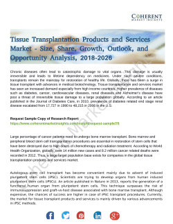
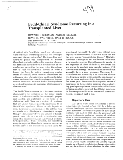
Fig. 1. Histological observations of germ cells in situ in gonads of Pleurodeles waltl larvae. (A) Transverse section
through the undifferentiated gonads and the mesonephnc region at stage 49. Germs cells are voluminous and display an
interphasic multilobed nucleus. (B) Detail of a gonad at stage 47 showing a germ cell, tangentially sectionned, with
cytoplasmic pigment granules, gc, germ cell; g, gonad; m, mesonephros; pg, pigment granules. Bar, 50fim.
Transfer of germ cell nuclei into eggs in Pleurodeles
Nuclei donor cells
Nuclei donor cells were diploid germ cells isolated from
larvae at stages 41 to 55 (Gallien & Durocher, 1957). The
gonads were either at the genital crest stage, the undifferentiated stage or at the differentiated stage. Control nuclei
were taken from mesonephric cells of larvae at stage 41.
Cell donor larvae were anaesthetized in 0002 % MS222
prior to dissection. As shown in Fig. 1A, gonads are located
on the dorsal side and extend along the mesonephros. They
append in the general cavity by a very short mesenter,
which was sectioned with a pair of fine forceps. The gonads
were placed into Steinberg's medium containing EDTA
(10~ 3 M), without calcium and magnesium (at pH7-4 for
about lOmin) to dissociate the cells.
Among the dissociated cells, germ cells were easily
recognizable since they are larger (30-40 um in diameter)
than somatic cells (10-15 um in diameter) and appear
brighter in artificial light (Fig. 2A). Examination of these
cells under a photonic microscope shows that only the
largest refringent ones present a multilobed nucleus (phase
contrast, Fig. 2C) and cytoplasmic pigment granules (transmitted light, Fig. 2B) which are specific histological characteristics for the identification of germ cells (Fig. 1A,B).
Mesonephros were removed and dissociated by the same
technique to obtain isolated mesonephric cells.
Nuclear transplantation
The nuclear transplantation was done by the classical
technique developed for the embryonic nuclei of Rana
pipiens by Briggs & King (1952) and adapted to Pleurodeles
waltl by Signoret & Picheral (1962). Since the germ cells, at
stages 41 to 55, are different (much smaller and without
vitellus) from the cells of early embryonic stages, the
operations were carried out using two binocular microscopes (removal of the nucleus at a magnification of x80
and implantation into the eggs at x25) and working
manually without a micromanipulator. The germ cell implantations are 'single transfer' types, nuclei taken directly
from the donor larvae.
Control experiments for the transfers
In order to confirm by cytological methods that development was directed by implanted nuclei, two experimental
series were performed using nonlethal chromosomal mutations as markers. A pericentric inversion of chromosome
6 (Jaylet, 1971) served as a marker for the recipient egg
nuclei and a reciprocal heterozygotic translocation between
chromosomes 6 and 11 (Labrousse, 1984) marked the donor
germinal nuclei. In each series, a caryotypic examination
was conducted on a larva killed at the feeding larval stage.
In Pleurodeles waltl, there are few viable strains with
chromosomal mutations and the mutant fertility is very low.
For these reasons, chromosomal markers could only be
used in two control series.
Moreover, in order to exclude the hypothesis of experimental gynogenetic development in adults obtained from
the transplantation of nuclei without marker, sexual genotype was identified by analysis of the expression of peptidase-1, a polymorphic sex-linked enzyme. This enzyme is
dimeric and depends on a pair of codominant alleles, Pep-
473
1A situated on the Z sex chromosome and Pep-1B located on
the W sex chromosome (Ferrier, Gasser, Jaylet & Cayrol,
1983).
Results
After nuclear transplantation, the development of
recipient eggs was observed. Table 1 summarizes the
fate of eggs up to the blastula stage as related to the
origin of donor nuclei (germ cells or mesonephric
cells) and the larval stage of the donor. Table 2 gives
the number of individuals surviving the successive
developmental stages after cleavage.
As shown in these tables, 35 % of the recipient eggs
with implanted germ cell nuclei cleaved and 9 %
reached the blastula stage. Similar percentages were
obtained for eggs that received mesonephric nuclei of
larval stage 41 (29 % cleaved, 8 % reached blastula
stage), but none of these embryos showed any development beyond the blastula stage. Thus, mesonephric nuclei from larval stages later than stage 41 were
not tested. In marked contrast, embryos from eggs
receiving germ cell nuclei developed beyond the
blastula stage; 3 % of complete blastulae (0-3 % of
total transfers) reached metamorphosis.
Six adults are presently being reared in the laboratory. All are diploids. Three are genotypic ZZ males
and three are genotypic ZW females. Previously, it
has been established that, in Pleurodeles, gynogenesis
results in 50% ZZ males and 50% WW females
(Ferrier et al. 1983; Jaylet & Ferrier, 1978). Therefore, since we have obtained the same number of ZZ
males and ZW females but no WW females, our
results demonstrate that each individual developed
from a single grafted germ cell nucleus. Actually, four
adults are 15 months old and the other two 21 and 29
months old, respectively. In February 1986, two
adults, obtained from the transplantation of nuclei
without marker, were mated to test their fertility. Out
of the 439 eggs spawned, 350 reached feeding larval
stage, a percentage of surviving animals comparable
to that obtained in standard spawning. The caryotype
of ten of these larvae was examined and found to
be normal, showing that the parents were diploid.
The offspring were normally metamorphosed. After
metamorphosis, 50 animals, taken at random, were
reared in the laboratory. They are now 11 months
old. 23 are phenotypic males and 27 are phenotypic females. The male/female ratio confirms
the ZW heterogamety of the mother (Dournon &
Houillon, 1984, 1985), because a WW mother would
474
M. Lesimple, C. Dournon, M. Labrousse and Ch. Houillon
have given unisexual progeny (Collenot, 1973; Jaylet
& Ferrier, 1978; Dournon & Houillon, 1984).
Concerning the two control series made with
chromosomal markers, the first larva killed was
obtained from a germ cell nucleus derived from a
standard larval donor and transplanted into an egg
from a female with a caryotype showing the pericentric inversion of chromosome 6. None of the mitoses
analysed showed this marker chromosome or any
other abnormality. The second larva killed came from
the transplantation of a germ cell nucleus carrying the
reciprocal translocation into an egg from a standard
female. The only chromosomal aberration observed
in all of the examined mitoses was the translocation.
In both cases, the caryotype of the larva was, as
expected, that of the transplanted nucleus.
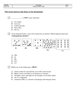
Fig. 2. In vivo observations of dissociated germ cells and somatic cells from a gonad of Pleurodeles waltl. (A) Cells
under the operating stereomicroscope before nuclear transplantation. (B) Cells under the phase-contrast microscope.
Germ cells are much larger than somatic cells and display a multilobed nucleus. (C) Cells under the light photonic
microscope. Pigment granules are visiblein the cytoplasm of germ cells, gc, germ cell; sc, somatic cell of the gonad;
mn, multilobed nucleus; pg, pigment granules. Bar, 50jum.
Transfer of germ cell nuclei into eggs in Pleurodeles
475
Table 1. Cleavage of eggs following nuclear transplantation
Segmentations
Cellular origin
of the donor
nuclei
Developmental stage
of the donor nuclei
(age and length of larval donor)
Number of
transplantations
Number of
aborted cleavages*
61 (71 %)
Number of
abnormal
segmentations!
18 (21 %)
Number of
blastulae
Mesonephric cells
41
(25 days, 14 mm)
Germ cells
41
(25 days, 14 mm)
237
161
26
50
45
123
96
21
6
148
93
36
19
117
41
66
10
31
25
4
2
78
68
3
7
460
320
102
38
271
192
57
22
431
321
71
39
354
268
66
20
658
308
307
43
78
48
23
7
86 (100 %)
7 (8 %)
(40 days, 18 mm)
46
(43 days, 18 mm)
47
(46 days, 19 mm)
48
(50 days, 20 mm)
49
(53 days, 21 mm)
50
(57 days, 23 mm)
51
(61 days, 24 mm)
52
(68 days, 27 mm)
53
(72 days, 30 mm)
54
(79 days, 38 mm)
55
(99 days, 60 mm)
Total:
2986 (100 %)
1941 (65 %)
782 (26 %)
263 (9 %)
* Decaying eggs that did not cleave or that extruded the transplanted nucleus.
t Irregular cleavage or regular cleavage that was limited to part of the egg (partial segmentation).
These results provide cytological evidence that the
development of the experimental animals was actually directed by the grafted germinal nuclei.
Discussion
The nuclear transplantation experiments performed
with different types of anuran and urodele amphibians have shown that, except in some cases in Xenopus laevis (Gurdon & Uehlinger, 1966; Brun & Kobel,
1972), somatic nuclei lose their ability to support
development during the embryonic period (see Gallien, 1966 and Gurdon, 1986 for review). Our experiments with mesonephric nuclei corroborate these
findings.
Study of the morphogenetic potencies of germ cell
nuclei in Rana pipiens has shown that nuclei of
primordial germ cells isolated from young tadpoles
are totipotent (Smith, 1965), but nuclei of spermatogonia isolated from juveniles and adults have lost
potencies to give normal animals (Di Berardino &
Hoffner, 1971).
We have examined the modifications of the morphogenetic potencies of germ cells during the larval
period in Pleurodeles waltl. Our results show, for the
first time, that, at least up to metamorphosis, some
germ cell nuclei are able to support larval development and also the development into adult and fertile
animals. Therefore, these nuclei are totipotent.
Nevertheless, we noted a decrease in the number of
hatching larvae obtained from the transplantation of
germinal nuclei taken from stage 50. This decrease
could be due, in a part of the germ cell population
from stage 50, to modifications in the structural
organization of nuclei. These modifications could
prevent the expression of the totipotency in nuclear
transplantation experiments. Another possibility is
476
M. Lesimple, C. Dournon, M. Labrousse and Ch. Houillon
Table 2. Development of blastulae arising from transplantation of nuclei
Number of surviving animals at:
Cellular origin
of the donor
nuclei
Developmental
stage of the
donor nuclei
Viesonephric
cells
41
Germ cells
41
45
46
47
48
49
50
51
52
53
54
55
Number of
blastulae
(st. 5 to 7)
7 100%
Total:
Number
Gastrula st.
(st. 8 to 13)
Neurula st. Tail-bud st. Hatching st.
(st. 14 to 21) (st. 22 to 32)
(st. 33)
Feeding st. Metamorphosis st.
(st. 38)
(st. 56)
0
0
0
0 0%
0
50
6
19
10
2
7
38
22
39
20
43
7
43
4
9
8
2
6
24
13
14
14
19
4
19
3
5
5
2
6
8
9
10
7
7
3
10
2
2
3
1
3
6
3
4
5
4
3
6 12%
2 33%
2 11%
2 20%
1 50%
1 14%
1 3%
2 9%
2 5%
1 5%
2 5%
0 0%
1
2*
2*
1
1
1
1
0
1
0
2
0
263 (9 %)
100%
160 (5 %)
84 (3 %)
46(1-5%)
61%
32%
17%
22 (0-7 %)
8%
12 (0-4%)
5%
of
adults
0
0
It
1
It
1
0
1
0
1
0
9 (0-3 %)
1
1
6 (0-2 %)
3%
Percentages in brackets were calculated from the total number of transfers.
Percentages in bold were calculated from the original number of blastulae obtained for each stage.
* One larva was sacrificed for caryotypic examination (control series).
t Mating animals which reproduced at 18 months for the female and 10 months for the male.
that modifications could occur at the level of the
genome itself and, therefore, lead to a restriction of
morphogenetic potencies of the nuclei. From stage 50
onwards, a modification of the rate of the germ cell
proliferation appears and the proliferation becomes
different in male and in female larvae (Dournon,
unpublished data). A differentiation of germ cells
into spermatogonia or oogonia could explain these
phenomena. However, gonocytes remained in the
gonads of young and adult animals and our successful
experiments at the later larval stages were perhaps
due to the transplantation of nuclei from these stem
germ cells, which lead us to suspect that a totipotent
germ cell population is maintained throughout the
entire lifetime of these amphibians.
We thank Drs C. Aimar, J. Lefresne and C. Pieau for
their help during the course of this work. We are particularly grateful to Dr J. Signoret for his valuable discussions
and constructive suggestions. We are obliged to Dr J. B.
Gurdon for the critical reading of the manuscript. We thank
J. Desrosiers for the illustration. The only support was
provided by University Pierre et Marie Curie.
References
BRIGGS, R. & KING, T. J. (1952). Transplantation of
living nuclei from blastula cells into enucleated frogs'
eggs. Proc. natn. Acad. Sci. U.S.A. 38, 455-463.
BRIGGS, R. & KING, T. J. (1954). Transplantation of
living nuclei of late gastrulae into enucleated eggs of
Rana pipiens. J. Embryol. exp. Morph. 2, 73-80.
BRIGGS, R. & KING, T. J. (1960). Nuclear transplantation
studies on the early gastrula of Rana pipiens (nuclei of
presumptive endoderm). Devi Biol. 2, 252-270.
BRUN, R. B. & KOBEL, H. R. (1972). Des grenouilles
m6tamorphos6es obtenues par transplantation nucl£aire
a partir du prosencephale et de l'6piderme larvaire de
Xenopus laevis. Rev. Suisse Zool. 79, 961-965.
COLLENOT, A. (1973). Obtention, par la m6thode des
greffes embryonnaires, d'une femelle a descendance
unisexu6e femelle, chez le triton Pleuwdeles waltlii
Michah. Experientia 29, 885-887.
Di BERARDINO, M. A. & HOFFNER, N. (1971).
Development and chromosomal constitution of nuclear
transplants derived from male germ cells. /. exp. Zool.
176, 61-72.
DOURNON, C. & HOUILLON, C. (1984). Demonstration
g6n£tique de l'inversion fonctionnelle du ph6notype
sexuel femelle sous l'action de la temperature d'61evage
chez l'Amphibien Urodele Pleurodeles waltlii Michah.
Reprod. Nutr. Develop. 24, 361-378.
DOURNON, C. & HOUILLON, C. (1985). Thermosensibilit6
de la differentiation sexuelle chez l'Amphibien
Urodele, Pleurodeles waltlii Michah. Conditions pour
obtenir l'inversion du ph£notype sexuel de toutes les
femelles g£n£tiques sous Faction de la temperature
d'elevage. Reprod. Nutr. Develop. 25, 671-688.
FERRJER, V., GASSER, F., JAYLET, A. & CAYROL, C.
(1983). A genetic study of various enzyme
polymorphisms in Pleurodeles waltlii (urodele
2%
Transfer of germ cell nuclei into eggs in Pleurodeles
amphibian). II-peptidases: demonstration of sex
linkage. Biochem. Genet. 21, 535-549.
FISCHBERG, M., GURDON, J. B. & ELSDALE, T. R. (1958).
Nuclear transfer in Amphibia and the problem of the
potentialities of the nuclei of differentiating tissues.
Expl Cell Res. (Suppl.) 6, 161-178.
GALLIEN, L. (1966). La greffe nucl6aire chez les
Amphibiens. Annie Biol. 5, 241-269.
GALLIEN, L. & DUROCHER, M. (1957). Table
chronologique du d6veloppement chez Pleurodeles
waltlii Michah. Bull. Biol, Fr. Belg. 91, 97-114.
GURDON, J. B. (1962). The developmental capacity of
nuclei from intestinal epithelium cells of feeding
tadpoles. J. Embryol. exp. Morph. 10, 622-640.
GURDON, J. B. & UEHUNGER, V. (1966). "Fertile"
intestine nuclei. Nature, Lond. 210, 1240-1241.
GURDON, J. B. (1986). Nuclear transplantation in eggs
and oocytes. J. Cell Sci. 4, 287-318.
JAYLET, A. (1971). Modification du caryotype par une
inversion pe'ricentrique a l'6tat homozygote chez
l'Amphibien Urodele Pleurodeles waltlii Michah.
Chromosoma (Berl.) 35, 288-299.
JAYLET, A. & FERRIER, V. (1978). Experimental
gynogenesis in the newt species Pleurodeles waltlii and
P. poireti. Chromosoma (Berl.) 65, 65-80.
LABROUSSE, M. (1984). Trisomisation par non-disjonction
des chromosomes impliqu^s dans une translocation
rdciproque, chez l'Amphibien Urodele Pleurodeles
waltlii Michah. Genetica 63, 195-202.
477
B. (1962). Capacity des noyaux de cellules
endodermiques embryonnaires a organiser un germe
viable chez l'Urodele Pleurodeles waltlii. C.r. hebd.
Sianc. Acad. Sci., Paris 255, 2509-2511.
PICHERAL,
SlGNORET, J., BRIGGS, R. & HUMPHREY, R. R. (1962).
Nuclear transplantation in the Axolotl. Devi Biol. 4,
134-164.
SIGNORET, J., DAVID, J. C , LEFRESNE, J. & HOUILLON, C.
(1983). Control of DNA ligase molecular forms in
nucleocytoplasmic combinations of axolotl and
Pleurodeles. Proc. natn. Acad. Sci. U.S.A. 80,
3368-3371.
SIGNORET, J. & FAGMER, J. (1962). Activation
exp^rimentale de l'oeuf de pleurodele. C.r. hebd.
Sianc. Acad. Sci., Paris 254, 4079-4080.
SIGNORET, J. & PICHERAL, B. (1962). Transplantation de
noyaux chez Pleurodeles waltlii Michah. C.r. hebd.
Sianc. Acad. Sci., Paris 254, 1150-1151.
SMITH, L. D. (1965). Transplantation of the nuclei of
primordial germ cells into enucleated eggs of Rana
pipiens. Proc. natn. Acad. Sci. U.S.A. 54, 101-107.
STEINBERG, M. (1957). In Carnegie Inst. Washington
Yearbook (Report by EBERT, J. D.) 56, 347.
WYLIE, C. C , HEASMAN, J., SNAPE, A., O'DRISCOLL, M.
& HOLWILL, S. (1985). Primordial germ cells of
Xenopus laevis are not irreversibly determined early in
development. Devi Biol. 112, 66-72.
{Accepted 9 March 1987)
© Copyright 2026 Paperzz