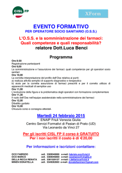
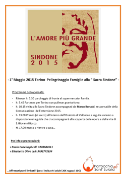
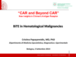
This article is from the
August 2005 issue of
published by
The American Phytopathological Society
For more information on this and other topics
related to plant pathology,
we invite you to visit APSnet at
www.apsnet.org
MPMI Vol. 18, No. 8, 2005, pp. 849–855. DOI: 10.1094 / MPMI -18-0849. © 2005 The American Phytopathological Society
An Endopolygalacturonase
from Sclerotinia sclerotiorum Induces
Calcium-Mediated Signaling and Programmed Cell Death
in Soybean Cells
Anna Zuppini,1 Lorella Navazio,1 Luca Sella,2 Carla Castiglioni,2 Francesco Favaron,2 and
Paola Mariani1
1
Dipartimento di Biologia, Università di Padova, via U. Bassi 58/B, 35131 Padova, Italy; 2Dipartimento Te. S.A.F., Sez.
Patologia Vegetale, Università di Padova, Viale dell’Università 16, 35020 Legnaro, Padova, Italy
Submitted 21 January 2005. Accepted 5 April 2005.
A basic endopolygalacturonase (PG) isoform, produced
early by Sclerotinia sclerotiorum when infecting soybean
seedlings, was used to examine the signaling role of the
enzyme in aequorin-expressing soybean cells. A cytosolic
Ca2+ elevation was induced, with a rapid increase (phase 1)
and a very slow decrease (phase 2) of Ca2+ concentration,
indicating the involvement of Ca2+ ions in PG signaling.
Within 1 h of PG-cell contact a remarkable level of cell
death was recorded, significantly higher than the control
cell culture turnover. The observed morphological and biochemical changes were indicative of the activation of programmed cell death; in particular, cytochrome c release in
the cytoplasm and activation of both caspase 9-like and
caspase 3-like proteases were found. When a polygalacturonase-inhibiting protein (PGIP) and the PG were simultaneously applied to cells, both the Ca2+ increase and cell
death were annulled. The possible roles of prolonged sustained cytosolic Ca2+ concentrations in inducing cell death
and of the PG-PGIP interaction in preventing PG signaling
are discussed.
Additional keywords: fungal hydrolytic enzyme.
In plant-microbe interactions, the plant cell wall plays several roles, as a physical barrier to the pathogen infection, as a
source of nutrients for the invaders, and as origin of signaling
molecules. To overcome the plant cell wall, early during
pathogenesis, most fungal pathogens secrete a wide array of
cell wall-degrading enzymes with different substrate specificity, which facilitate the penetration of the pathogens into the
host. The activity of pectin-degrading enzymes, in particular
endopolygalacturonases (PGs) and endopectate lyases, is crucial for the colonization of plant tissues (Prade et al. 1999),
and these enzymes can be determinants of pathogenicity or
virulence (Willats et al. 2001). PGs hydrolyze internal α(1-4)
glycosidic bonds in deesterified regions of the middle lamella
and primary cell wall homogalacturonans and, when produced
in relevant amounts, as by some necrotrophic fungi, determine
A. Zuppini and L. Navazio contributed equally to this work.
Corresponding authors: P. Mariani; E-mail: [email protected];
Telephone: +39.049.8276277; Fax: +39.049.8276280; and F. Favaron; Email: [email protected]; Telephone: +39.049.8272892; Fax:
+39.049.8272890.
the maceration of infected tissues. PGs are often encoded by
multigene families and produced in several molecular forms
with different specificity and regulation. This multiplicity can
confer adaptative flexibility to pathogens when infecting different hosts or plant organs (De Lorenzo et al. 2001).
Biologically active mixtures of oligogalacturonides (OGs)
with various degrees of polymerization (from 2 to about 20
galacturonic acid residues) are released from the cell wall
pectins by PGs (Ridley et al. 2001). OGs participate as endogenous signal molecules in the dialogue between host and microbe, activating in plants a cascade of physiological events
that are at least partially Ca2+-mediated (Navazio et al. 2002)
and elicit defense responses (Coté and Hahn 1994).
Plants synthesize a class of proteins that specifically counteract the fungal PG activity. These polygalacturonase-inhibiting proteins (PGIPs) are located in the plant apoplast, in
which, by complexing PGs, they reduce the homogalacturonan
breakdown rate and the depolymerization of active OGs into
shorter, inactive fragments (De Lorenzo and Ferrari 2002).
Sclerotinia sclerotiorum is a necrotrophic fungus causing
soft rots in a number of host plants, and PG and oxalic acid,
abundantly secreted by S. sclerotiorum in the host tissue, contribute to disease symptoms (Godoy et al. 1990; Marciano et
al. 1983).
In this paper, we demonstrate that a basic PG isoform expressed early by S. sclerotiorum during soybean infection
(Favaron and Marciano 1992; Favaron et al. 2004) induces, in
aequorin-expressing soybean cells, a rapid and sustained cytosolic Ca2+ elevation that, in turn, leads to cell death exhibiting
several hallmarks of programmed cell death (PCD). Both the
Ca2+ increase and cell death induced by the PG are prevented
by the simultaneous administration to cells of the PG-inhibitor
PGIP, suggesting that PG and PGIP protein interaction alters
the sensing of the fungal signaling molecule by the plant cells.
RESULTS
PG induces a sustained increase
of intracellular Ca2+ concentration in soybean cells.
The PG purified from S. sclerotiorum was tested for its potential ability to elicit a Ca2+ response in soybean suspension
cell cultures stably expressing aequorin. At a 0.5 enzyme reducing unit (RU) per milliliter dose, after a delay of 40 s, a
rapid increase in cytosolic free-Ca2+ concentration ([Ca2+]cyt)
was observed, reaching a peak of about 1.2 µM Ca2+ after
Vol. 18, No. 8, 2005 / 849
about 2 min (phase 1) and slowly decreasing to a concentration
of 0.6 µM within 10 min (phase 2) (Fig. 1). [Ca2+]cyt remains at
this value without declining to the basal level (approximately
100 nM) during the next hour (data not shown). The kinetic
parameters of both phases of the Ca2+ transient were found to
depend on the amount of PG applied (ranging from 0.05 to 1.0
RU/ml) (Fig. 1).
PG triggers cell death in soybean cells.
The viability of soybean cells treated with PG (0.5 RU/ml)
was tested using the Evans blue method (Levine et al. 1996).
The PG treatment was found to induce a twofold increase in
the percentage of dead cells after 1 h of treatment (22.8 ±
0.4%) in comparison with untreated cells (10.2 ± 2.1%) (Fig.
2). Monitoring cell death over 12 h of PG treatment, we detected a progressive increase in the number of dead cells that
reached nearly 100% after 12 h of PG incubation (Fig. 2).
Loss of cell viability was accompanied by changes in nuclear morphology, as examined by double staining of DNA
with Hoechst 33342 (HO) and propidium iodide (PI) fluorescent dyes. In vivo, HO is a cell membrane–permeant dye that
has access into slightly damaged as well as altered apoptotic
Fig. 1. Effect of endopolygalacturonase (PG) purified from Sclerotinia
sclerotiorum on cytosolic Ca2+ concentration ([Ca2+]cyt) in soybean cells.
At the time indicated (arrow) soybean cells stably expressing cytosolic
aequorin were challenged with different doses of PG and the changes in
[Ca2+]cyt were continuously recorded. These and the following traces are
representative of three independent experiments, which gave very similar
results.
Fig. 2. Viability of soybean cells treated with endopolygalacturonase (PG).
Exponentially growing cells were incubated with PG (0.5 RU/ml) for
different times (gray bars). Cells cultured without PG were taken as a
control (white bar). The 100% value corresponds to cells treated for 10
min at 100°C. Data are means ± standard deviation of three independent
experiments. Statistically significant at P < 0.005 (three asterisks).
850 / Molecular Plant-Microbe Interactions
nuclei, whereas PI is a cell-impermeant dye that can enter only
in late apoptotic and necrotic cultured cells. These fluorochromes allow the discrimination of healthy (HO–/PI–), apoptotic (HO+/PI–), and necrotic (HO+/PI+) cells in an asynchronized cell population (Hamatake et al. 2000). After 1 h of PG
treatment, about 15% HO+/PI– nuclei (Fig. 3E and F) and 20 to
30% HO+/PI+ nuclei (Fig. 3H and I) were observed, indicating
chromatin condensation (HO positivity) and suggesting the simultaneous occurrence of both early and late apoptotic-like
stages. In comparison, nuclei of untreated cells showed mainly
the absence of HO and PI fluorescence, only about 5% of the
cell population displayed positivity to both dyes (Fig. 3B and
C).
Morphological observations under light microscopy revealed,
in PG-treated cells, a gradual shrinkage of the nucleus and detachment of the plasma membrane from the cell wall as the
cell death program proceeds (Fig. 3D and G). The detachment
of the plasma membrane from the cell wall is considered one
of the hallmarks indicating the occurrence of a PCD process in
plant cells (Beers and McDowell 2001). Nuclei of control cells
appeared larger and rounder compared with PG-treated cells,
and the plasma membrane was completely adherent to the cell
wall (Fig. 3A).
In order to investigate the mode of PG-induced cell death,
all further analyses were carried out after 1 h of treatment. At
this timepoint, in fact, the cell population is in a dynamic state
toward PCD.
PG-induced cell death is associated with
cytochrome c release and caspase-like protease activation.
To clarify the mechanism of cell death induced by PG,
attention was focused on the cytochrome c/caspase 9 PCD
pathway and downstream targets such as caspase 3. Cytochrome
c release from mitochondria is an early event of apoptosis in
animal cells. It is required for the activation of the caspase protease cascade as well as downstream events (Desagher and
Martinou 2000). Cytosolic extracts from soybean cells were
separated on sodium dodecyl sulfate-polyacrylamide gel electrophoresis (SDS-PAGE) and were analyzed by Western blot
with an anti-cytochrome c monoclonal antibody. In cytosolic
Fig. 3. Nucleus staining of endopolygalacturonase (PG)-treated cells.
Untreated cells and cells treated for 1 h with PG (0.5 RU/ml) were stained
with Hoechst (HO) and propidium iodide (PI) and were processed for
fluorescence microscopy. A through C, untreated cells; D through I, cells
treated with 0.5 RU/ml PG; E and F, early programmed cell death (PCD)
nucleus; H and I, late PCD nucleus. Pictures represent typical examples.
cc = chromatin condensation, cw = cell wall, nu = nucleus, and pm =
plasma membrane. The arrow indicates the detachment of the plasma
membrane from the cell wall.
extracts from cells treated with PG (0.5 RU/ml) for 1 h, the
presence of cytochrome c was detected; control cells did not
show any cytochrome c protein band (Fig. 4A). This result
suggests the likely activation of a mitochondrial PCD pathway
during PG treatment. To directly investigate whether PGinduced cell death is associated with the activation of caspaselike proteases, hydrolysis of the specific caspase 9 synthetic
substrate LEHD and of the caspase 3 substrate DEVD, both
conjugated with p-nitroaniline (pNA), was assayed in protein
extracts from soybean cells. Caspase 9-like activity in cells
treated with 0.5 RU/ml PG for 1 h was 1.86 ± 0.12 times
greater than that in the control (Fig. 4B). Furthermore, incubation of cells with PG also heightened caspase 3-like activity by
about 1.3 ± 0.03 times as compared with untreated cells (Fig.
4C). Although both analyzed caspase substrates were cleaved
upon PG treatment, there was a quantitative difference between
the two enzymatic reactions. The activity of caspase 9-like
protease seemed to be about 1.4-fold higher than that of caspase
3-like protease (Fig. 4B and C). Both activities of caspase 9like and caspase 3-like proteases were significantly decreased
by the Ac-LEHD-CHO and Ac-DEVD-CHO inhibitors of animal caspase 9 and 3, respectively (Fig. 4B and C). Thus, the
mechanism of cell death induced by PG seems to involve cytochrome c release, caspase 9-like and caspase 3-like activation,
suggesting the occurrence of a PCD process.
PGIP blocks both the PG-induced Ca2+ signaling and PCD.
PGIP is constitutively present in the tissues of soybean seedlings (Favaron et al. 1994), and it is secreted in the culture medium by soybean cells (data not shown). However, the washing
procedure carried out before the PG treatment freed the cell of
any detectable PGIP activity. Therefore, exogenous PGIP (2
inhibitor units [IU] per milliliter) was administered to cell suspensions simultaneously with the PG treatment (0.5 RU/ml).
The simultaneous administration to cells of PG and its inhibitor PGIP resulted in the almost complete annulment of the PGinduced Ca2+ transient, with a remarkable reduction of phase 1
amplitude and the complete abolishment of the phase 2 plateau
(Fig. 5A). It has to be noted that PGIP by itself could not elicit
any significant [Ca2+]cyt elevation over the basal level. Indeed,
Fig. 4. Cytochrome c release and measurements of caspase-like activities
in soybean cells incubated with endopolygalacturonase (PG) (0.5 RU/ml)
for 1 h. A, Cytochrome c accumulation in the cytosolic extracts from
untreated (control) and PG-treated cells was assessed by Western blot
analysis. B, Caspase 3-like activity in untreated cells (white bar), PGtreated cells (gray bar), and PG-treated cells in the presence of the caspase
3 inhibitor Ac-DEVD-CHO (3 µM) (striped bar). C, Caspase 9-like
activity in untreated cells (white bar), PG-treated cells (gray bar), and PGtreated cells in the presence of the caspase 9 inhibitor Ac-LEHD-CHO (3
µM) (striped bar). Data are means ± standard deviation of three independent experiments. Statistically significant at P < 0.05 (one asterisk).
Fig. 5. Polygalacturonase inhibiting protein (PGIP) effectively blocks
endopolygalacturonase (PG)-induced Ca2+ signaling. A, Cells were treated
(arrow) with either PG (0.5 RU/ml) alone (black trace) or PG (0.5 RU/ml)
and PGIP (2 inhibitor units per milliliter) administered simultaneously
(gray trace) and their effect on cytosolic free-Ca2+ concentration [Ca2+]cyt
was monitored. B, Cells were challenged (arrow) with PGIP (black trace)
or culture medium containing the same percentage of the solvent as PGIPtreated cells (gray trace).
Vol. 18, No. 8, 2005 / 851
the PGIP-induced Ca2+ trace appeared almost superimposable
to that of control cells challenged with culture medium (Fig.
5B).
Figure 6 shows that PGIP effectively blocks the activation of
the PG-induced PCD program, as demonstrated by the Evans
blue cell viability assay (Fig. 6A) and double staining with HO
and PI (Fig. 6B, panels e and f). Cell treatment with only PGIP
did not induce either a decrease of cell viability (Fig. 6A) or
positivity to HO and PI (Fig. 6B, panels b and c).
The cytosolic Ca2+ elevation is a key determinant
of the PG-induced cell death.
To investigate the role of Ca2+ in PG-activated signaling, the
Ca2+ channel blocker La3+, which has been previously demonstrated to effectively abolish the Ca2+ transient elicited by OGs
(Navazio et al. 2002), was applied to soybean cells 10 min before treatment with PG. The inhibitor markedly reduced both
phase 1 and 2 of the Ca2+ transient (Fig. 7A). The annulment
3+
of the Ca2+ signal in La -pretreated cells abolished both caspase 3-like activation (Fig. 7B) and cell death (Fig. 7C), which
did not significantly differ from that of control cells. Moreover, the number of HO/PI positive cells was comparable to
the control (Fig. 7D), highlighting the absence of chromatin
condensation in PG-treated cells after incubation with La3+.
Fig. 6. Polygalacturonase inhibiting protein (PGIP) effectively blocks
endopolygalacturonase (PG)-induced cell death. A, Exponentially growing
cells were incubated for 1 h with PG (0.5 RU/ml) (gray bar), PGIP (2
inhibitor units [IU] per milliliter) (striped bar), or a mixture of PG (0.5
RU/ml) plus PGIP (2 IU/ml) (black bar). Cells cultured without PG and
PGIP were taken as a control (white bar). Data are means ± standard
deviation of three independent experiments. Statistically significant at P <
0.005 (three asterisks). B, Cells treated for 1 h with PGIP (2 IU/ml) and
with PG (0.5 RU/ml) plus PGIP (2 IU/ml) were stained with Hoechst (HO)
and propidium iodide (PI) dyes. a through c, PGIP-treated cells; d through
f, PG plus PGIP-treated cells. Pictures represent typical examples. nu =
nucleus.
852 / Molecular Plant-Microbe Interactions
DISCUSSION
In this paper, we show that administering to soybean cell
cultures a PG produced by Sclerotinia sclerotiorum when infecting soybean plants triggers a rapid elevation in [Ca2+]cyt,
which is suggestive of Ca2+-mediated signaling induced by a
secreted fungal enzyme. This enzyme in vivo degrades the
pectic components of the plant cell wall, generating oligosaccharide fragments, which in turn, act as endogenous signaling
molecules (Coté and Hahn 1994). The mixture of OGs deriving from PG enzymatic activity is known to activate a Ca2+mediated signal pathway (Navazio et al. 2002). Nevertheless,
the temporal kinetics of the OG-elicited Ca2+ transient significantly differs from that induced by treatment with PG. In particular, although both elicitors evoke a single immediate Ca2+
spike, the OG-activated Ca2+ transient is rapidly dissipated
within a few minutes (Navazio et al. 2002), whereas the PGactivated Ca2+ signal persists for at least 1 h, maintaining, in
the cell, Ca2+ levels higher than the physiological ones. In both
animal and plant cells, perturbations of Ca2+ homeostasis have
been associated with cytotoxic events (Kass and Orrenius
1999; White and Broadley 2003).
Here, we provide evidence for the occurrence of PCD induced
by PG in soybean cells. It must be noted that the products of
PG enzymatic activity, namely OGs, do not by themselves induce cell death in either soybean cells (data not shown) or
other experimental systems (Binet et al. 2001; Lecouriex et al.
2002; Shibuya and Minami 2001). Thus, PG seems to be, per
se, the causative agent of the cell death, which could depend
on the cell sensing of the protein as a virulence factor and may
be unrelated to its enzymatic activity. Likewise, the elicitor activity of other fungal hydrolytic enzymes has been previously
shown by various experimental approaches not to be necessarily
due to the release of biologically active oligosaccharide
fragments (Carden and Felle 2003; Martinez et al. 2001;
Poinsott et al. 2003; Rouet-Mayer et al. 1997; Yano et al. 1998).
The elevation of cytosolic Ca2+ level seems to be essential
for the induction of cell death. Indeed, the Ca2+ channel
blocker La3+ abolishes both phases of the PG-induced Ca2+
transient, caspase 3-like activation, chromatin condensation,
and cell death. A relationship between sustained [Ca2+]cyt and
activation of pathogen defense responses has been previously
reported (Grant et al. 2000).
The sequence of events starting from signal perception to
cell death in PG-treated cells has been partially unraveled in
this paper. Based on previous findings by several authors
(Boudart et al. 2003; Cotton et al. 2002; Poinsott et al. 2003),
it can be argued that, downstream of the Ca2+ transient, an
extracellular H2O2 accumulation can occur. Both [Ca2+]cyt elevation (Viroleinen et al. 2002) and production of reactive oxygen species (Tiwari et al. 2002) may be cocausative agents in
triggering PCD through the mediation of mitochondria. Our
data provide evidence for the release, in the cytoplasm, of cytochrome c as well as activation of caspase-like proteases in
PG-induced cell death. Nevertheless, the involvement of other
proteases, such as the newly characterized serin proteases
named saspases (Coffeen and Wolpert 2004), cannot be ruled
out.
The complete block of both PG-induced Ca2+ transient and
PCD following the interaction of PG with its inhibitor PGIP is
consistent with an interference of PGIP with signaling by PG.
PGIP, by complexing PG, besides blocking the substrate admittance to the enzyme-active site (Di Matteo et al. 2003), might
prevent the ligand access to the putative receptor, hampering
its recognition. Consequently, any cellular receptor-mediated
response to the elicitor is annulled. The inhibition of H2O2 production shown by Poinsott and associates (2003), consequent
to the simultaneous administration of PG and PGIP to grapevine cells, gives support to this hypothesis. An open question
remains whether the PGIP role in the modulation of PG functioning is really dual (i.e., block of PG recognition and of its
enzymatic activity) or if, rather, all PG-induced cell responses
are prevented by the failed PG interaction with its receptor.
Poinsott and associates (2003) speculated that the PG catalytic
domain and the recognition motif might correspond. Others
have also suggested (De Lorenzo et al. 1997; Esquerré-Tugayé
2000; James and Dubery 2001) that PGIP might act as a cellsurface receptor or, at least, be part of a receptor complex.
In conclusion, a PG produced early during plant infection by
S. sclerotiorum is a candidate factor for triggering pathogeninduced PCD. As recently reported, this necrotrophic fungus
seems to co-opt PCD response in host cells to produce disease
symptoms (Dickman et al. 2001; Govrin and Levine 2000),
and the expression of animal antiapoptotic genes in transgenic
tobacco plants prevents fungal colonization and disease development (Dickman et al. 2001).
The plant PGIP blocking the PCD induced by this S. sclerotiorum PG may be a successful instrument to improve plant
resistance to this pathogen.
MATERIALS AND METHODS
Plant material.
Soybean (Glycine max L.) suspension cell cultures stably
expressing cytosolic aequorin (Mithöfer et al. 1999) were
provided by G. Neuhaus (Freiburg, Germany). Cells were
cultivated in Murashige and Skoog liquid medium supplemented with 0.5% sucrose, 10 µg naphthaleneacetic acid per
milliliter, 2 µg of kinetin per milliliter, 1 µg of thiamine-HCl
per milliliter, and 10 µg of kanamycin per milliliter on a rotary shaker (80 rpm) at 24°C under a 16-h photoperiod and
were subcultured every three weeks with a 10% (vol/vol) inoculum.
Fungus and plant infection.
The B-24 isolate of S. sclerotiorum (Lib. De Bary) was
grown on potato dextrose agar medium to obtain mycelium
for soybean inoculation. Six-day-old etiolated soybean plants
were obtained at 25°C in sterilized moist vermiculite. Soybean seedlings were inoculated by placing plugs (10 × 2 mm)
of mycelium-colonized agar, cut from marginal zones of
colonies, along the middle region of hypocotyls. The trays
Fig. 7. La3+ inhibition of endopolygalacturonase (PG)-triggered cytosolic Ca2+ concentration [Ca2+]cyt elevations, caspase 3-like activity, chromatin
condensation, and cell death. A, Cells were incubated with 3 mM La3+ (gray trace) for 10 min prior to the addition of PG (0.5 RU/ml) (arrow), and the Ca2+
response was monitored. Control cells were treated only with PG (black trace). B, Caspase 3-like activity and C, viability of soybean cells treated for 1 h
with PG (0.5 RU/ml) alone (black bar) or 3 mM La3+ (striped bar) or La3+ plus PG (gray bar). Cells cultured without PG and La3+ were taken as a control
(white bar). Data are means ± standard deviation of three independent experiments. Statistically significant at P < 0.05 (one asterisk) and P < 0.005 (three
asterisks). D, Cells treated for 1 h with PG (0.5 RU/ml) after a 10-min pretreatment with 3mM La3+ were stained with Hoechst and propidium iodide dyes. a
through c, Control cells; d through f, cells treated with La3+; g through i, cells treated with La3+ plus PG. Pictures represent typical examples. Cells cultured
without PG and La3+ were taken as a control.
Vol. 18, No. 8, 2005 / 853
containing inoculated seedlings were sealed with plastic film
and were incubated at 25°C in the dark. Infected tissue was
harvested for PG extraction and purification 48 h after inoculation.
PG extraction, purification and assay.
A basic PG isoform of S. sclerotiorum was extracted and
purified from infected soybean tissue as reported (Favaron et
al. 1997). Briefly, the PG was purified by two successive
preparative isoelectrofocusing (IEF) runs in the range of pH
3 to 10 and 6 to 9, respectively, and by a MonoS cation-exchange column. PG activity was determined as an increase in
reducing end groups over time. Reducing end groups were
measured using the method described by Milner and Avigad
(1967), with D-galacturonic acid as a standard. The incubation mixture contained 200 µl of 0.5% polygalacturonic acid
(PGA, from orange, code P3889, lot 85C-0121; Sigma-Aldrich, St. Louis) in 100 mM sodium acetate buffered at pH
4.6 and 0.1 mg of bovine serum albumin per milliliter. One
RU was defined as that producing 1 µmol of reducing end
groups per minute at 30°C and corresponds to about 1.4 µg
of PG protein.
PGIP production and assay.
PGIP was obtained from leaves of 14-day-old plants of
Phaseolus vulgaris L. (cv. Borlotto). Proteins were extracted
and PGIP was purified by preparative IEF (pH 3 to 10), PGSepharose affinity chromatography, and MonoS cation-exchange chromatography as previously reported (Favaron et al.
1994). PGIP activity was determined against the purified PG
of S. sclerotiorum, by adding the inhibitor to the enzyme mixture described in the PG-reducing end groups assay. One IU
was defined as that required to reduce by 50% the activity of
one PG unit. About 4 IU (approximately 3.2 µg of PGIP) were
used in order to abolish completely 1 PG unit.
PG treatment.
Cell treatments with the S. sclerotiorum PG were performed
three weeks after reinoculation, during the exponential growth
phase of the cells. Cells were washed three times with 10 vol
of fresh hormone-free culture medium and were used after 30
min of equilibration. Control cells were incubated in the same
conditions without the elicitor.
Aequorin-dependent Ca2+ measurements.
Reconstitution of the Ca2+-sensitive photoprotein aequorin
was carried out by incubating soybean cells with 5 µM coelenterazine overnight, in the dark. All measurements were
performed at room temperature in a final volume of 50 µl,
containing approximately 5 mg (fresh weight) of reconstituted cell-suspension culture. Treatment with PG was carried
out by injecting, with a light-tight syringe through the luminometer port, an equal volume of twofold-concentrated stock
solutions (dissolved in the basal cell culture medium) into
the cell suspension culture. All experiments were terminated
by discharging the remaining aequorin pool with 0.33 M
CaCl2 in 10% ethanol. Luminescence data were collected and
elaborated into [Ca2+] values by a computer algorithm based
on the Ca2+ response curve of aequorin (Brini et al. 1995).
Cell viability and treatments.
Cell viability was determined by incubation with 0.05%
Evans blue (Levine et al. 1996). Cells were then extensively
washed with deionized water to remove excess and unbound
dye. Dye bound to dead cells was solubilized in methanol/SDS
(50:1) for 30 min at 50°C and was quantified by absorbance at
600 nm.
854 / Molecular Plant-Microbe Interactions
Nucleus staining.
To analyze nuclear morphological changes, cells were
stained with fluorescence dyes HO and PI. Suspension cells
(3-week-old) were incubated in darkness with 8 µg of HO per
milliliter (Sigma-Aldrich) and 5 µg of PI per milliliter (SigmaAldrich) at room temperature for 10 min and were observed
under fluorescence microscopy, by using an excitation light of
350 nm and 570 nm, respectively, for the two dyes.
Detection of cytochrome c release.
Cytosolic protein extracts were obtained by incubating cells
(15 min at 4°C) with 75 mM NaCl, 1 mM NaH2PO4, 8 mM
Na2HPO4, 250 mM sucrose, and digitonin (380 µg/ml). Cytosolic fractions were then separated by centrifugation (5 min,
14,000 rpm, 4°C), were loaded (10 µg) on SDS-PAGE (12%
acrylamide gel) (Laemmli 1970), and were transferred to Immobilon-P transfer membrane (Millipore, Bedford, MA,
U.S.A.) for chemiluminescent detection according to Towbin
and associates (1979). Filters were incubated with 5% skim
milk in Tris-buffered saline (TBS) for 1 h and were probed
with a mouse monoclonal cytochrome c antibody (4 µg/ml)
(BioSource International, Camarillo, CA, U.S.A.) for 2 h. After
incubation, filters were washed two times with a 0.05% Tween/
TBS solution, followed by one wash in TBS before incubation
with the secondary antibody. Labeled proteins were detected
using CDP-star (Biolabs, Hitchin, U.K.) and autoradiography
films (Sigma-Aldrich).
Measurements of caspase-like activities.
Caspase 9- and caspase 3-like activities were measured using
the Caspase-9 and Caspase-3 colorimentric activity assay kits
(Chemicon International, Inc., Temecula, CA, U.S.A.), respectively, as described by the manufacturer. The assays are based on
spectrophotometric detection of the chromophore pNA after the
caspase-dependent cleavage from the labeled substrates DEVDpNA or LEHD-pNA by active caspase 3 or 9, respectively. Cytosolic protein extracts from soybean cells (15 µg) were incubated
for 2 h at 37°C with DEVD-pNA or LEHD-pNA, in the presence or absence of 3 µM caspase inhibitors Ac-DEVD-CHO
(for caspase 3) or Ac-LEHD-CHO (for caspase 9). Caspase-like
activity was determined by spectrophotometric quantification of
the free pNA (λem = 405 nm).
Statistical analysis.
Data are presented as mean plus or minus standard deviation
of three independent experiments, and the differences between
groups were assessed by Student’s t-test.
ACKNOWLEDGMENTS
We thank G. Neuhaus (Freiburg, Germany) for kindly providing the
transgenic soybean cells expressing cytosolic aequorin. This work was
supported by grants from Progetti di Ricerca di Interesse Nazionale 2001
(prot. 2001055885) and Fondo per gli Investimenti della Ricerca di Base
2002 (prot. RBNE01K2E7).
LITERATURE CITED
Beers, E. P., and McDowell, J. M. 2001. Regulation and execution of programmed cell death in response to pathogens, stress and developmental
cues. Curr. Opin. Plant Biol. 4:561-567.
Binet, M.-N., Humbert, C., Lecouriex, D., Vantard, M., and Pugin, A.
2001. Disruption of microtubular cytoskeleton induced by cryptogein,
an elicitor of hypersensitive response in tobacco cells. Plant Physiol.
125:564-572.
Boudart, G., Charpentier, M., Lafitte, C., Martinez, Y., Jauneau, A.,
Gaulin, E., Esquerré-Tugayé M.-T., and Dumas, B. 2003. Elicitor activity of a fungal polygalacturonase in tobacco requires a functional catalytic site and cell wall localization. Plant Physiol. 131:93-101.
Brini, M., Marsault, R., Bastianutto, C., Alvarez, J., Pozzan, T., and
Rizzuto, R. 1995. Transfected aequorin in the measurements of cytosolic Ca2+ concentration ([Ca2+]c). J. Biol. Chem. 270:9896-9903.
Carden, D. E., and Felle, H. H. 2003. The mode of action of cell-wall degrading enzymes and their interference with Nod factor signaling in
Medicago sativa root hairs. Planta 216:993-1002.
Coffeen, W. C., and Wolpert, T. J. 2004. Purification and characterization
of serin proteases that exhibit caspase-like activity and are associated
with programmed cell death in Avena sativa. Plant Cell 16:857-873.
Cotton, P., Rascle, C., and Fevre, M. 2002. Characterization of PG2, an
early endoPG produced by Sclerotinia sclerotiorum, expressed in yeast.
FEMS (Fed. Eur. Microbiol. Soc.) Microbiol. Lett. 213:239-244.
Coté, F., and Hahn, M. G. 1994. Oligosaccharins: Structures and signal
transduction. Plant Mol. Biol. 26:1379-1411.
De Lorenzo, G., and Ferrari, S. 2002. Polygalacturonase-inhibiting proteins in defense against phytopathogenic fungi. Curr. Opin. Plant Biol.
5:1-5.
De Lorenzo, G., Castoria, R., Bellincampi, D., and Cervone, F. 1997. Fungal invasion enzymes and their inhibition. Pages 61-83 in: The Mycota
V Part A. Plant Relationships. G. C. Carrol and P. Tudzynski, eds.
Springer-Verlag, Berlin.
De Lorenzo, G., D’Ovidio R., and Cervone, F. 2001. The role of polygalacturonase-inhibiting proteins (PGIPs) in defense against pathogenic
fungi. Annu. Rev. Phytopathol. 39:313-325.
Desagher, S., and Martinou J. C. 2000. Mitochondria as the central control
point of apoptosis. Trends Cell Biol. 10:369-377.
Di Matteo, A., Federici, A., Mattei, B., Salvi, G., Johnson, K. A., De
Lorenzo, G., Tsernoglou, D., and Cervone, F. 2003. The crystal structure of polygalacturonase-inhibiting protein (PGIP), a leucin-rich repeat
protein involved in plant defense. Proc. Natl. Acad. Sci. U.S.A.
100:10124-10128.
Dickman, M. B., Park, Y. K, Oltersdorf, T., Li, W., Clemente, T. and French,
R. 2001. Abrogation of disease development in plants expressing animal
antiapoptotic genes. Proc. Natl. Acad. Sci. U.S.A. 98:6957-6962.
Esquerré-Tugayé, M.-T., Boudart, G., and Dumas, B. 2000. Cell wall degrading enzymes, inhibitory proteins, and oligosacchairdes participate
in the molecular dialogue between plants and pathogens. Plant Physiol.
Biochem. 38:157-163.
Favaron, F., and Marciano, P. 1992. Polygalacturonase regulation in Sclerotinia sclerotiorum: Effect of carbon source on the isoenzymatic pattern. Riv. Patol. Veg. S. V. 2:111-123.
Favaron, F., D’Ovidio, R., Porceddu, E., and Alghisi, P. 1994. Purification
and molecular characterization of a soybean polygalacturonase-inhibiting protein. Planta 195:80-87.
Favaron, F., Castiglioni, C., D’Ovidio, R., and Alghisi, P. 1997. Polygalacturonase inhibiting proteins from Allium porrum L. and their role in
plant tissue against fungal endo-polygalacturonases. Physiol. Mol. Plant
Pathol. 50:403-417.
Favaron, F., Sella, L., and D’Ovidio, R. 2004. Relationships among endopolygalacturonase, oxalate, pH, and plant polygalacturonase-inhibiting
protein (PGIP) in the interaction between Sclerotinia sclerotiorum and
soybean. Mol. Plant-Microbe Interact. 17:1402-1409.
Godoy, G., Steadman, J. R., Dickman, M. B., and Dam, R. 1990. Use of
mutants to demonstrate the role of oxalic acid in pathogenicity of Sclerotinia sclerotiorum on Phaseolus vulgaris. Physiol. Mol. Plant Pathol.
37:179–191.
Govrin, E. M., and Levine, A. 2000. The hypersensitive response facilitates plant infection by the necrotrophic pathogen Botrytis cinerea.
Curr. Biol. 10:751-757.
Grant, M., Brown, I., Adams, S., Knight, M., Ainslie, A., and Mansfield, J.
2000. The RPM1 plant disease resistance gene facilitates a rapid and
sustained increase in cytosolic calcium that is necessary for the oxidative burst and hypersensitive cell death. Plant J. 23:441-450.
Hamatake, M., Iguchi, K., Hirano, K., and Ishida, R. 2000. Zinc induces
mixed types of cell death, necrosis, and apoptosis in molt-4 cells. J
Biochem. 128:933-939.
James, J. T., and Dubery, I. A. 2001. Inhibition of polygalacturonase from
Verticillium daliae by a polygalacturonase inhibiting protein from cotton. Phytochemistry 57:149-156.
Kass, G. E. N., and Orrenius, S. 1999. Calcium signaling and cytotossicity.
Environ. Health Perspect. 107:25-35.
Laemmli, U. K. 1970. Cleavage of structural proteins during the assembly
of the head of bacteriophage T4. Nature 227:680-685.
Lecourieux, D., Mazars, C., Pauly, N., Ranjeva, R., and Pugin, A. 2002.
Analysis and effects of cytosolic free calcium increases in response to
elicitors in Nicotiana plumbaginifolia cells. Plant Cell 14:2627-2641.
Levine, A., Pennell, R. I., Alvarez, M. E., Palmer, R. and Lamb, C. 1996.
Calcium-mediated apoptosis in a plant hypersensitive disease resistance
response. Curr. Biol. 6:427-437.
Marciano, P., Di Lenna, P. and Magro, P. 1983. Oxalic acid, cell walldegrading enzymes and pH in pathogenesis and their significance in the
virulence of two Sclerotinia sclerotiorum isolates on sunflower. Physiol.
Plant Pathol. 22:339–345.
Martinez, C., Blanc, F., Le Claire, E., Besnard, O., Nicole, M., and Bacou,
J.-C. 2001. Salycilic acid and ethylene pathways are differentially activated in melon cotyledons by active or heat-denaturated cellulase from
Trichoderma longibrachiatum. Plant Physiol. 127:334-344.
Milner, Y., and Avigad, G. 1967. A copper reagent for the determination of
hexuronic acids and certain ketohexoses. Carbohydr. Res. 4:359-361.
Mithöfer, A., Ebel, J., Bhagwat, A. A., Boller, T., and Neuhaus-Url, G.
1999. Transgenic aequorin monitors cytosolic calcium transients in soybean cells challenged with β-glucan or chitin elicitors. Planta 207:566574.
Navazio, L., Moscatiello, R., Bellincampi, D., Baldan, B., Meggio F.,
Brini, M., Bowler, C., and Mariani, P. 2002. The role of calcium in oligogalacturonide-activated signaling in soybean cells. Planta 215:596605.
Poinsott, B., Vandelle, E., Bentéjac, M:, Adrian, M., Levis, C., Brygoo, Y.,
Garin, J., Sicilia, F., Coutos-Thévenot, P., and Pugin, A. 2003. The
endopolygalcturonase 1 from Botrytis cinerea activates grapevine defense reactions unrelated to its enzymatic activity. Mol. Plant-Microbe
Interact. 16:553-564.
Prade, R. A., Zhan, D., Ayoubi, P., and Mort, A. J. 1999. Pectins, pectinases and plant-microbe interactions. Biotech. Genet. Eng. Rev.
16:361-391.
Ridley, B. L., O’Neill, M. A., and Mohnen, D. 2001. Pectins: Structure,
biosynthesis and oligogalcturonide-related signaling. Phytochemistry
57:929-967.
Rouet-Mayer, M.-A, Mathieu, Y., Cazalé, A.-C., Guern, J., and Laurière C.
1997. Extracellular alkalinization and oxidative burst induced by fungal
pectin lyase in tobacco cells are not due to the perception of oligogalacturonide fragments. Plant Physiol. Biochem. 35:321-330.
Shibuya, N., and Minami, E. 2001. Oligosaccharide signaling for defence
responses in plant. Physiol. Mol. Plant Pathol. 59:223-233.
Tiwari, B. S., Belenghi, B., and Levine, A. 2002. Oxidative stress increased
respiration and generation of reactive oxygen species, resulting in ATP
depletion, opening of mitochondrial permeability transition, and programmed cell death. Plant Physiol. 128:1271-1281.
Towbin, H., Staehelin, T., and Gordon, J. 1979. Electrophoretic transfer of
proteins from polyacrylamide gels to nitrocellulose sheets: Procedure
and some applications. Proc. Natl. Acad. Sci. U.S.A. 76:4350-4354.
Virolainen, E., Blokhina, O., and Fagerstedt, K. 2002. Ca2+-induced high
amplitude swelling and cytochrome c release from wheat (Triticum aestivum L.) mitochondria under anoxis stress. Ann. Bot. 90:509-516.
White, P. J., and Broadley, M. R. 2003. Calcium in plants. Ann. Bot.
92:487- 511.
Willats, W. G. T., McCartney, L., Mackie, W., and Knox, J. P. 2001.
Pectins: Cell biology and prospects for functional analysis. Plant Mol.
Biol. 47:9-27.
Yano, A., Suzuki, K., Uchimiya, H., and Shinshi, H. 1998. Induction of
hypersensitive cell death by a fungal protein in cultures of tobacco
cells. Mol. Plant-Microbe Interact. 11:115-123.
Vol. 18, No. 8, 2005 / 855
© Copyright 2026 Paperzz