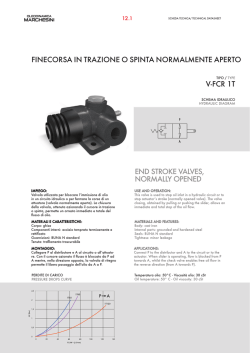
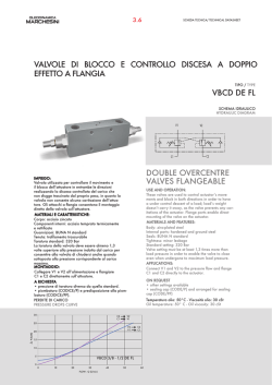
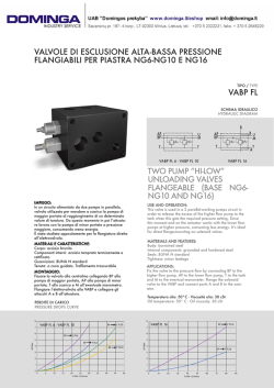
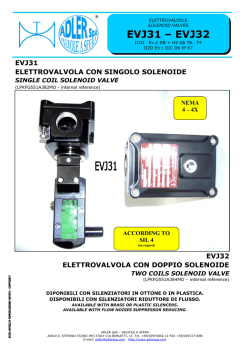
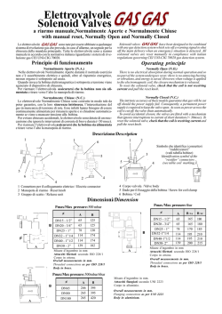
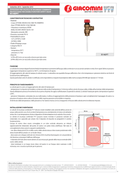
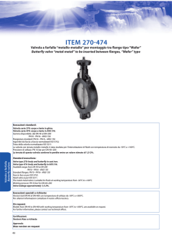
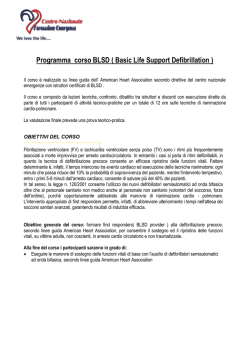
research from animal testing to clinical experience 178 Ann Ist Super Sanità 2007 | Vol. 44, No. 2: 178-186 Electrospun bioresorbable trileaflet heart valve prosthesis for tissue engineering: in vitro functional assessment of a pulmonary cardiac valve design Costantino Del Gaudio(a, b), Alessandra Bianco(b) and Mauro Grigioni(a) (a) (b) Dipartimento di Tecnologie e Salute, Istituto Superiore di Sanità, Rome, Italy Dipartimento di Scienze e Tecnologie Chimiche, Università degli Studi “Tor Vergata”, Rome, Italy Summary. Currently implanted prosthetic heart valves, both mechanical or biological ones, are used to restore the proper blood hemodynamics when the native valves fail. However, these medical devices are not free from drawbacks, such as hemolysis or calcification, also presenting the relevant disadvantage of being unable to growth, repair and remodel. An improvement could be represented by bioresorbable polymeric tissue-engineered heart valves. In this paper a poly(ε-caprolactone) (PCL) heart valve prosthesis, realized by means of electrospinning, and its in vitro functional characterization in a pulse duplicator, resembling pulmonary conditions, is presented. Morphological examination revealed polymeric micrometric fibers randomly oriented with an average porosity of about 90%. Pulse duplicator testing highlighted that leaflets opened synchronously and showed a correct coaptation in the diastolic phase, even if a slight rotation of the leaflets was visualized. In silico study by numerical simulation of the closed phase predicted the stress distribution within the leaflet, showing that peak levels are reached at the commissures and sustained by the structure without failure. The present study highlighted the technical feasibility to produce polymeric bioresorbable functional heart valves by means of electrospinning. Further studies and design changes are needed in order to optimize the final scaffold to bear arterial hemodynamic conditions. Key words: bioresorbable heart valve, electrospinning, functional in vitro testing. Riassunto (Valvola cardiaca bioriassorbibile elettrofilata per ingegneria dei tessuti: valutazione funzionale in vitro di un modello polmonare). Le protesi valvolari cardiache attualmente impiantate, sia meccaniche che biologiche, sono in grado di ristabilire la corretta emodinamica e garantire una qualità di vita soddisfacente quando le valvole native non possono più assolvere alla propria funzione. Tuttavia questi dispositivi presentano diversi limiti (emolisi, calcificazione) e non possono rimodellarsi in risposta alle modificazioni dell’organismo ospite. Un miglioramento in tal senso può essere rappresentato dalle valvole cardiache in polimero bioriassorbibile. In questo lavoro si presenta la realizzazione e caratterizzazione funzionale, parametri del sito polmonare, di una protesi valvolare in policaprolattone (PCL) prodotta mediante elettrofilatura (electrospinning). L’esame morfologico ha evidenziato una struttura composta di fibre polimeriche casualmente orientate con una porosità del 90%. Le prove funzionali in un duplicatore di impulsi hanno mostrato l’apertura sincrona dei lembi valvolari e una corretta apposizione in diastole, anche se una leggera deformazione della valvola è stata evidenziata. La simulazione numerica della distribuzione degli sforzi nei lembi valvolari ha evidenziato che i valori più alti sono raggiunti a livello delle commissure. Questo studio ha mostrato la possibilità di produrre valvole cardiache in polimero bioriassorbibile mediante elettrofilatura. Ulteriori studi sono necessari per ottimizzarne la struttura al fine di sopportare il carico pressorio arterioso. Parole chiave: valvola cardiaca bioriassorbibile, elettrofilatura, caratterizzazione funzionale in vitro. INTRODUCTION Mechanical or biological heart valves are the most common medical devices currently used to restore the failure of native valves. However both of them are affected from several drawbacks that limit the long-term efficacy. To date a nonthrombogenic, non- calcific prosthesis, which maintains mechanical and hemodynamic characteristics and exhibits sufficient fatigue properties, has not been designed [1]. Tissue engineering could be an alternative approach to move towards a heart valve equivalent. A number of studies investigated the possibility to produce tissue engi- Address for correspondence: Mauro Grigioni, Dipartimento di Tecnologie e Salute, Istituto Superiore di Sanità, Viale Regina Elena 299, 00161 Rome, Italy. E-mail: [email protected]. Electrospun bioresorbable trileaflet heart valve prosthesis for tissue engineering neered prostheses, derived from polymers, decellularized scaffolds or biological scaffolds of non-valvular origin. Synthetic polymeric scaffolds present several attractive characteristics including the possibility to deal with bioresorbable biomaterials, to have a number of assembling techniques available, to control the repeatability of the resulting scaffold and to have greater control over mechanical properties. However, possible limitations related to these biomaterials can also be highlighted. Long term mechanical properties can change as the polymer degrades and the reaction products can potentially induce a toxic response [1]. Moreover, a number of difficulties in the regulation of cell adhesion and tissue organization can be reported: extracellular matrix proteins, such as specific ligands to promote cell attachment to the matrix, are not present in synthetic polymers [2]. The use of decellularized scaffolds can overcome some disadvantages over synthetic materials [2, 3], but the development of an effective technique for decellularizing heart valves and removing cellular debris is a key-point to be addressed for a successful scaffold, that is not often achieved [4-6]. Finally, biological scaffolds of non-valvular origin can be regarded as a valid alternative. These materials (e.g., collagen) contains natural cellular adhesion sites and are less likely to initiate toxic immune response. Being acellular materials the decellularization process is not required, thus preventing the consequences of an incomplete decellularization. However, also these materials have not fulfilled all the requirements for a proper tissue-engineered heart valve scaffold (e.g., mechanical properties) [1]. Moreover, collagen type I can elicit platelet adhesion and aggregation; several studies investigated the role of glycoprotein VI in the interaction with collagen to promote platelet activation and thrombus formation [7-9]. A brief survey of studies on tissue-engineered heart valves is resumed in Table 1. Material This study presents the early results of a functional poly(ε-caprolactone) (PCL) heart valve made by electrospinning. PCL is a bioresorbable aliphatic polyester, subjected to enzymatic degradation through hydrolysis of ester bonds [22]. The slow degradation period, due to its semicrystalline nature, allows a) seeded cells to create extracellular matrix (ECM), b) provide prolonged mechanical reliability [22, 23], c) elicits low inflammatory response within the surrounding tissue, due to the lower concentration of released acidic products [24]. Specifically, PCL degradation is a two-stage process; first, the molecular weight reduces according approximately to an exponential law, then polymer fragments are processed by cellular phagocitosis. PCL is finally excreted by biliar or gastrointestinal route [25, 26]. Moreover taking into account mechanical proper- ties, PCL is supposed to be an eligible biomaterial for cardiovascular tissue engineering applications, compared to other polyesters (e.g., polyglycolic acid, polylactic acid) characterized by higher stiffness and lower compliance. Production process Electrospinning is an efficient technique to produce polymeric fibers with diameters ranging from nano- to micrometers. It is realised by applying high voltage between a capillary, through which a polymeric solution flows, and a grounded collecting target [27]. Electric field induces charges on the surface of the pendant drop at the tip of the capillary and mutual charge repulsion causes a force directly opposite to the surface tension [27]. As the intensity of the electric field increases, the formation of the so-called Taylor cone from the polymeric drop occurred and when the electrostatic force overcomes surface tension and viscoelastic forces (for a critical value of the external electric field) a charged jet is forced from its tip [27, 28]. The polymeric jet is then subjected to instability that determines the stretching of the polymeric jet itself and the evaporation of the solvent, this process leads to the formation of a series of dry fine fibers [28]. Fibers are randomly collected onto a fixed metal grounded target in form of nonwoven mat or as a fibrous aligned mat on a rotating target. Electrospun PCL heart valve was functionally characterized by means of pulse duplicator in physiological condition resembling the pulmonary site. High speed cinematography was also carried out to investigate leaflet dynamic behaviour and to asses whether a proper leaflet coaptation occurred in the diastolic phase. Finally a structural numerical simulation was performed to highlight stress distribution within the leaflet, in the closed position. MATERIALS AND METHODS Valve prosthesis experimental procedure Trileaflet stentless PCL (Sigma-Aldrich, Mn=80000) valve was realized by means of electrospinning. The polymer was solved in chloroform (14% w/v) and successively collected into a glass syringe fitted with a metallic blunt tip needle (22G). PCL was electrospun on a rotating custom-made aluminium trileaflet heart valve-shaped target at 10 cm from the needle; due to its complex geometry, the rotating speed was 0.3-0.4 rpm in order to favour a homogeneous polymer deposition. Briefly, the valve prosthesis was designed according to the following technical procedure, resembling several choices already used on the marketed animal tissue derived prostheses, such as the sewing procedure made by using three fixation points where the posts are located in stented prostheses. The coaptation region was modelled by straight lines linked with an arc, then each leaflet was realized extruding the above mentioned region along a spline trajectory, in the 179 180 Costantino Del Gaudio, Alessandra Bianco and Mauro Grigioni Table 1 | Overview of tissue-engineered heart valves Scaffold Production technique Cell seeding Testing Ref. PGA-P4HB Heat application welding technique Human marrow stromal cells Bioreactor [10] PHOH-P4HB Stereolithography Bioreactor [11] Vascular cells from ovine carotid artery and jugular vein. Lamb model Pulmonary site [12] PHO PGA-P4HB Heat application welding technique Autologous ovine myofibroblasts and endothelial cells Bioreactor conditioning Lamb model Pulmonary site [13] PCL Electrospinning Human myofibroblast Bioreactor [14] PGA-P4HB Molding Cells from human vena saphena magna Bioreactor [15] PGA-PLLA Melt extrusion to have flat nonwoven sheets to be assembled in heart valve scaffolds MSC from ovine bone marrow Sheep model Pulmonary site [16] Decellularized ovine pulmonary valves Autologous jugular veins endothelial cells Bioreactor conditioning Lamb implantation [17] Decellularized porcine pulmonary valves Canine bone marrow-derived cells Dog implantation [18] Decellularized porcine pulmonary valves Sheep vascular endothelial cells Sheep implantation [19] Evaluation of the residual potential to attract monocytic cells depending on the origin of the scaffold [4] Decellularized porcine and human pulmonary valve conduits Decellularized porcine heart valve conduits HUVEC Evaluation of endothelial cells to abolish platelet adhesion and activation (decellularized porcine matrix acts as a plateletactivating surface) [5] Acellularized allogenic lamb heart valve conduits Lamb myofibroblasts Reseeded and acellularized heart valve conduits were implanted into lambs [2] Decellularized human pulmonary valve allogarfts Endothelial progenitor cells Implantation into two pediatric patients [3] Bovine type-I collagen Rapid prototyping Human aortic valve interstitial cells In vitro evaluation of cell response to collagen concentration (discshaped scaffolds) in static conditions [20] Fibrin Moulding Carotid artery-derived cells Bioreactor [21] PGA: polyglycolic-acid; PHOH: poly-3-hydroxyoctanoate-co-3-hydroxyhexanoate; P4HB: poly-4-hydroxybutyrate; PHO: polyhydroxyoctanoate; PCL: Poly(ε-caprolactone); HUVEC: human umbilical vein endothelial cells; MSC: mesenchymal stem cells. sagittal plane, from the top of the valve to the annulus. Table 2 summarizes the design parameters. A steady state flow rate of the polymeric solution was achieved by means of a syringe pump (KD Scientific, USA) running at 0.6 ml/h, while a high voltage power supply (Spellman, UK) assured the tension of 12 kV for the electrospinning process. Polymer deposition time was fixed to 1.5 h. Structural characterization Morphology of the heart valve prosthesis was investigated by scanning electronic microscopy (SEM). Thickness measurements of the two commissural regions of each leaflet were performed to check if uniform polymer deposition occurred. For this aim a measuring pressure of 10 g/cm2 was imposed to the samples, as prescribed by ISO 7198. Electrospun bioresorbable trileaflet heart valve prosthesis for tissue engineering Table 2 | Heart valve design parameters Tissue annulus diameter 19 mm Valve inner diameter 14.5 mm Valve height 11.5 mm Arc radius of central leaflet coaptation area to reduce peak stress 3 mm Porosity (ε) was estimated according to the following relationship [29, 30]: ( ) ρ 0 ⋅ 100 ε = 1 − ρ (Eq. 1) where ρ is the density as calculated from the weight to volume ratio. Circular samples were cut out from the leaflets of each valve to estimate ε. Thickness was evaluated as previously described, while weight was evaluated by means of analytical balance (Sartorius CP124S, resolution 10-4 g) Density value of 1.145 g/ ml (as-purchased polymer) was considered for ρ0. Functional testing Electrospun trileaflet stentless heart valve prosthesis was tested in the VSI pulse duplicator (Vivitro Systems, Inc., Canada), properly modified with a thin glass window at the top of the testing site to monitor the valve function, in the opening and in the closing phases, with a camera [31]. The electrospun valve was located in the testing site by means of custom-made retention PVC ring. Valve commissures were loosely sutured to three posts at 120°, fixed on the ring, in order to prevent a possible collapse in the diastolic phase (Prolene 7-0, Ethicon). A mechanical no-leakage reference valve was inserted in the tricuspid site. Functional signals (ventricular, atrial, arterial pressures and arterial and tricuspid Fig. 1 | PCL electrospun heart valve. flows) were acquired. Hydrodynamic behaviour of the prosthetic valve was investigated using saline solution (0.9% NaCl), as test fluid. A sine waveform was selected to drive the pump at 60 bpm. Due to the small size of the prosthesis a specific investigating protocol was considered. Three stroke volumes were selected (20, 30 and 40 ml) for a mean pulmonary pressure of 30 mmHg. Each setting condition was averaged on 16 cardiac cycles and repeated three times. Mean transvalvular pressure drop, cardiac output and energy loss were evaluated for all the conditions investigated. Energy loss was computed as the integral over time of the product of instantaneous transvalvular pressure and flow rate [32]. Kinematics of the prosthetic valve leaflets was studied using the Kodak Ektapro camera (sampling rate 250 frames/s), located on top of the testing site. Numerical study Prediction of stress distribution on valve leaflet was determined by means of numerical simulation (Comsol, Sweden). Because of valve symmetry only one leaflet was modelled in the closed position (diastolic phase). Due to the particular retention system adopted (see “Functional testing” subsection), leaflet surface relative to valve stent was considered fixed while on the arterial side of the leaflet the transvalvular diastolic pressure was imposed (35 mmHg corresponding to the imposed stroke volume of 40 ml and mean pressure of 30 mmHg, respectively). The model was discretized by means of tetrahedral elements (about 11000). In this preliminary stage the material was considered isotropic, being a reasonable assumption due to the randomly orientation of polymeric fibers, with an elastic modulus of 6.4 MPa (estimated by means of uniaxial tensile test on dog-bone shaped specimens) and a Poisson coefficient of 0.45. RESULTS The electrospun valve is showed in Figure 1. Measured thickness of leaflets was 0.84 ± 0.14 mm, while estimated porosity was 89.13 ± 2.50%. SEM micrographs showed a random arrangement of polymeric fibers without beads defect (Figure 2), with an average fiber diameter in the micrometric range (3.11 ± 0.43 µm). The average cardiac cycle for the electrospun valve, acquired in the experimental session with the pulse duplicator, is reported in Figure 3, while Figure 4 showed the measured functional valve parameters. Mean transvalvular pressure drop was in the range 7-12 mmHg while the measured cardiac output was in the range 1.18-2.35 l/min, suggesting that the valve was affected by unsignificant leakage flow. The estimate energy loss was in the range 30-100 mJ, corresponding about to 28% of the ventricle energy within a cardiac cycle. Valve dynamic behaviour was recorded by camera acquisition. The opening behaviour at the ejection 181 Costantino Del Gaudio, Alessandra Bianco and Mauro Grigioni Fig. 2 | SEM micrograph of PCL electrospun heart valve. peak is reported in Figure 5a. Leaflets opened synchronously, even if a suboptimal opening occurred, due to the mild setting conditions imposed by the pulse duplicator. In the diastolic period the valve did not maintain the correct shape showing a slight rotation of the leaflets (Figure 5b). The predicted stress distribution in the diastolic phase is reported in Figure 6 for maximum loading conditions of the experimental protocol (stroke volume of 40 ml and mean pressure of 30 mmHg). Quite uniform stress distribution was predicted within the leaflet showing an increase in the central region of the free edge (about 100 kPa). Highest stresses occurred at the intersection of the free edge of the leaflet with the resembling stent structure, being in the range 200-250 kPa. DISCUSSION Valid alternatives to currently used heart valve prostheses represent a relevant issue to be addressed by means of tissue engineering applications. This study reported the early results of the hydrodynamic characterization of a trileaflet stentless electrospun PCL heart valve. The most immediate need for tissue engineered heart valves and regenerative technology is in the pediatric and young adult patients since results of valve replace- 60 150 40 100 20 50 0 0 -20 0 0.1 0.2 0.3 0.4 0.5 0.6 Time (s) 0.7 0.8 0.9 1 Valve flow (ml/s) Pressure (mmHg) 182 -50 Fig. 3 | Average cardiac cycle curves (pressures and valve flow) for the PCL electrospun heart valve for a stroke volume of 40 ml. Energy loss (mJ) Cardiac output (l/min) Mean pressure drop (mmHg) Electrospun bioresorbable trileaflet heart valve prosthesis for tissue engineering 14 12 10 8 6 15 20 25 30 35 40 45 15 20 25 30 35 40 45 2.5 2 1.5 1 150 100 50 0 15 20 25 30 Stroke volume (ml) 35 ment are not as favourable as in older adults [33, 34]. Children undergone to heart valve replacement can have a positive outcome thanks to prosthetic devices made by bioresorbable polymer. On this basis, small size heart valve prosthesis was selected for a pivotal study and infant pulmonary system [35] conditions were replicated by means of the pulse duplicator. Moreover, it has been also reported that a mild conditioning in bioreactors is the first step for a successful tissue-engineered heart valve [13, 17]. Thus moderate pulsatile circulation can promote a confluent monolayer of endothelial cells on valve cusps, playing an important role in the long-term durability and functionality of tissue engineered heart valve prostheses [17]. The investigated condition also resembled these settings, promoting a step of growing of native structured valve directly in the patient with the capability to regenerate during its own life while the PCL structure is bioresorbed. SEM investigation revealed PCL fibers randomly arranged; this is the typical result of the electrospinning process when a fixed or slowly rotating target A B 40 45 Fig. 4 | Electrospun functional parameters in terms of measured transvalvular pressure drop, cardiac output and energy loss for all the stroke volumes investigated (20, 30, 40 ml). is used to collect polymeric fibers. In particular, the low speed imposed to the metallic target was selected to favour a homogenous deposition of the polymer to resemble the very complex three-dimensional geometry of a trileaflet heart valve. Moreover, the high estimated porosity represents a valuable feature for a tissue-engineered scaffold, promoting easy diffusion of nutrients to and waste products from the implant and vascularization as well [36]. The experimental session in the pulse duplicator highlighted a proper functioning of the herein proposed device also showing a good repeatability for the test conditions, as suggested by the small standard deviations calculated. Cinematographic analysis revealed synchronously opening of the leaflets during the ejection period and a good apposition in the diastolic period, even if a slight rotation was detected which prevented to maintain the correct valve shape. However, this promising result needs to be validated under more severe loading conditions, e.g. resembling the aortic site or fatigue testing, and according Fig. 5 | Opening frame at the systolic peak (a) and in the diastolic phase (b). 183 184 Costantino Del Gaudio, Alessandra Bianco and Mauro Grigioni 250 200 150 100 50 Fig. 6 | Numerical stress distribution within the leaflet in the diastolic phase (system pressure 30 mmHg) [kPa]. to specific protocols for tissue engineering applications (cell-to-scaffold response). Hydrodynamic assessment of the heart valve prosthesis is only the preliminary step, because it should be pointed out that artificial heart valves do not resemble the complex architecture and organization of native valve leaflets. Semilunar heart valves are microscopically composed of three layers: ventricularis (rich in radially aligned elastin fibers), spongiosa (largely composed of glycosoaminoglycans) and fibrosa (primarily composed of dense packed collagen fibers, arranged parallel to the cuspal free edge). Each layer contributes to the definition of a biomechanical characteristic that enables the heart valve to its specific function. Structural elements within each layer provide the high anisotropic properties [34, 37]. Thus the ultimate goal of a functional tissue engineered heart valve prosthesis can be reached replicating the structure of a native valve. For this aim mechanical conditioning in bioreactors of seeded scaffolds seems to promote anisotropy, suggesting that repetitive changes in strains can induce desired organization in the leaflets, enhances tissue formation and thereby improve mechanical strength [15]. The reproducibility in fiber deposition was evaluated by means of uniaxial tensile test on several specimens (at least four per test type) cut out from electrospun mats collected with a similar experimental set-up. Average tensile modulus and tensile strength were 6.4 ± 0.2 MPa and 0.84 ± 0.07 MPa, respectively, highlighting standard deviations obtained in the production process to be within 10%. Nevertheless the evaluation of repeatability of hydrodynamic characteristics of electrospun valves is an issue to be addressed, it should be pointed out that the in vitro assessment cannot give complete insights on the in vivo functioning. Due to the nature of the device, i.e. stentless valve with particular sewing procedure, hemodynamic features will depend on the implantation criteria adopted by the surgeon to fit to patient’s anatomy. The latter possibility is generally appreciated by surgeon to adapt the geometry to the pathological aortic root to be treated. Numerical simulation showed the stress distribution within the leaflet in the closed position, assuming the measured diastolic pressure difference as input variable. Numerical simulations were carried out under simplified assumptions not strictly resembling the structural characteristics of the electrospun leaflet (the fibrous morphology was neglected) or the application of the correct experimental boundary conditions (leaflet surface connected to the stent was considered fixed in the numerical simulation). Although the influence of anisotropy in heart valve leaflets should be considered for a more realistic stress analysis, in this study numerical simulation run under the assumption of isotropy. The aim was to predict the peak stress locations depending on the shape given to the leaflet, before cell seeding and culturing: highest values were found where the free edge is connected to the resembling stent structure. The obtained results are to be intended as a starting point in view of design optimization of leaflet geometry in order to minimizing critical stress concentrations. As previously reported, the anisotropic nature of tissue engineered heart valve is generally developed by means of dynamic culture systems, i.e. bioreactors. For instance, Driessen et al. [38] presented the prediction of the evolution of stress distribution at selected time points after culturing non-woven polyglycolic acid, coated with poly-4hydroxybutyrate, heart valve scaffold in a diastolic pulse duplicator, finding a monotonically stress increase caused by the decrease of construct thickness. Numerical analysis represents an improvement in the prediction of stress distribution or in the design of a novel device, but it should be also underlined that the obtained results need to be validated with experimental data in order to deal with a realistic model. Differences arise comparing numerical results of tissue engineered leaflets and native porcine leaflets, showing that the mechanical behaviour of engineered leaflets is less nonlinear, less anisotropic and lower coaptation occurred due the absence of large radial strain [38]. However, computational analysis of the mechanical response of a closed valve is a relevant issue to be addressed, because peak stresses in the fully closed position contribute to tears and perforations of the leaflets [39]. Electrospinning was previously implemented as a multistep procedure to realize a heart valve with the Electrospun bioresorbable trileaflet heart valve prosthesis for tissue engineering aortic root. Experimental parameters differed from the ones here considered and a not homogenous deposition of the polymer onto the target was obtained. It has been reported that the scaffold failed due to weakness of the bellies of the leaflets and the aortic ring, where the construct was very thin and tore easily in in vitro testing session by means of pulse duplicator [14]. Results here presented showed the technical feasibility to produce a functional bioresorbable heart valve by means of electrospinning. A correct functional response was shown to the testing protocol by means of pulse duplicator. Nevertheless under the mild setting conditions considered, the valve did not undergone to structural damage. Future development of this study is represented by the optimization of the electrospinning parameters, valve design criteria and the selection of the most suitable polymer, co-polymer or blend to be used for tissue engineering heart valve application. Acknowledgements The authors wish to thank Dr. F. Nanni (Dipartimento di Scienze e Tecnologie Chimiche, Università degli Studi “Tor Vergata”, Rome, Italy) for SEM analysis. Received on 28 October 2007. Accepted on 19 February 2008. References 1. Brody S, Pandit A. Approaches to heart valve tissue engineering scaffold design. J Biomed Mater Res B Appl Biomater 2007;83:16-43. 2. Steinhoff G, Stock U, Karim N, Mertsching H, Timke A, Meliss RR, Pethig K, Haverich A, Bader A. Tissue engineering of pulmonary heart valves on allogenic acellular matrix conduits: in vivo restoration of valve tissue. Circulation 2000;102(Suppl 3):50-5. 3.Cebotari S, Lichtenberg A, Tudorache I, Hilfiker A, Mertsching H, Leyh R, Breymann T, Kallenbach K, Maniuc L, Batrinac A, Repin O, Maliga O, Ciubotaru A, Haverich A. Clinical application of tissue engineered human heart valves using autologous progenitor cells. Circulation 2006;114(Suppl 1):32-37. 4. Rieder E, Seebacher G, Kasimir MT, Eichmair E, Winter B, Dekan B, Wolner E, Simon P, Weigel G. Tissue engineering of heart valves: decellularized porcine and human valve scaffolds differ importantly in residual potential to attract monocytic cells. Circulation 2005;111:2792-7. 5. Kasimir MT, Weigel G, Sharma J, Rieder E, Seebacher G, Wolner E, Simon P. The decellularized procine heart valve matrix in tissue engineering. Platelet adhesion and activation. Thromb Haemost 2005;94:562-7. 6. Conconi MT, Rocco F, Spinazzi R, Tommasini M, Valfre C, Busetto R, Polesel E, Albertin G, Dei Tos A, Iacopetti I, Cecchetto A, Zussa C, Grigioni M, Parnigotto PP, Nussdorfer GG. Biological fate of tissue-engineered porcine valvular conduits xenotransplanted in the sheep thoracic aorta. Int J Mol Med 2004;14:1043-8. 7. Badimon L, Badimon JJ, Turitto VT, Vallabhajosula S, Fuster V. Platelet thrombus formation on collagen type I. A model of deep vessel injury. Influence of blood rheology, von Willebrand factor, and blood coagulation. Circulation 1988;78(6):1431-42. 8. Savage B, Ginsberg MH, Ruggeri ZM. Influence of fibrillar collagen structure on the mechanisms of platelet thrombus formation under flow. Blood 1999;94(8):2704-15. 9. Goto S, Tamura N, Handa S, Arai M, Kodama K, Takayama H. Involvement of glycoprotein VI in platelet thrombus formation on both collagen and von Willebrand factor surfaces under flow conditions. Circulation 2002;106(2):266-72. 10. Hoerstrup SP, Kadner A, Melnitchouk S, Trojan A, Eid K, Tracy J, Sodian R, Visjager JF, Kolb SA, Grunenfelder J, Zund G, Turina MI. Tissue engineering of functional trileaflet heart valves from human marrow stromal cells. Circulation 2002;106(Suppl 1):143-50. 11. Sodian R, Loebe M, Hein A, Martin DP, Hoerstrup SP, Potapov EV, Hausmann H, Lueth T, Hetzer R. Application of stereolithography for scaffold fabrication for tissue engineered heart valves. ASAIO J 2002;48:12-16. 12. Sodian R, Hoerstrup SP, Sperling JS, Daebritz S, Martin DP, Moran AM, Kim BS, Schoen FJ, Vacanti JP, Mayer JE Jr. Early in vivo experience with tissue-engineered trileaflet heart valves. Circulation 2000;102(19 Suppl.3):III22-9. 13. Hoerstrup SP, Sodian R, Daebritz S, Wang J, Bacha EA, Martin DP, Moran AM, Guleserian KJ, Sperling JS, Kaushal S, Vacanti JP, Schoen FJ, Mayer JE Jr. Functional living trileaflet heart valves grown in vitro. Circulation 2000;102(19 Suppl III):III44-9. 14. van Lieshout MI, Vaz CM, Rutten MC, Peters GW, Baaijens FP. Electrospinning versus knitting: two scaffolds for tissue engineering of the aortic valve. J Biomater Sci Polym Ed 2006;17(1-2):77-89. 15. Mol A, Rutten MC, Driessen NJ, Bouten CV, Zund G, Baaijens FP, Hoerstrup SP. Autologous human tissue-engineered heart valves: prospects for system application. Circulation 2006;114(Suppl I):I152-8. 16. Sutherland FW, Perry TE, Yu Y, Sherwood MC, Rabkin E, Masuda Y, Garcia GA, McLellan DL, Engelmayr GC Jr, Sacks MS, Schoen FJ, Mayer JE Jr. From stem cells to viable autologous semilunar heart valve. Circulation 2005;111:2783-91. 17. Lichtenberg A, Cebotari S, Tudorache I, Sturz G, Winterhalter M, Hilfiker A, Haverich A. Flow-dependent re-endothelialization of tissue-engineered heart valves. J Heart Valve Dis 2006;15(2):287-93. 18. Kim SS, Lim SH, Hong YS, Cho SW, Ryu JH, Chang BC, Choi CY, Kim BS. Tissue engineering of heart valves in vivo using bone marrow-derived cells. Artif Organs 2006;30:554-7. 19. Dohmen PM, Ozaki S, Nitsch R, Yperman J, Flameng W, Konertz W. A tissue engineered heart valve implanted in a juvenile sheep model. Med Sci Monit 2003;9:BR97-BR104. 20.Taylor PM, Sachlos E, Dreger SA, Chester AH, Czernuszka JT, Yacoub MH. Interaction of human interstitial cells with collagen matrices manufactured using rapid prototyping. Biomaterials 2006;27:2733-7. 21. Flanagan TC, Cornelissen C, Koch S, Tschoekea B, Sachwehb JS, Schmitz-Rode T, Jockenhoevel S. The in vitro development of autologous fibrin-based tissue-engineered heart valves through optimised dynamic conditioning. Biomaterials 2007;28:3388-97. 22.Ciardelli G, Chiono V, Vozzi G, Pracella M, Ahluwalia A, Barbani N, Cristallini C, Giusti P. Blends of poly(ε-caprolactone) and polysaccharides in tissue engineering applications. Biomacromlecules 2005;6:1961-76. 23. Martin DP, Williams SF. Medical applications of poly-4hydroxybutyrate: a strong flexible absorbable biomaterial. Biochem Eng J 2003;16:97-105. 185 186 Costantino Del Gaudio, Alessandra Bianco and Mauro Grigioni 24. Sung HJ, Meredith C, Johnson C, Galis ZS. The effect of scaffold degradation rate on three-dimensional cell growth and angiogenesis. Biomaterials 2004;25:5735-42. 25. Pitt CG, Chasalow FI, Hibionada YM, Klimax DM, Schindler A. Aliphatic polyesters I. The degradation of poly(ε-caprolactone) in vivo. J Appl Polymer Sci 1981;26:3779-87. 26. Sun H, Mei L, Song C, Cui X, Wang P. The in vivo degradation, absorption and excretion of PCL-based implant. Biomaterials 2006;27:1735-40. 27. Frenot A, Chronakis IS. Polymer nanofibers assembled by electrospinning. Curr Opinion Coll Interface Sci 2003;8:64-75. 28. Murugan R, Ramakrishna S. Nano-featured scaffolds for tissue engineering: a review of spinning methodologies. Tissue Eng 2006;12:435-47. 29. Vaz C, van Tuijl S, Bouten CVC, Baaijens FPT. Design of scaffolds for blood vessel tissue engineering using a multi-layering electrospinning technique. Acta Biomaterialia 2005;1:575-82. 30. Ma Z, Kotaki M, Inai R, Ramakrishna S. Potential of nanofiber matrix as tissue-engineering scaffolds. Tissue Eng 2005;11:101-9. 31. Grigioni M, Daniele C, Romanelli C, Morbiducci U, D’Avenio G, Del Gaudio C, Barbaro V. Pathological patient in protocol definition for bench testing of mechanical cardiac support system. Int J Artif Organs 2003;26(1):64-72. 32. VSI Manual, Vivitro Systems Inc., Victoria, Canada. 33. Vesely I. Heart valve tissue engineering. Circ Res 2005;97:743-55. 34. Mendelson K, Schoen FJ. Heart valve tissue engineering: concepts, approaches, progress, and challenges. Ann Biomed Eng 2006;34(12):1799-819. 35. Goodwin JA, van Meurs WL, Sa Couto CD, Beneken JE, Graves SA. A model for educational simulation of infant cardiovascular physiology. Anesth Analg 2004;99(6):1655-64. 36. Yang S, Leong KF, Du Z, Chua CK. The design of scaffolds for use in tissue engineering. Part I Traditional factors. Tissue Eng 2001;7:679-89. 37. Breuer CK, Mettler BA, Anthony T, Sales VL, Schoen FJ, Mayer JE. Application of tissue-engineering principles toward the development of a semilunar heart valve substitute. Tissue Eng 2004;10:1725-36. 38. Driessen NJB, Mol A, Bouten CVC, Baaijens FPT. Modeling the mechanics of tissue-engineered human heart valve leaflets. J Biomech 2007;40:325-34. 39. Liu Y, Kasyanov V, Schoephoerster RT. Effect of fiber orientation on the stress distribution within a leaflet of a polymer composite heart valve in the closed position. J Biomech 2007;40:1099-106.
© Copyright 2026 Paperzz