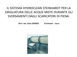
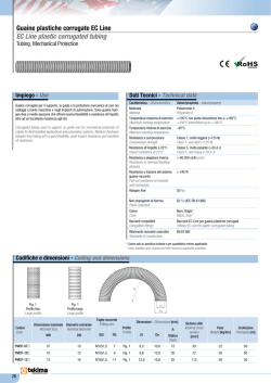
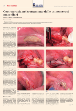
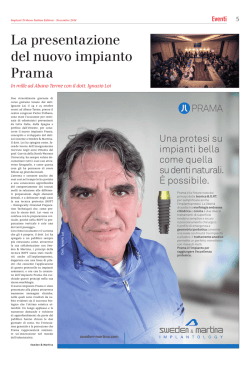
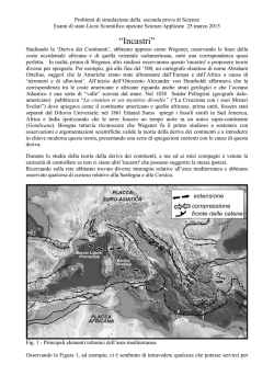
Mar Biol (2007) 152:969–979
DOI 10.1007/s00227-007-0747-4
R ES EA R C H A R TI CLE
Sexual reproduction, larval development and release in Spongia
oYcinalis L. (Porifera, Demospongiae) from the Apulian coast
R. Baldacconi · C. Nonnis-Marzano · E. Gaino ·
G. Corriero
Received: 14 November 2006 / Accepted: 4 June 2007 / Published online: 3 July 2007
© Springer-Verlag 2007
Abstract The reproductive cycle of the bath sponge
Spongia oYcinalis L. has been studied over 1 year on 11
tagged specimens of diVerent sizes (from 82 to 886 ml, in
volume) from Ionian coasts of Apulia (SE Italy). According
to literature data, the sponge is viviparous. All the monitored specimens showed sexual reproduction, even if the
process usually involved small portions of the sponge tissue. Ten specimens were gonochoric (sex ratio 1:1), one
specimen showed successive hermaphroditism, with alternate production of oocytes and spermatic cysts in the same
reproductive season. Young oocytes occur almost all year
round, whereas large mature eggs show a peak in October–
November, concomitantly with the appearance of spermatic
cysts. No relationships were observed between the sponge
size and the presence of sexual elements within the range of
the sponge size considered in this research. Embryo development occurs in patches of choanosomal tissue, which
contain four or more elements. Cleavage is total and equal;
it starts in November and in May leads to a solid stereoblastula, which develops into a parenchymella larva, from May
to July. The stereoblastula lacks Xagella and its surface is
delimited by elongated cells well segregated from the internal ones. Parenchymella larvae are released from June to
July, asynchronously, either at the individual or population
Communicated by R. Cattaneo-Vietti.
R. Baldacconi · C. Nonnis-Marzano · G. Corriero (&)
Dipartimento di Zoologia, Università degli Studi di Bari,
Via Orabona, 4, 70125 Bari, Italy
e-mail: [email protected]
E. Gaino
Dipartimento di Biologia Cellulare e Ambientale,
Università degli Studi di Perugia, Via Elce di Sotto,
06123 Perugia, Italy
level, with a few days of de-phasing. Up to 523 larval
elements/48 h for a sponge specimen were counted. The
free-swimming larvae are ovoid and uniformly Xagellated.
Flagella are longer at the posterior region, than on the rest
of the larval body. Flagellated cells form a pseudo-stratiWed
epithelial layer, delimiting the outermost larval surface, and
are Wlled with electron-lucent vesicles showing a homogenous content. No choanocyte chambers, pinacocytes or
skeletal elements are present in the newly released larvae.
Introduction
Spongia oYcinalis L. is a sponge known worldwide and
much utilized for cosmetic purposes. Prolonged over-Wshing and some recent epidemics have caused its disappearance from wide areas of the West Mediterranean Basin
(Gaino et al. 1992; Pronzato et al. 1996; Cerrano et al.
2000), thus determining the inclusion of this bath sponge
among the marine endangered species requiring speciWc
management measures (Annex 3 of the Bern Convention on
the Protection of Wildlife). In this context, knowledge on
the reproductive pattern and larval behavior are essential
for understanding the biological and ecological needs of
this species, and may yield information useful for proper
management, here including conservation plans (Kaye and
Reiswig 1991).
Data on the reproductive biology of S. oYcinalis are due
to Gaino et al. (1984), who investigated sperm cyto-diVerentiation at the ultra-structural level. Some aspects of the sexual cycle are reported in a former work in which about 400
specimens were examined, collected over 3 years at diVerent
sites from the Adriatic and Ionian Seas (Scalera Liaci et al.
1971). Reproductive processes of some West Indian species
of Spongia have also been investigated (Kaye 1990; Kaye
123
970
and Reiswig 1991). On the whole, the studies were based on
specimens randomly collected within diVerent populations.
This sampling technique does not allow the determination of
the ratio between gonochoric versus hermaphroditic specimens or whether reproduction is an age/size dependent event
or not. By contrast, Ayling (1980), Wapstra and van Soest
(1987), Witte et al. (1994), Usher et al. (2004) and Sidri
et al. (2005) report valuable information on the sexual cycle
of some demosponges obtained from investigating tagged
individuals over time. This method, which requires large
sponges and experienced SCUBA divers, has several advantages with respect to the traditional technique, such as a
stricter deWnition of the sex ratio and of the reproductive
periodicity of a population. In addition, such a technique
makes it possible to ascertain the possible relationships
between the biological features of a sponge (age, size, morphology) and its reproductive process.
The present work deals with the reproductive cycle of 11
specimens of S. oYcinalis investigated in the same area for
1 year. The investigation was carried out by utilizing the
technique of tagging each individual with a PVC stick and
cutting oV tissue samples from each of them every month
for histological and ultra-structural analyses. Larval release
was also investigated.
Materials and methods
Study site
The study was carried out at a depth of 6–7 m, on the rocky
bottom of La Pierta, a large embayment oV the coast of
Porto Cesareo (SE Italy, Ionian Sea-N 40°16⬘312⬙ E
17°51⬘593⬙). The rocky bottom consists of a horizontally
exposed wide carbonatic platform. The rocky surface is
aVected by high water movement and exposed to high solar
radiation. The benthic community is characterized by a
very rich sponge population with the dominance of Sarcotragus spinosulus, Chondrilla nucula and Spongia oYcinalis,
with a few frondose algae (Corriero et al. 2004b).
Species studied
Spongia oYcinalis (Demospongiae, Dictyoceratida, Spongiidae) displays a wide variety of massive forms (Pronzato
et al. 2003). The specimens studied in the present work
show a recurrent shape, characterized by a very large base
and a limited thickness, as a result of high mechanical
stress due to water movement conditions. The consistency
of the specimens is elastic, the surface smooth and Wnely
conulose, with a few oscules irregularly scattered. The
external color is black, owing to high light exposure, and
the internal color is brownish-yellow.
123
Mar Biol (2007) 152:969–979
Sampling protocol
Within an area of about 400 m2, eleven specimens of S.
oYcinalis of diVerent volume were selected and tagged
with numbered plastic labels. The volume of these specimens was estimated at the beginning of the study by measuring in situ, with a caliper, the height of each sponge (Wve
replicas for each measurement) and estimating the area of
each sponge upper surface by analyzing underwater photographs (Nikonos V system) using a KURTA IS/ADB digitizer connected to a Macintosh PC.
From April 2004 to March 2005, with monthly frequency, small sponge fragments (about 3 cm3 on the whole)
were carefully collected from each specimen by scuba
divers, to minimize damage. Samples were Wxed in a solution of formaldehyde in sea water (4%) and preserved in
ethanol 70%. To detect the presence of sexual reproductive
elements, the samples were dehydrated in ethanol and
embedded in Paraplast for routine histological examination.
Sections 7 m-thick were then stained with toluidine blue.
Sections were observed under a light microscope to
determine the monthly percentage of specimens with
oocytes, spermatic cysts, embryos and larvae, the mean
diameter (considering them spherical) and the density (number/mm3) of reproductive elements. In evaluating the phases
of the embryonic development of S. oYcinalis, the elements
in which a clear segregation between the superWcial layer
and the internal cells was evident were counted as larvae and
all the previous developmental phases as embryo.
The quantitative evaluation of the reproductive elements
was carried out using the Abercrombie (1941) formula as
suggested by Elvin (1976).
Univariate statistical analysis was carried out considering
only reproductive specimens. The relationship between
reproductive parameters (frequency of reproductive specimens, number of sexual elements, percentage of reproductive tissue) and the size of the sponge specimens were
calculated using the Spearman (rs) coeYcient of correlation.
For transmission electron microscopy (TEM), samples
Wxed in a solution of glutaraldehyde in sea water (4%,
pH 7.4) for 24 h were rinsed in artiWcial sea water, used as a
buVer, and post-Wxed for 30 min in osmium tetroxide (1%
in sea water). Afterwards they were repeatedly rinsed in the
same buVer, dehydrated in a graded ethanol series and
embedded in an epon-araldyte mixture. Ultra-thin sections
were obtained with an LKB ultramicrotome, contrasted
with uranyl acetate and lead citrate, and examined with a
Philips EM 208 transmission electron microscope. For
scanning electron microscopy (SEM), Wxed material was
dehydrated and critical point dried in a Pabish CPD 750
apparatus and coated with gold palladium (20 nm). Some
paraYn embedded samples, after the cutting of a face view
of the tissue including embryos and/or larvae, were treated
Mar Biol (2007) 152:969–979
971
to remove the embedding medium and then processed using
the above reported technique for SEM analysis. Specimens
were observed under a Philips XL 30 scanning electron
microscope at an accelerating voltage of 18 kV.
In the month of June, concomitantly with the occurrence
of larvae in the sponge tissue, traps were employed to collect
the larvae released in the Weld. The trap consisted of a conical
net (base diameter = 20 cm; height = 30 cm), the base of
which was provided with a closure system, made of a plastic
clamp. The cloth used in the net was high capacity nylon
monoWlament screen with a mesh of 250 m. The sponges
which had been previously found to be reproductively active
(specimens no. 2, 4, 10) were enclosed in the collecting traps,
which, in turn, were closed at the base of the sponge by the
plastic clamp. The traps were placed four times at 48 h intervals, with weekly frequency. After collection, larvae were
placed at 20°C in Wltered seawater and immediately preserved for histological and ultra-structural investigations.
Results
Sponge size
Sponge thickness never exceeded the value of 6 cm in any
of the studied specimens. The area of the sponge upper surface varied between 81.7 and 393.9 cm2. The estimated volumes varied between 81.7 and 886.2 ml, with a highest
frequency around 200–400 ml. These data were coherent
with unpublished observations referring to about 100 specimens studied from the study site.
Description of sexual elements
Female lineage
In the early phase of diVerentiation, the oocytes of S. oYcinalis, measuring about 15 m in diameter, were fairly triangular shaped (Fig. 1a). Growing oocytes acquired a
spherical shape and showed an eccentric nucleolated
nucleus and cytoplasm with scarce inclusions (Fig. 1b).
They were found in low densities throughout the choanosomal region, separated from one another by mesohyl elements and collagen. In the successive steps of development,
the cytoplasm was gradually enriched with yolk stored
material (Fig. 1c) up to its complete Wlling (Fig. 1d). In the
last phase of development, they reached their maximum
diameter (up to 200 m).
Male lineage
Spermatic cysts (Fig. 1e) occurred in the choanosomal
region. Germinal cell maturation occurred synchronously in
Fig. 1 a Oocytes of S. oYcinalis in the early phase of diVerentiation
(bar = 20 m). b Growing oocytes showing a spherical shape and
eccentric nucleolated nucleus (bar = 20 m). c Oocyte with nucleolated nucleus and cytoplasm in its initial phase of yolk accumulation
(bar = 50 m). d An oocyte whose cytoplasm is Wlled with yolk stored
material (bar = 50 m). e Spermatic cyst (SC) in the choanosomal region. DiVerent cysts contain sperm in various phases of diVerentiation
(bar = 50 m)
the same cysts, whereas it was asynchronous in diVerent
cysts of the same specimen.
Sexual reproductive cycle
Sexual elements (oocytes, spermatic cysts, embryos, larvae) were found throughout the year, with a monthly frequency of reproducing sponges gradually increasing from
September (63.6%) to November (100%) (mean annual
value 58.3 § 5.4%) (Fig. 2a).
Specimens with oocytes occurred almost all year round
(mean frequency 44.7 § 2.1%) (Fig. 2b). The density trend
showed some Xuctuations during the study year, with the
maximum value recorded in June (6.0 § 2.8 oocytes/mm3)
123
972
Mar Biol (2007) 152:969–979
a
Reproductive specimens
100
% frequency
80
60
40
20
0
Ap Ma Ju
Jul Au Se Oc No De
2004
Specimens with oocytes
Specimens with embryos
b
Ja
Fe Mar
2005
Specimens with sperm cysts
Specimens with larvae
60
% frequency
50
40
30
20
10
0
Ap Ma Ju
Jul Au Se Oc No De
2004
Ja
Fe Mar
2005
Fig. 2 Sexual cycle trend of S. oYcinalis in the study site. a Monthly
frequency of reproducing specimens. b Monthly frequency of specimens with oocytes, sperm cysts, embryos and larvae
(Fig. 3a). Oocytes grew in size from July (50.9 § 6.6 m in
diameter) to October (84.8 § 22.2 m) (Fig. 3a), then they
fell. Concomitantly with the maximum size of the oocytes,
spermatic cysts became evident. They were found in
autumn and early winter (mean frequency = 10.6 § 5.2%)
(Fig. 2b) with density values about 100 times higher than
those estimated for female elements. In contrast with
oocytes, the size of the spermatic cyst showed moderate
oscillations (from 50 to 60 m) (Fig. 3b).
Fertilized eggs showed total and equal cleavage. The
process started in November, giving rise to a solid morula
stage measuring 361.0 § 12.4 m in means, which consisted of few cells (4–16 cells). In December embryos consisted of numerous cells (Fig. 4a). The ensuing
development took place up to April and led to embryos
measuring 429.8 § 21.2 m as a maximum (Fig. 3c). In
May, the cleavage resulted in a solid white-translucent stereoblastula and parenchymellae measuring 402.4 §
16.9 m. Larvae grew up to a Wnal size of 480 m in July
(Fig. 3d).
In electron microscopy, the cells of the embryo possessed a cytoplasm packed with both the large electrondense and small electron-translucent inclusions (Fig. 4b).
123
The former contained dark granular bodies variously
dispersed. The nucleolate nucleus (Fig. 4c) tended to be
slightly eccentric. Just outside the nucleus, Golgi-derived
vesicles with electron-dense granules were evident
(Fig. 4d). Although embryonic cells tend to maintain a certain distance from one another, on occasions a cell establishes contact by means of membrane bridges (Fig. 4e) that
interact with the membrane of an adjacent cell (Fig. 4f).
Cleavage led to an early solid stereoblastula (Fig. 5a)
presenting a large amount of inclusions, mainly accumulated in the central region of the embryo. Under TEM they
revealed their heterogeneity, being the result of electrondense granules, varying in size and content (Fig. 5b). The
largest granules consisted of diVerent types of material
(Fig. 5c), among which even bacteria (Fig. 5c, inset).
Small cells with micro-granular electron-dense inclusions
and short microvilli, protruding from the outer cell surface, were evidently dispersed among the inclusions
(Fig. 5d).
The peripheral region of the early stereoblastula showed
numerous cells whose morphology was consistent with a
migration activity. They were elongated in shape with electron-dense inclusions, an eccentric nucleolated nucleus and
pseudopodia surrounding the bacteria dispersed in the mesohyl matrix (Fig. 5e). Some granules were minute and
encased in large vacuoles.
Images of sectioned specimens appropriately processed
for SEM analysis, after having removed the embedding paraYn (see “Materials and Methods” section), revealed that
the developing embryos and the ensuing larvae tended to
gather together to form clumps (Fig. 6a). Such brood chambers (Leys and Degnan 2002) were scattered in the mesoyl
of the sponge. The development of the embryos was dephased in such a way that in some clumps late stereoblastulae coexisted with mature parenchymellae (Fig. 6a).
Embryos and larvae were individually included in a cavity
(Fig. 6b), the wall of which were delimited by a monolayer
of Xattened cells (Fig. 6c).
The solid late stereoblastula showed a clear segregation
between peripheral non-Xagellated cells, showing an elongated shape and the more-rounded internal cells (Fig. 6d).
Mature parenchymellae (up to 600 m in size along the
main axis), were totally Xagellated and their outermost surface protruded in numerous folds (Fig. 7a). The Xagella
extended towards the cavity walls in such a way that they
crossed the space in between (Fig. 7b). The cavities were
separated from one another by a narrow collagen region
(Fig. 7c). The parenchymellae maintained the solid organisation of the stereoblastula, but their superWcial layer
showed a Xagellated epithelium (Fig. 7d).
The body of released larvae can be approximated to an
oval (Fig. 8a), whose posterior pole had a slightly
depressed region encircled by an uplifting ring (Fig. 8b),
Mar Biol (2007) 152:969–979
Fig. 3 Sexual cycle trend
of S. oYcinalis in the study site:
monthly mean size and number
of oocytes (a), sperm cysts (b),
embryos (c) and larvae (d) per
mm3 of sponge tissue (§standard error). Arrows indicate the
larval release
973
a
n°/mm³
10
Oocytes
Number
Size
c
µm
120
Embryos
n°/mm³
3
Number
100
8
Size
2.5
80
2
60
1.5
40
1
20
0.5
6
µm
500
400
300
4
2
0
0
A M J
200
0
J A S O N D J F M
2004
2005
b
Sperm cysts
n°/mm³
500
Number
100
0
A M J
d
µm
100
Size
n°/mm³
3
J A S O N D J F M
2004
2005
Larvae
Number
400
80
300
60
µm
500
Size
2.5
400
2
300
1.5
40
200
200
1
20
100
0
0
A M J
J A S O N D J F M
2004
2005
made of cells whose Xagella were longer than those of the
cells delimiting the rest of the larval surface.
TEM images gave evidence of the arrangement of the
Xagellated cells. They formed a pseudo-stratiWed continuous layer, were elongated in shape and possessed cytoplasmic extensions surrounding the single Xagellum (Fig. 8c).
Most of the cell body was Wlled with electron-lucent inclusions showing a homogeneous content, whereas mitochondria and electron-dense granules tended to accumulate in
the apical region (Fig. 8d).
Numerous bacteria were dispersed in the mesohyl matrix
and several TEM images proved that they are taken up
along the peripheral border of the cells with electron-dense
microgranules. These cells showed an elongated shape, an
eccentric nucleolated nucleus, scarce cytoplasm and numerous protrusions (Fig. 8e), which tended to branch and come
back to the original surface to surround bacteria. On occasions, bacteria were seen within blind ended pockets
formed by lobose pseudopodia wrapped around themselves
(Fig. 8f). No skeletal elements, choancocyte chambers or
pinacocytes were observed.
The embryo annual mean density in the mother sponge
was 1.3 § 0.6 no./mm3 (Fig. 3c). The progressive larval
development took place from May (mean annual density:
0.6 § 0.4 no./mm3) (Fig. 3d); the parenchymella was ready
to be released from June to July. The mean monthly fre-
100
0.5
0
0
A M J
J A S O N D J F M
2004
2005
quency of specimens with embryos and larvae was
30.3 § 6.6% and 6.1 § 3.6%, respectively (Fig. 2b). From
August to October neither embryos nor larvae were
detected (Fig. 3c, d).
The reproductive activity of each of the eleven monitored specimens is shown in Fig 9. During the study period,
ten specimens were found to be gonochoric (sex ratio 1:1)
and one hermaphrodite (specimen no. 6). In the six sponges
producing oocytes (Wve plus the hermaphrodite), the reproductive eVort and periodicity were diVerent: specimens no.
1, 2, 4 and 10 sustained oogenesis nearly all year round
(11 months), while no. 5 and 6 showed female gametes for
10 and 5 months, respectively. Among the six sponges producing spermatic cysts, the longest period of spermatogenesis was detected in specimen no. 7 (from September to
December), in which the highest values of density were
also observed, while no. 3 and 6 produced spermatic cysts
for 1 month (November and October, respectively). Sponge
no. 9 showed the lowest values of cyst density. As far as
embryos and larvae are concerned, the general behavior
observed was coherent with the above trend described for
oocytes: specimens no. 1, 2, 4 and 10 were the most
involved in embryo and larval production, a process that
took place in 8/9 months.
No relationships were observed between sponge size and
density of sexual elements (rs = 0.028; P > 0.05).
123
974
Mar Biol (2007) 152:969–979
Fig. 4 Embryogenesis of Spongia oYcinalis. a Morula stage
(bar = 150 m). b Electron
micrograph showing the variety
of inclusions packed in the morula cells (bar = 10 m). c Detail
of a nucleolated nucleus of a
morula cell showing the surrounding electron-dense (ED)
and electron-lucent (EL) inclusions (bar = 2.5 m). d A
zoomed view of the region
squared in (c). Note some Golgi
vesicles with membrane-bounded electron-dense granules (arrows) (bar = 250 nm). e Two
adjacent cells (C1–C2) showing a
contact by a membrane bridge
protruding from C1 (arrow)
(bar = 1 m). f Detail of the contact region (arrows)
(bar = 250 nm)
Larval release
Larvae were released from June to July (Fig. 3d), asynchronously, either at the individual or at the population level,
with few days of de-phasing. All the three monitored specimens of S. oYcinalis relased larvae during the third week
of June with values variyng from 50 (specimen no. 4) to
211 (no. 10) and 523 (no. 2) elements/48 h of monitoring.
A severe and unexpected storm in the last week of June
removed the trap and hampered further observations.
Discussion
The technique used in the present investigation, based on
tagged individual (Mercurio et al. 2006), allowed us to conWrm a prevalent gonochoric condition of S. oYcinalis. In
addition, a low percentage of specimens capable of sexual
reverse, showing a successive hermaphroditism. Sexual
reverse leads to successive hermaphroditism. Successive
123
hermaphroditism has been suggested for several sponge species (Sarà 1961; Diaz 1973) even if, so far, changes of sex
have only been demonstrated in tagged specimens of freshwater demosponges consecutively sampled (Van de Vyver
and Willenz 1975; Gilbert and Simpson 1976). Moreover,
the occurrence of Wve males out of ten gonochoric specimens
refutes a sex ratio in favour of the females, a feature that
seems to be fairly common among Porifera (Scalera Liaci
et al. 1971; Wapstra and van Soest 1987; Corriero et al.
1996, 1998; Mercurio et al. 2000; Meroz-Fine et al. 2005).
The general pattern of oogenesis and embryo development in S. oYcinalis is similar to that reported for other
viviparous demosponges (Saller and Weissenfels 1985;
Saller 1988; Leys and Ereskovsky 2006; Maldonado 2006)
and particularly for the Caribbean species of Spongia (Kaye
1990; Kaye and Reiswig 1991). Oogenesis begins with the
diVerentiation of primary oocytes, about 15 m in diameter,
probably diVerentiated from archaeocytes. They grow up to
200 m in diameter and acquire a round shape. Their cytoplasm becomes gradually Wlled with yolk granules.
Mar Biol (2007) 152:969–979
975
Fig. 5 Embryonic development of Spongia oYcinalis.
a Two solid stereoblastulae
(bar = 100 m). b Electron
micrograph showing the variety
of inclusions internally positioned in the stereoblastula
(bar = 10 m). c Detail of a large
inclusion showing the diVerent
components and an encased bacterium (inset) (bar = 1 m; bar
of inset = 100 nm). d A microgranular cell with evident microvilli-like marginal extensions
(arrows) (bar = 1.5 m). e Microgranular cells gathered along
the outermost surface of the solid stereoblastula. Note the bacteria amidst the cell cytoplasmic
extensions (arrows)
(bar = 1.5 m)
Fig. 6 Scanning electron microscopy view of stereoblastula and parenchymella stages of Spongia oYcinalis. a Steroblastula and parenchymella (P) grouped to form a clump within brood chambers
scattered in the mesohyl of the sponge (bar = 500 m). b A stereoblastula included in its chamber (bar = 500 m). c Zoomed view of the epithelium (E) of the chamber wall in which the stereoblastula develops
(bar = 25 m). d A stereoblastula showing a clear segregation between
the non Xagellated peripheral (A) and the internal (B) cells. Note the
solid mass of the internal cells (bar = 25 m)
Spermatogenesis inside spermatic cysts, deriving from
choanocyte chambers (Scalera Liaci and Sciscioli 1969;
Gaino et al. 1984), does not occur synchronously either in
individuals or in a population level and it is limited to
4 months as a maximum. This temporal lag overlaps the
presence of the largest oocytes, thereby corroborating the
hypothesis of Kaye (1990) that these elements can be
zygotes, even if a fertilization process has never been
detected.
The development of the parenchymella of S. oYcinalis
follows the typical stages of the formation of a solid, nonXagellated stereoblastula followed by the diVerentiation of
a Xagellated epithelium (see review in Simpson 1984). A
general monitoring of the parenchymella architecture
shows that the cell component is similar to that described
by Kaye and Reiswig (1991) for caribbean Spongia species,
where amoeboid cells prevailed, whereas pinacocytes, skeletal elements, choanocytes and other components of the
aquiferous system were lacking. This feature diVers from
other observations on parenchymellae of haplosclerid
sponges (Saller and Weissenfels 1985; Saller 1988; Ereskovsky 1999; Leys and Degnan 2002; Leys and Ereskovsky
2006), thus emphasising the striking diVerence in the
sponges belonging to this order, in which choanocyte
chambers, a pinacocyte lined cavity and siliceous spicules
diVerentiate precociously. According to Maldonado et al.
(2003), the cytoplasmic extensions protruding from the apical region of the Xagellated layer could be involved in functional beating.
The occurrence of bacteria inside the large electron-dense
inclusions of the early stereoblastula as well as along the
peripheral border of its electron-dense microgranular cells,
which come to surround bacteria by cytoplasmic extensions
and store them within intracellular vacuoles, proves that the
uptake of such micro-organisms already takes place in the
123
976
Fig. 7 Scanning electron micrographs of parenchymella of Spongia
oYcinalis. a Section of a mature larva showing the folds of its outermost Xagellated surface (bar = 25 m). b Detail of the Xagellated epithelium. Note the Xagella (arrows) crossing the space interposed
between the larva and the chamber wall (bar = 10 m). c The chamber
walls of two adjacent larvae (1, 2) are separated by a narrow collagencontaining region (CL) (bar = 100 m). d Portion of the Xagellated surface (A) and internal region (B) of the larva (bar = 25 m)
initial phase of larval development. This process is particularly evident in the newly released parenchymella. We can
suppose that the bacteria, derived from the mother sponge,
constitute the main food source for the free-swimming larva
owing to the lack of an aquiferous system able to support its
Wlter-feeding activity. Likewise, the accumulation of the
large inclusions in the central region of the stereoblastula, as
a result of cell disintegration, could be a way of providing
nutrients, a phenomenon already reported for the larval
development of other sponge species (Lévi 1956; Reiswig
1976). The bulk of data are in agreement with the fact that
most sponge larvae are lecithothrophic (Maldonado 2006)
and therefore self-sustaining by utilizing stored material.
This mechanism has also been observed in asexually produced bodies described in Tethya species where an aquiferous system was lacking (Gaino et al. 2006a).
Bacteria of the parenchymella escaping phagocytosis
can pass to the new generation, a feature supporting a verti-
123
Mar Biol (2007) 152:969–979
Fig. 8 A newly released parenchymella of Spongia oYcinalis. a Scanning electron microscopy of a whole larva (bar = 50 m). b Detail of
the posterior larval pole showing the slight depression (D) delimited by
an uplifted ring (R) (bar = 25 m). c Transmission electron microscopy
of the posterior larval ring showing the arrangement of the Xagellated
cells elongated in shape (bar = 5 m). d Detail of the Xagellated cells
showing their cytoplasmic organelles (mitochondria M; electron-dense
inclusions (I) in the apical cell region, and the numerous electron-lucent vesicles (V) with homogeneous content (bar = 1 m). e Microgranular cells with long branching cytoplasmic extensions surrounding
bacteria (arrows) (bar = 10 m). f Detail of a microgranular cell whose
pseudopods converge to form a pocket that includes bacteria. Note the
presence of bacteria within membrane-bounded vacuoles located along
the cell margin (arrow) (bar = 1 m)
cal transmission of symbiotic bacteria throughout sexually
produced elements (Kaye 1990). A similar mechanism has
also been observed in the buds of Tethya orphei where Wlamentous cyanobacteria located in the mother sponge enter
these asexually produced elements (Gaino et al. 2006b).
The homogenous material included in the vacuoles of the
parenchymella Xagellated epithelial cells could be extruded
from the larva at settlement, thus acting as a matrix substance able to assure the adhesion to the substratum at metamorphosis. Indeed, matrix substances are doubtlessly
involved in determining to a large extent the successful
adhesion allowing cell movement, repositioning and growth
processes, which take place in newly formed sponges.
Current literature reports that the annual reproductive
index in sponges is usually low, with only a limited number
Mar Biol (2007) 152:969–979
977
N°1
ln (n°/mm³)
N°2
ln (n°/mm³)
ln (n°/mm³)
1000.00
1000.00
1000.00
100.00
100.00
100.00
10.00
10.00
10.00
1.00
1.00
1.00
0.10
0.10
0.10
0.01
0.01
A M J
J A S O N D J F M
2004
2005
N° 4
ln (n°/mm³)
0.01
A M J
J A S O N D J F M
2004
2005
N°5
ln (n°/mm³)
A M J
ln (n°/mm³)
1000.00
1000.00
1000.00
100.00
100.00
100.00
10.00
10.00
10.00
1.00
1.00
1.00
0.10
0.10
0.10
0.01
0.01
A M J
J A S O N D J F M
2004
2005
N°7
ln (n°/mm³)
J A S O N D J F M
2004
2005
N°8
ln (n°/mm³)
ln (n°/mm³)
1000.00
100.00
100.00
100.00
10.00
10.00
10.00
1.00
1.00
1.00
0.10
0.10
0.10
0.01
J A S O N D J F M
2004
2005
N° 10
ln (n°/mm³)
N°6
//
A M J
1000.00
A M J
J A S O N D J F M
2004
2005
0.01
A M J
1000.00
0.01
N°3
0.01
A M J
J A S O N D J F M
2004
2005
A M J
J A S O N D J F M
2004
2005
N°11
ln (n°/mm³)
1000.00
100.00
100.00
10.00
10.00
1.00
1.00
Embryos
0.10
0.10
Larvae
0.01
0.01
J
A S O N D J F M
2004
2005
//
N° 9
1000.00
A M J
//
J A S O N D J F M
2004
2005
Oocytes
Sperm cysts
A M J
J A S O N D J F M
2004
2005
Fig. 9 Sexual abundance trend (mean monthly number of oocytes, sperm cysts, embryos and larvae per mm3 of sponge tissue, §standard error)
for each monitored individual of S. oYcinalis
of specimens, generally not exceeding 50%, able to diVerentiate gametes (Scalera Liaci et al. 1973; Corriero et al.
1996, 1998). Scalera et al. (1971) reported for S. oYcinalis
a mean reproductive index of 49.4% with a peak of reproductive specimens in December (87.5%). It is quite surpris-
ing that all the individuals of S. oYcinalis, monitored in the
present study reproduce all year round. Likewise, the prolonged presence of embryos within the same specimen
corroborates previous observations carried out on some
tropical and subtropical sponges, which may brood within
123
978
large chambers scattered in the mesohyl of the sponge
(Leys and Degnan 2002), of variable amounts of embryos
at any time during the year, as reported by Whalan et al.
(2005) and Maldonado (2006). It should be emphasized that
our technique of tagging specimens proved the storage of
embryos in the same individual, thus giving a better opportunity to investigate the reproductive strategies in sponges.
In S. oYcinalis the density of spermatic cysts is higher
than that of the oocytes (343.9 § 89.7 no./mm3 and
2.7 § 1.4 no./mm3, respectively), a pattern previously
observed in other viviparous species, such as Tedania anhelans (Nonnis-Marzano et al. 2000) and Pellina semitubulosa (Mercurio et al. 2000). Moreover, if we compare
viviparous and oviparous demosponges, the percentage of
volume occupied by female gametes in the latter is much
higher, likewise observed in Tethya aurantium (up to 1%)
and T. citrina (up to 3.5%) (Corriero et al. 1996).
The mechanisms controlling the onset of reproduction,
its duration and the growth of gametes are so far scarcely
documented. It is well known that in sponges gamete diVerentiation varies from individual to individual, probably
according to age and size (see Simpson 1980 for a review).
Indeed, it has been frequently observed that the relative
amount of resources put into reproduction increases with
age (Graham 1985). According to Storr (1964) in the bath
sponge Hippospongia lachne the minimum reproductive
size corresponds to about 14 cm in maximum diameter.
Our data prove full sexual activity even in specimens of
S. oYcinalis with a volume of 82 ml, corresponding to
about 10 cm in maximum diameter. On the other hand,
despite their phylogenetic aYnity, these two species exhibit
remarkable diVerences in other aspects of their life strategy,
more rapid growth in the tropical species H. lachne (Storr
1964) and slower in the temperate S. oYcinalis (Corriero
et al. 2004a). Nevertheless, among demosponges, the pattern of sexual activity is strongly variable, in Suberites
Wcus, for instance, oogenesis usually occurs only in specimens no larger than about 5 cm in size (Lévi 1951).
References
Abercrombie M (1941) Estimation of nuclear populations from microtomic sections. Anat Rec 94:239–247
Ayling AL (1980) Patterns of sexuality, asexual reproduction and
recruitment in some subtidal marine Demospongiae. Biol Bull
Mar Biol Lab, Woods Hole 158:271–281
Cerrano C, Bavestrello G, Nike-Bianchi C, Cattaneo-Vietti R, Bava S,
Moganti C, Morri C, Picco P, Sarà G, Schiaparelli S, Siccardi A,
Sponga F (2000) A catastrophic mass-mortality episode of gorgonians and other organisms in the Ligurian Sea (North-western
Mediterranean Sea), in summer 1999. Ecol Lett 3:284–293
Corriero G, Sarà M, Vaccaro P (1996) Sexual and asexual reproduction
in two species of Tethya (Porifera, Demospongiae) from a Mediterranean coastal lagoon. Mar Biol 126:175–181
123
Mar Biol (2007) 152:969–979
Corriero G, Scalera Liaci L, Nonnis Marzano C, Gaino E (1998)
Reproductive strategies of Mycale contarenii (Porifera: Demospongiae). Mar Biol 131:319–327
Corriero G, Longo C, Mercurio M, Nonnis Marzano C, Lembo G, Spedicato MT (2004) Rearing performance of Spongia oYcinalis on
suspended ropes oV the Southern Italian Coast (Central Mediterranean Sea). Aquaculture 238:195–205
Corriero G, Gherardi M, Giangrande A, Longo C, Mercurio M, Musco
L, Nonnis Marzano C (2004) Inventory and distribution of hard
bottom fauna from the marine protected area of Porto Cesareo
(Ionian Sea): Porifera and Polychaeta. Ital J Zool 71:237–245
Diaz JP (1973) Cicle sexuel de deux Démosponge de l’étang de Thau:
Suberites massa Nardo et Hymeniacidon caruncula Bowerbank.
Bull Soc Zool Fr 98:145–156
Elvin DW (1976) Seasonal growth and reproduction of an intertidal
sponge, Haliclona permollis (Bowerbank). Biol Bull mar biol
Lab, Woods Hole 151:108–125
Ereskovsky AV (1999) Development of Sponges of the Other Haplosclerida. Rus J Mar Biol 25:361–371
Gaino E, Burlando B, Zunino L, Pansini M, BuVa P (1984) Origin of
male gametes from choanocytes in Spongia oYcinalis (Porifera,
Demospongiae). Int J Inv Rep Dev 7:83–93
Gaino E, Pronzato R, Corriero G, BuVa P (1992) Mortality of commercial sponges: incidence in two Mediterranean areas. Boll Zool
59:79–85
Gaino E, Scalera Liaci L, Sciscioli M, Corriero G (2006) Investigation
of the budding process in Tethya citrina and Tethya aurantium
(Porifera, Demospongiae). Zoomorphology 125:87–97
Gaino E, Sciscioli M, Lepore E, Rebora M, Corriero G (2006) Association of the sponge Tethya orphei (Porifera, Demospongiae) with
Wlamentous cyanobacteria. Invertebr Biol 125(4):281–287
Gilbert JJ, Simpson TL (1976) Sex reversal in a freshwater sponge. J
Exp Zool 195:145–151
Graham E (1985) Reproductive pattern of marine invertebrates. Oceanogr Mar Biol Ann Rev 23:273–398
Kaye HR (1990) Sexual reproduction in four Caribbean commercial
sponges. II. Oogenesis and transfer of bacterial symbionts. Invertebr Reprod Dev 19:13–24
Kaye HR, Reiswig HM (1991) Sexual reproduction in four Caribbean
commercial sponges. III. Larval behaviour, settlement and metamorphosis. Invertebr Reprod Dev 19:25–35
Lévi C (1951) L’Oviparité chez les spongiaires. C R Hebd Seances
Acad Sci 233:272–275
Lévi C (1956) Étude des Halisarca de RoscoV. Embryologie et systématique des Démosponges. Archs Zool Exp Gen 93:1–181
Leys SP, Degnan B (2002) Embryogenesis and methamorphosis in a
haplosclerid demosponge: gastrulation and transdiVerentiation of
larval ciliated cells to choanocytes. Invertebr Biol 121:171–189
Leys SP, Ereskovsky AV (2006) Embryogenesis and larval diVerentiation in sponges. Can J Zool 84:262–287
Maldonado M (2006) The ecology of the sponge larva. Can J Zool
84:175–194
Maldonado M, Durfort M, McCarthy D, Young CM (2003) The cellular basis of photobehavior in the tufted parenchymella larva of
demosponges. Mar Biol 143:427–441
Mercurio M, Corriero G, Scalera Liaci L, Gaino E (2000) Silica content and spicule size variations in Pellina semitubulosa (Porifera:
Demospongiae). Mar Biol 137:87–92
Mercurio M, Corriero G, Gaino E (2006) Sessile and non-sessile morphs of Geodia cydonium (Jameson) (Porifera, Demospongiae) in
two semi-enclosed Mediterranean bays. Mar Biol 148(3):489–
501
Meroz-Fine E, Shefer S, Ilan M (2005) Changes in morphology and
physiology of an East Mediterranean sponge in diVerent habitats.
Mar Biol 147:243–250
Mar Biol (2007) 152:969–979
Nonnis Marzano C, Mercurio M, Scalera Liaci L (2000) Il ciclo sessuale di Tedania anhelans Lieberkühn (Porifera, Demospongiae)
nell’insenatura della Strea di Porto Cesareo. Biol Mar Medit
7(1):703–706
Pronzato R, Rizzello R, Dessy E, Corriero G, Scalera Liaci L (1996)
Distribuzione e pesca di Spongia oYcinalis (L.) (Porifera, Demospongiae) lungo il litorale ionico pugliese. Boll Mus Ist Biol Univ
Genova 60–61:79–89
Pronzato R, Dorcier M, Sidri M, Manconi R (2003) Morphotypes of
Spongia oYcinalis (Demospongiae, Dictyoceratida) in two Mediterranean populations. Ital J Zool 70:327–332
Reiswig HM (1976) Natural gamete release and oviparity in Caribbean
Demospongiae. In: Harrison FW, Cowden RW (eds) Aspects of
sponge biology. Academics, New York pp 99–112
Saller U (1988) Oogenesis and larval development of Ehydatia Xuviatilis (Porifera, Spongillidae). Zoomorphology 108:23–28
Saller U, Weissenfels N (1985) The development of Spongilla lacustris from the oocyte to the free larva (Porifera, Spongillidae). Zoomorphology 105:367–374
Sarà M (1961) Ricerche sul gonocorismo ed ermafroditismo nei Poriferi. Boll Zool XXVIII:47–61
Scalera Liaci L, Sciscioli M (1969) La riproduzione sessuale di alcuni
Tetractinellidi (Porifera). Boll Zool 36:61–70
Scalera Liaci L, Sciscioli M, Matarrese A (1971) La riproduzione sessuale di alcuni Tetractinomorpha (Porifera). Atti della Società Peloritana di Scienze Fisiche Matematiche e Naturale 14:235–245
Scalera Liaci L, Sciscioli M, Matarrese A (1973) RaVronto tra il
comportamento sessuale di alcune Ceractinomorpha. Riv Biol
66:135–162
979
Sidri M, Milanese M, Bümmer F (2005) First observations on egg release in the oviparous sponge Chondrilla nucula (Demospongiae,
Chondrosida, Chondrillidae) in the Mediterranean Sea. Invertebr
Biol 124(2):91–97
Simpson TL (1980) Reproductive processes in sponges: a critical evaluation of current data and views. Int J Invertebr Reprod Dev
2:251–269
Simpson TL (1984) The cell biology of sponges. Springer, Berlin
Storr JF (1964) Ecology of the Gulf of Mexico commercial sponges
and its relation to the Wshery. Spec scient Rep US (Fish) Wildl
Serv (Fish) 466:1–73
Usher KM, Sutton D, Toze S, Kuo J, Fromont J (2004) Sexual reproduction in Chondrilla australiensis (Porifera: Demospongiae).
Mar Freshwater Res 55:123–134
Van de Vyver G, Willenz P (1975) An experimental study of the life
cycle of the freshwater sponge Ephydatia Xuviatilis in its natural
surroundings. Willhelm Roux Archiv 177:41–52
Wapstra M, van Soest RWM (1987) Sexual reproduction, larval morphology and behaviour in demosponges from the Southwest of
the Netherlands. In: Vacelet J, Boury-Esnault N (eds) Taxonomy
of Porifera. Springer, Heidelberg, pp 281–307
Whalan S, Johnson MS, Harvey E, Battershill C (2005) Mode of reproduction, recruitment, and genetic subdivision in the brooding
sponge Haliclona sp. Mar Biol 146:425–433
Witte U, Barthel D, Tendal OS (1994) The reproductive cycle of the
sponge Halichondria panicea Pallas (1766) and its relationship to
temperature and salinity. J Exp Mar Biol Ecol 183:41–52
123
© Copyright 2026 Paperzz