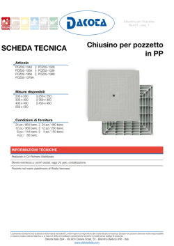
Surgical Neurology International
OPEN ACCESS
SNI: Unique Case Observations, a supplement to Surgical Neurology International
For entire Editorial Board visit :
http://www.surgicalneurologyint.com
Editor:
S. A. Enam, MD
Aga Kahn University; Karachi,
Sindh, Pakistan
Combined supra‑transorbital keyhole approach for treatment of
delayed intraorbital encephalocele: A minimally invasive approach
for an unusual complication of decompressive craniectomy
Lucia di Somma, Maurizio Iacoangeli, Davide Nasi, Paolo Balercia1, Ettore Lupi1, Riccardo Girotto1,
Gabriele Polonara2, Massimo Scerrati
Departments of Neurosurgery, and 1Oral and Head‑Neck Surgery, Umberto I General Hospital, Polytechnic University of Marche, 2Department of Radiology,
Section of Neuroradiology, Umberto I General Hospital, Polytechnic University of Marche, Ancona, Italy
E‑mail: Lucia di Somma ‑ [email protected]; *Maurizio Iacoangeli ‑ [email protected]; Davide Nasi ‑ [email protected];
Paolo Balercia ‑ [email protected]; Ettore Lupi ‑ [email protected]; Riccardo Girotto ‑ [email protected];
Gabriele Polonara ‑ [email protected]; Massimo Scerrati ‑ [email protected]
*Corresponding author
Received: 04 September 15 Accepted: 20 October 15 Published: 07 January 16
Abstract
Background: Intraorbital encephalocele is a rare entity characterized by the
herniation of cerebral tissue inside the orbital cavity through a defect of the orbital
roof. In patients who have experienced head trauma, intraorbital encephalocele
is usually secondary to orbital roof fracture.
Case Description: We describe here a case of a patient who presented an intraorbital
encephalocele 2 years after severe traumatic brain injury, treated by decompressive
craniectomy and subsequent autologous cranioplasty, without any evidence of
orbital roof fracture. The encephalocele removal and the subsequent orbital roof
reconstruction were performed by using a modification of the supraorbital keyhole
approach, in which we combine an orbital osteotomy with a supraorbital minicraniotomy
to facilitate view and access to both the anterior cranial fossa and orbital compartment
and to preserve the already osseointegrated autologous cranioplasty.
Conclusions: The peculiarities of this case are the orbital encephalocele without
an orbital roof traumatic fracture, and the combined minimally invasive approach
used to fix both the encephalocele and the orbital roof defect. Delayed intraorbital
encephalocele is probably a complication related to an unintentional opening of
the orbit during decompressive craniectomy through which the brain herniated
following the restoration of physiological intracranial pressure gradients after
the bone flap repositioning. The reconstruction of the orbital roof was performed
by using a combined supra‑transorbital minimally invasive approach aiming at
achieving adequate surgical exposure while preserving the autologous cranioplasty,
already osteointegrated. To the best of our knowledge, this approach has not been
previously used to address intraorbital encephalocele.
Access this article online
Website:
www.surgicalneurologyint.com
DOI:
10.4103/2152-7806.173561
Quick Response Code:
Key Words: Decompessive craniectomy, intraorbital encephalocele, minimally
invasive surgery, piezosurgery
This is an open access article distributed under the terms of the Creative Commons Attribution-NonCommercial-ShareAlike 3.0 License, which allows others to remix, tweak, and
build upon the work non-commercially, as long as the author is credited and the new creations are licensed under the identical terms.
For reprints contact: [email protected]
How to cite this article: di Somma L, Iacoangeli M, Nasi D, Balercia P, Lupi E, Girotto R, Polonara G, Scerrati M. Combined supra-transorbital keyhole approach for treatment
of delayed intraorbital encephalocele: A minimally invasive approach for an unusual complication of decompressive craniectomy. Surg Neurol Int 2016;7:S12-6.
http://surgicalneurologyint.com/Combined-supra‑transorbital-keyhole-approach-for-treatment-of-delayed-intraorbital-encephalocele:-A-minimally-invasive-approach-for-anunusual-complication-of-decompressive-craniectomy/
S12
© 2016 Surgical Neurology International | Published by Wolters Kluwer - Medknow
SNI: Unique Case Observations 2016, Vol 7: Suppl 1 - A Supplement to Surgical Neurology International
INTRODUCTION
Intraorbital encephalocele is a rare entity characterized
by the herniation of cerebral tissue inside the orbital
cavity through a defect of the orbital roof.[1,3,11] The
most common causes reported in literature are trauma,
skull base malformations, and tumors.[3,4] In patients
who have experienced head trauma, intraorbital
encephalocele is usually secondary to orbital roof
fracture.[1‑4,8‑12] Early recognition and treatment are
very important since the raised intraorbital pressure
may irreversibly damage the optic nerve and causes
pulsating exophthalmos.[8] Excision of the herniated
brain tissue, duraplasty and reconstruction of the orbital
roof are the key steps of surgery.[10‑12] In almost all the
reported cases, the fronto‑basal approach with wide
craniotomy is the preferred approach.[1‑4,8‑12] We describe
here a case of a patient who presented an intraorbital
encephalocele 2 years after severe traumatic brain injury,
treated by decompressive craniectomy and subsequent
autologous cranioplasty, without any evidence of orbital
roof fracture. The encephalocele removal and the
subsequent orbital roof reconstruction were performed
by using a modification of the supraorbital keyhole
a
b
d
e
approach, in which we combine an orbital osteotomy
with a supraorbital minicraniotomy to facilitate view
and access to both the anterior cranial fossa and orbital
compartment. The peculiarities of this case are the
orbital encephalocele without an orbital roof traumatic
fracture, and the combined minimally invasive approach
used to fix both the encephalocele and the orbital roof
defect.
CASE REPORT
A 40‑year‑old female was admitted at our institution
following a motor vehicle accident. The patient was
unresponsive with a Glasgow Coma Scale (GCS) score of
6 (E1V1M4) and was put on mechanical ventilation. The
pupils were bilaterally equal and reactive. She had no
periorbital hematoma and no proptosis. Visual acuity and
extraocular muscle motility of both eyes were not assessed
because of the reduced level of consciousness of the
patient. The immediate postinjury computed tomography
(CT) scan showed only a mild, diffuse brain swelling
[Figure 1a]. Thin‑slice CT scan with three‑dimensional
(3D) reconstruction and coronal and sagittal sections did
not show skull base fractures, in particular, posttraumatic
c
f
Figure 1: (a and b) The immediate postinjury thin-slice computed tomography scan with coronal reconstruction showed generalized
brain edema without left orbital roof fractures. (c) Computed tomography scan 24 h after trauma revealed a left-sided frontal contusion
causing midline shift and increased diffuse brain swelling. (d and e) Postoperative computed tomography scan showing left decompressive
craniotomy and evacuation of frontal contusion with a small bony defect in the roof of the orbit (see arrow) probably related to an
unintentional opening of the orbit during keyhole burr hole without evidence of encephalocele. (f) Computed tomography scan after
cranioplasty confirming the small bone opening on the orbital roof (see arrow)
S13
SNI: Unique Case Observations 2016, Vol 7: Suppl 1 - A Supplement to Surgical Neurology International
bony defects on the left orbital roof [Figure 1b]. The
patient was then transferred to the Intensive Care Unit,
and an intracranial pressure (ICP) probe was implanted
for ICP monitoring. After 24 h, despite maximal medical
treatment, the patient developed intractable ICP with
stable values up to 30 mmHg. Hence, she repeated
immediately a CT scan that revealed a massive increment
of the brain swelling, and a left‑sided frontal contusion
causing contralateral midline shift [Figure 1c]. She
underwent left decompressive craniotomy and evacuation
of frontal contusion. Immediately after decompressive
surgery, the ICP decreased significantly. The immediate
postoperative CT scan confirmed evacuation of left
posttraumatic hemorrhage and resolution of the mass
effect [Figure 1d]. At a deeper examination, this
exam also showed a small bony defect on the orbital
roof, undetected until the occurrence of the orbital
encephalocele (see the arrow in Figure 1e). During the
following days, the ICP remained stable within normal
limits. The patient recovered well and was discharged
from the hospital on the 25th posttraumatic day when she
was conscious with a GCS score of 15 and with only a
mild right hemiparesis. After 2 months, the patient was
submitted to autologous bone flap repositioning. There
were no complications during the cranioplasty procedure.
Wound healing was uneventful, and postoperative
neuroimaging showed accurate fitting of cranioplasty,
along with the undetected bony defect of the orbital
roof [Figure 1f]. After cranioplasty, the patient presented
full neurological recovery, included the hemiparesis, and
returned to the previous occupation.
Two years following the last operation, the patient
complained left fronto‑orbital headache without any
others ocular symptoms and/or signs, such as visual
disturbance, proptosis, or pulsatile exophthalmos. 3D
CT scan demonstrated the complete bone fusion of
autologous cranioplasty, and no others intracranial
complication. However, a thin‑slice orbital CT sections
with coronal reconstruction revealed the enlargement
of the supero‑lateral orbital roof defect with intraorbital
encephalocele [Figure 2a‑c]. Magnetic resonance
imaging (MRI) confirmed the herniation of brain
parenchyma into the left orbit through that bony defect
[Figure 2d]. Accurate anamnestic investigation excluded
a history of congenital anomalies, bone dysplasia,
or neurofibromatosis (that could be associated with
congenital skull base encephalocele). Hence, the patient
underwent a combined left supra‑transorbital minimally
invasive approach aiming at achieving adequate surgical
exposure without compromising the previously implanted
autologous cranioplasty, already osteointegrated. A left
superior blepharoplasty skin incision was performed to
expose the orbital rim. The piezoelectric bone scalpel
(piezosurgery) was used to perform a small supraorbital
S14
a
b
c
d
Figure 2: (a-c) Thin-slice orbital computed tomography sections
with three-dimensional and coronal reconstruction (2 years after
the cranioplasty) revealed the enlargement of the lateral orbital
roof bony defect associated with intraorbital encephalocele. The
arrow indicates the burr hole, filled by autologous bone dust mixed
with a bone substitute, too close to the orbital wall. (d) Magnetic
resonance imaging image confirming the herniation of brain matter
into the left orbit
keyhole craniotomy including the supero‑lateral orbital
rim in one piece using the defect of the orbital roof
as the posterior edge of the orbitotomy [Figure 3a‑e].
This combined minimally invasive approach gave access
to both the superior orbital cavity and the intracranial
supraorbital anterior skull base without the need for
cranioplasty to be removed [Figure 3e]. The periorbital
and orbital contents were identified and separated
from the herniated cerebral tissue. Herniated brain
parenchyma was resected with microsurgical technique
revealing the clear margin of dural and bony defect of
the orbital roof. The dura was re‑approximated with
a locked running stitch, and water‑tight closure was
achieved. Then, the orbital roof was reconstructed
with autologous bone, obtained from the outer bone
parietal layer contralateral to the cranioplasty through
a small linear skin incision and fixed by titanium
low‑profile miniplate and screws [Figure 3c, d, and f‑h].
The thin bone cutting line, performed by piezoelectric
scalpel, allowed a perfect realignment of bone stumps
[Figure 3c]. The postoperative period was uneventful,
and she was discharged from the hospital 5 days after
surgery. The esthetic results were excellent and patient
was able to achieve complete eye closure within 2 weeks.
Postoperative thin‑slice 3D CT scan showed the stable
reconstruction of the orbital roof and good restoration
of roof contour and normal orbital volume, excluding
intracranial complications [Figure 3f‑h]. MRI images
2 months after surgery confirmed the complete reduction
of encephalocele [Figure 3i]. At the last follow‑up
after 3 years, the patient was in good health with no
neurological deficits.
SNI: Unique Case Observations 2016, Vol 7: Suppl 1 - A Supplement to Surgical Neurology International
b
a
d
g
e
h
c
f
i
Figure 3: Abcdefghi: Intraoperative images and postoperative thin-slice computed tomography scan with three-dimensional reconstruction.
(a) Left superior blepharoplasty incision. (b and c) Supra-transorbital keyhole approach with the combination of an orbital osteotomy
with a supraorbital minicraniotomy.The use of piezoelectric scalpel allows to realize precise and thin osteotomies, for better future bone
healing and better aesthetic result. (c and d) The one-piece bone flap includes the frontal bone, and the orbital rim using the defect of the
orbital roof as the posterior edge of the orbitotomy.The one-piece bone flap was then fixed by titanium low-profile miniplate and screws.
(e) Intraoperative working area with the exposure of both orbital and intracranial compartments. (f-i) Postoperative thin-slice computed
tomography scan with three-dimensional reconstruction and magnetic resonance imaging showing left orbital roof reconstruction with
autologous bone, obtained from the split calvarial parietal bone contralateral to the cranioplasty
DISCUSSION
Orbital encephalocele is a rare entity, and it is
characterized by the herniation of part of the brain
through a congenital, traumatic, neoplastic, or
iatrogenic orbital roof defect.[1‑4,11] The herniation
of brain parenchyma can also occur through natural
foramina such as the superior orbital fissure or the optic
foramen. Orbital encephaloceles may be congenital or
acquired with congenital encephaloceles more likely
to occur in early childhood.[3] In the adult population,
acquired encephaloceles are reported in patients who
have experienced head trauma with associated orbital
roof fracture.[3,9,11] However, even traumatic orbital
encephaloceles are a quite rare evenience. There are
<25 reported cases in literature thus far, including
both pediatric and adult population.[1‑4,8‑12] In all these
cases, there was a radiological or direct intraoperative
evidence of an orbital roof fracture associated with the
encephalocele. The incidence of orbital roof fractures is
higher in children because an impact to the orbital rim
before the age of 7 years, cannot be dissipated by the
frontal sinus (not yet completely pneumatized), and the
traumatic force is transmitted directly to the orbital roof.
In a pediatric series, orbital roof fractures were found in
7%, 1% of patients who suffered head injuries, and 13% of
those patients developed intraorbital encephalocele.[3] An
orbital encephalocele due to trauma may develop acutely
or may develop many years later as the arachnoid and
tissue gradually herniate, or as the fracture in the orbital
wall grows over time.[1‑4,7] Growing fractures of the
orbital roof have been reported several times, especially
in pediatric population.[3] Gradual herniation of the
arachnoid into the fracture, the physiologic growth of
the cranium and the brain (in children), continuous
pulsation of the CSF, and the absence of bony counter
compression are the accepted mechanisms of growth and
expansion of the fracture.[1] Our patient developed the
intraorbital encephalocele without any evidence of orbital
roof fracture at the initial thin‑slice CT scan with 3D
and coronal reconstructions. The first evidence of bony
defect in the orbital roof was undetected but present at
the immediate CT scan after decompressive craniectomy,
probably related to an unintentional opening of the
S15
SNI: Unique Case Observations 2016, Vol 7: Suppl 1 - A Supplement to Surgical Neurology International
orbit by the keyhole burr hole. The development of
the intraorbital encephalocele could be related to the
restoration of a positive pressure gradient between the
intracranial compartment, and the orbital compartment
after cranioplasty similar to the etiopathological
mechanism of the growing fracture in children. To the
best of our knowledge, this is the first case of intraorbital
encephalocele secondary to decompressive craniectomy
and subsequent bone replacement.
The main symptoms and signs of intraorbital encephalocele
are diplopia, exophthalmos, orbital edema, subconjunctival
hemorrhage, restricted movements of the eye, and loss
of vision. Among these, pulsatile exophthalmos is the
most reported symptom after traumatic intraorbital
encephalocele in the literature.[8] Our patient complained
only left fronto‑orbital headache. Early recognition and
treatment are very important since the raised intraorbital
pressure may irreversibly damage the optic nerve.[8,11]
Thin‑slice CT scan with 3D reconstruction is the gold
standard imaging modality to assess orbital roof fractures
or bony defects. However, MRI imaging is essential to
detect and evaluate the herniated brain tissue and for
differential diagnosis with others intraorbital lesions such
as tumors or vascular malformations.[1‑3]
Excision of the herniated brain tissue, duraplasty, and
reconstruction of the orbital roof are the key steps of
surgery.[10,12] There are two main routes to the orbital
roof: The transcranial and the extracranial transorbital
route.[10] The transcranial approach is commonly
performed through a bicoronal incision and a uni‑ or
bi‑frontal craniotomy. This approach can also allow to
address concomitant intracranial lesions in the same
session. The extracranial approaches to the orbit are
generally performed through a superior blepharoplasty
skin incision or through a preexisting skin laceration. In
all but one cases described to date in the literature, the
herniated brain parenchyma into the orbit was accessed
through a wide frontal craniotomy followed by the removal
of fractured bony fragments inside the orbit.[1‑4,8‑12] The
main limitation of this approach is the extensive brain
exposure and retraction that could result in cerebral
parenchyma contusion, infection, and epilepsy.[10,12] In our
case, to preserve the already osteointegrated autologous
cranioplasty, we adopted a modification of the supraorbital
keyhole approach, by combining an orbital osteotomy
with a supraorbital minicraniotomy. This combined
minimally invasive approach was used to facilitate both
views and access to the anterior cranial fossa and orbital
compartment. The one‑piece bone flap includes the
frontal bone, and the orbital rim using the defect of the
orbital roof as the posterior edge of the orbitotomy. The
use of piezoelectric scalpel enhance safety by preventing
injuries to nonosseous structures and allows to realize
precise, thin osteotomies, so facilitating the subsequent
bone healing with better esthetic result.[5‑7]
S16
CONCLUSIONS
The peculiarities of this case are the development of
intraorbital encephalocele without evidence of traumatic
orbital roof fracture and the combined supra‑transorbital
minimally invasive approach. The delayed intraorbital
encephalocele is probably a complication related to an
unintentional opening of the orbit during decompressive
craniectomy through which the brain herniated
following the restoration of physiological ICP gradients
after the bone flap repositioning. The reconstruction
of the orbital roof was performed by using a combined
supra‑transorbital minimally invasive approach aiming
at achieving adequate surgical exposure while preserving
the autologous cranioplasty, already osteointegrated. To
the best of our knowledge, this approach has not been
previously used to address intraorbital encephalocele.
Financial support and sponsorship
Nil.
Conflicts of interest
There are no conflicts of interest.
REFERENCES
1.
Antonelli V, Cremonini AM, Campobassi A, Pascarella R, Zofrea G, Servadei F.
Traumatic encephalocele related to orbital roof fractures: Report of six cases
and literature review. Surg Neurol 2002;57:117‑25.
2. Bruzek A, Shepherd D, Van Gompel J, Jentoft M. Pilocytic astrocytoma
presenting as an orbital encephalocele: A case report. Case Rep Neurol
2015;7:90‑4.
3. Cayli SR, Kocak A, Alkan A, Kutlu R, Tekiner A, Ates O, et al. Intraorbital
encephalocele: An important complication of orbital roof fractures in
pediatric patients. Pediatr Neurosurg 2003;39:240‑5.
4. Gazioglu N, Ulu MO, Ozlen F, Uzan M, Ciplak N. Acute traumatic orbital
encephalocele related to orbital roof fracture: Reconstruction by using
porous polyethylene. Ulus Travma Acil Cerrahi Derg 2008;14:247‑52.
5. Iacoangeli M, Di Rienzo A, di Somma LG, Moriconi E, Alvaro L, Re M, et al.
Improving the endoscopic endonasal transclival approach: The importance
of a precise layer by layer reconstruction. Br J Neurosurg 2014;28:241‑6.
6. Iacoangeli M, Di Rienzo A, Nocchi N, Balercia P, Lupi E, Regnicolo L, et al.
Piezosurgery as a further technical adjunct in minimally invasive supraorbital
keyhole approach and lateral orbitotomy. J Neurol Surg A Cent Eur Neurosurg
2015;76:112‑8.
7. Iacoangeli M, Neri P, Balercia P, Lupi E, Di Rienzo A, Nocchi N, et al.
Piezosurgery for osteotomies in orbital surgery: Our experience and review
of the literature. Int J Surg Case Rep 2013;4:188‑91.
8. Jaiswal M, Sundar IV, Gandhi A, Purohit D, Mittal RS. Acute traumatic orbital
encephalocele: A case report with review of literature. J Neurosci Rural
Pract 2013;4:467‑70.
9. Mohindra S, Mukherjee KK, Chhabra R, Gupta R. Orbital roof growing
fractures: A report of four cases and literature review. Br J Neurosurg
2006;20:420‑3.
10. Mokal NJ, Desai MF. Titanium mesh reconstruction of orbital roof fracture
with traumatic encephalocele: A case report and review of literature.
Craniomaxillofac Trauma Reconstr 2012;5:11‑8.
11. Morihara H, Zenke K, Shoda D, Fujiwara S, Suehiro S, Hatakeyama T.
Intraorbital encephalocele in an adult patient presenting with pulsatile
exophthalmos. Case report. Neurol Med Chir (Tokyo) 2010;50:1126‑8.
12. Sahoo SK, Salunke PS, Ghuman MS. Traumatic orbital encephalocele in an
infant: Using the fracture line to our advantage. Acta Neurochir (Wien)
2014;156:1357‑9.
© Copyright 2026 Paperzz