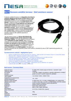
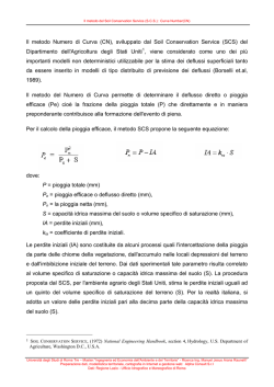
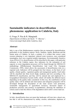
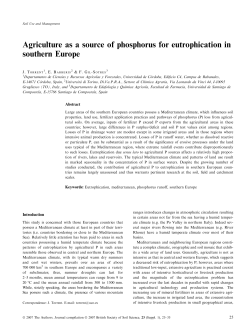
Dottorato di ricerca in Scienze Ambientali Scuola di dottorato in Scienze e Tecnologie Ciclo XXIV (A.A. 2008 - 2009) BIOGEOCHIMICA DEGLI ELEMENTI POTENZIALMENTE TOSSICI: DAI SUOLI ALLE PIANTE ED ALLA CATENA ALIMENTARE. ELEMENTI PER UNA VALUTAZIONE DEL RISCHIO PER LA SALUTE UMANA SETTORE SCIENTIFICO DISCIPLINARE DI AFFERENZA: AGR/14 Tesi di dottorato di Mohammad Ahmad Mutlak Wahsha, Matricola: 955571 Coordinatore del Dottorato Tutore del dottorando Prof. Bruno Pavoni Prof. Claudio Bini Dedicated to… My beloved father Ahmad, who has always been supporting me with his love My mother Ahlam, for her loving care, My brothers: Heider & Yarub My Sisters: Majd, Reem Abeer & Ghadeer, whom I have always had near me. I ACKNOWLEDGMENTS First of all I would like to thank God for giving me the ability to work hard and the strength to carry on even in difficult times. My deep thanks go to my Professor Claudio Bini for his encouragement, his precious advice and support. Without his help and supervision this work would not have been possible. Thanks also to Professor Laura Maleci for her help and her kindness. I would like to express my gratitude to Professor Gabriella Buffa, Professor Giampietro Ravagnan and Professor Gabriele Capodaglio for their time and for their advice. I am grateful to my colleagues, Diana Zilioli and Silvia Fontana who were always ready to help me. Thanks so much to Nicola Miotello, Gabriella Ceciliati, Rossella Vianello, and Lorena Gobbo for their kind help and their friendship and to Professor Emanuele Argese, who was so kind as to let me work in his lab. I would like to extend my thanks and to express my gratitude to my friends: Lisa Vannuzzo, Andrea Ferrarini, Eliana Grappiolo, Paolo Ravera, Francesco Giorgianni, Ettore Boer Bront, Andrea Mattioli and Sabrina Faggian. Finally, my appreciation and deep gratitude go to Dr. Silvia Bertapelle for supporting me with good advice, continuous help and sincere friendship. “Infine come non ringraziare questa città, davvero unica al mondo, che è stata la mia città per oltre tre anni? Venezia, un luogo straordinario e accogliente, dove mi sono sempre sentito a casa, e di cui conserverò sempre il ricordo. Sono stato fortunato a poter condividere con gli amici veneziani la loro quotidianità così particolare e diversa dal resto del mondo. Grazie amici. Grazie Venezia”. Mohammad Wahsha II List of Contents SECTION ONE PAGE NUMBER INTRODUCTION 1 1.1 PURPOSE OF STUDY 4 1.2 LITERATURE REVIEW 5 SECTION TWO MATERIALS AND METHODS 17 2.1 SITE DESCRIPTION 17 2.2 SAMPLE COLLECTION: 22 2.3 SOIL SAMPLING 23 2.4 PLANT SAMPLING 25 2.5 ANALYTICAL METHODS 26 2.5.1 SOIL PH 26 2.5.2 SOIL ORGANIC CARBON 28 2.5.3 CATION EXCHANGE CAPACITY 30 2.5.4 MEASUREMENT OF TOTAL CARBONATES 33 2.5.5 SOIL PARTICLE SIZE DISTRIBUTION 34 2.6 S OIL SAMPLES PREPARATION FOR HEAVY METALS 39 ANALYSIS 2.7 PLANT SAMPLES PREPARATION FOR HEAVY METALS 39 ANALYSIS 2.8 ESTIMATION OF LIPID PEROXIDATION (MDA CONTENT) 39 2.9 SOIL ENZYMATIC ASSAYS 41 2.9.1 ARYLESTERASE ASSAY 41 2.9.2 LEUCYL AMINOPEPTIDASE, Β -GLUCOSIDASE, 42 CHITINASE AND ALKALINE PHOSPHATASE ASSAYS 2.10. BIOLOGICAL QUALITY OF SOIL: MICRO-ARTHROPOD 44 METHOD (QBS-AR) 2.11 STATISTICAL ANALYSIS PROCEDURE 49 SECTION THREE RESULTS AND DISCUSSION 50 0 3.1 SITES DESCRIPTIONS 50 III 3.2 CHEMOPHYSICAL PROPERTIES OF SOIL SAMPLES 72 3.3 HEAVY METALS ACCUMULATION IN SOILS 74 3.4 HEAVY METALS ACCUMULATION IN PLANTS 76 3.5 LIPID PEROXIDATION QUANTIFICATION 81 3.6 SOIL ENZYMES ANALYSIS 84 3.7 BIOLOGICAL SOIL QUALITY INDEX 86 SECTION FOUR CONCLUSIONS 96 SECTUION FIVE REFERENCES 98 IV SECTION ONE INTRODUCTION Soil is a part of the earth’s surface which forms an active "unique environment" between the atmosphere and the lithosphere. It is both an ecosystem in itself, and a critical part of the larger terrestrial ecosystem, contains elements of water, air and mineral matter, together with a high diversity of living organisms which utilize the soil as a habitat, a source of energy and contribute to its formation (Lavelle and Spain, 2003). Over the past decades, due to a wide range of industrial, agricultural and mining activities, a large number of contaminants are released into the environment, causing a significant concern regarding potential toxicity (Bini, 2011). Heavy metals are considered one of the major sources of soil pollution1. Extraction of metals from sulphides minerals usually results in large amounts of waste materials which often contain elevated concentrations of harmful metals such as Cu, Zn, Cd, and Pb (Jian-Min et al., 2007; Lee et al., 2001). The natural background concentrations of heavy metals in soil depend mainly on the bedrock type from which the soil parent material was derived. Anthropogenic activities may increase metal concentrations in soils, and the most abundant metallic pollutants introduced into soil by anthropogenic activities are lead, cadmium, tin, and mercury (Maldonado el al., 2008). Heavy metals can be transported from contaminated soils to surface and ground water. This contamination threats water supply resources as well as the economic and environmental health of surrounding communities (Selim and Sparks, 2001). 1 Pollution occurs when a substance is present in greater than natural concentration as a result of human activities and having a net detrimental effect upon the environment and its components. Contamination occurs when the soil composition deviates from the normal composition. In their natural state contaminants may not be classified as pollutants unless they have some detrimental effects to the organisms (Bini, 2011). 1 The degree of heavy metal contamination around mines varies depending upon geochemical characteristics and degree of mineralization of the tailing (Navarro et al., 2008). Yun-Guo et al. (2006) reported that abandoned mining sites represent significant sources of metal pollutants in water and soils, effecting the functioning in both plants and soil biota. These metals can be transported, dispersed and accumulated in plants and then passed through the food chain to human beings as the final consumer. Metal solubility in soils is predominantly controlled by pH, amount of metals cations exchange capacity (CEC), organic carbon content and oxidation state of the system (Ghosh and Singh, 2005). Human health risk assessment has been used to determine if exposure to a chemical, at any dose, could cause an increase in the incidence of adverse effect to human health (Lim et al., 2008). There are several former mixed sulphides (mainly Cu, Fe, Pb, Zn) mining sites in Italy. Soils and plants growing on contaminated mine sites have been studied to determine the ability of these plants to accumulate metals and to grow on mine waste in the perspective of an ecological restoration of sites (Bini, 2011; Wahsha et al., 2012; Fontana et al., 2010; Mascaro et al., 2000). Recently, preliminary studies in the Department of Environmental Sciences of Ca' Foscari University of Venice have focused on the toxicity and influence of heavy metals in contaminated soils in food and wild plants (Fontana et al., 2011; Wahsha et al., 2011; Bini et al., 2010). However, such remediation technique strategies such as immobilization or extraction by physico-chemical techniques are too expensive and generally impractical (Zabludowska et al., 2009). Therefore, a need is required to develop in situ low cost technologies to have effective surface metal stabilization. The use of wild flora can be a useful tool, since there are some native plant species that can colonize parts of these polluted sites (Conesa et al., 2006). An interesting review article on phytoremediation of metal enriched mine waste has recently been published (Mukhopadhyay and Mait, 2010). 2 This review highlights the use of plants to reduce, remove, degrade, or immobilize environmental pollutants. Taraxacum officinale (the common dandelion), is a herbaceous perennial plant of the family Asteraceae. It has been reported by many authors to be capable of accumulating heavy metals (Bini et al., 2010; Fontana et al., 2010). In the last few years many studies have focused on the potential use of trees as a suitable vegetation cover for phytoremediation (Jensen et al., 2009; French et al., 2006; Dickinson, 2000). A very suitable tree for use in phytoremediation is willow (Landberg and Greger, 2002). Pulford and Watson (2003) detailed the phytoremediation potential of willow in heavy metal contaminated areas. Willows have not been included in the group of hyperaccumulators of heavy metals, but on the other hand they provide potential bioindicator of pollution (Mleczek et al., 2009). However, metal concentrations in willows depend on species, growth performance, root density, distribution within the soil profile and sampling period (Bedell et al., 2009; Chehregani, et al., 2009). And recently willow is being recognized as a good accumulator of heavy metals (Meers et al., 2007). 3 The significances and purposes of this study were to: 1. Assess the concentration and bioavailability of the following metals: Cd, Cr, Cu, Pb, Zn and Fe in soils of a mining area. 2. Test the ability of selected species of plants to accumulate heavy metals in their tissues. 3. Compare heavy metal accumulation in various parts of the plant species. 4. Investigate the relationship between the biological soil quality, lipid peroxidation assay, soil enzymatic activities and the metal contents of the plant and soil using statistical analysis. 5. Provide useful information for decision makers on the conditions of the studied sites with respect to metal pollution, and the best method to monitor the environment. 4 LITERATURE REVIEW Heavy metals are considered one of the major sources of soil pollution and it has become a serious environmental issue in many parts of the world (Wahsha et al., 2012). 1.1 Soil heavy metals Heavy metals are defined as chemical elements having density more than 5 g cm-3. Metal concentrations in soil range from 1 to 100000 mg kg-1. Metals are often closely bound to soil particles. Cations of heavy metals are generally bound to soil particles due to soil cations exchange capacity (Sinha and Sinha, 2008). Because soils are heterogeneous, many studies have focused on the interaction of several heavy metals with different soil parameters. Heavy metals in soils can be involved in a series of biochemical reactions, volatilization, and surface and solution phase (Selim and Sparks, 2001). In soils, heavy metals are distributed over the different soil particles in different chemical forms and they vary considerably in their relative reactivity (Fig 1). The soil system play an important role through which various forms of elements interact with the soil solid phase and soil biological activity (Hooda, 2010). 5 Fig 1: The multi reaction pools for heavy metals reactivity in soils. Source: Hooda, 2010. 1.2 Sources of heavy metal contamination in soil Unfortunately, soils are presently contaminated, in particular by heavy metals, which can negatively affect soil’s chemical, physical and biological properties worldwide. In this context, heavy metals added to the soil through diverse natural or / and anthropogenic sources. The major natural sources of heavy metals in soils are: weathering (such as soil erosion and deposition of windblown dust and running water); volcanic eruptions and bushfire. While the major anthropogenic sources of heavy metals input to soils are: Atmospheric deposition, result from energy and fuel production, power generation emissions, metal mining, smelting of metalliferous ores and manufacturing, waste and wood burning; sewage sludge, municipal and organic wastes and co-products from agriculture and food industries; Land disposal of industrial waste; Fertilizer and pesticide (insecticides, herbicides, fungicides and soil heavy metal pollution through chemical and biological warfare (Hooda, 2010; Alloway, 1995). 6 1.3 Biochemical effects of heavy metals in plants Some heavy metals are essential for nutrients cycle within biological metabolisms, manganese (Mn), chlorine (Cl), zinc (Zn), iron (Fe), boron (B), copper (Cu) and molybdenum (Mo). Cl and Fe within plants and soils are not considered as trace elements because their average concentration is generally greater than 100 mg kg-1. These elements are necessary for maintaining the life processes in plants and animals and therefore they are essentially micronutrients. At higher concentrations, however, they become harmful and toxicity damage may occur. For non essential metals, cobalt (Co), fluorine (F), chromium (Cr), nickel (Ni), iodine (I) and selenium (Se), they have a single function such as Ni in hydrogenases; and their signs of toxicity are realized above a range of tolerance. There is little evidence to suggest that arsenic (As), cadmium (Cd), lead (Pb) and mercury (Hg) play a nutritive role in higher plants and animals (Hooda, 2010; Bradl, 2005; He el al., 2005). The rest of the heavy metals are always toxic such as Cd, Pb, U, Tl, Cr, Ag and Hg. As and Se are non heavy metals. However, with respect to their toxicity effects, they are referred to as “metalloids” in the literature (Sherameti and Varma, 2010a). 1.4 Heavy metal contamination in the food chain As plants constitute the foundation of the food chain, some concerns have been raised about the possibility of toxic concentrations of certain elements being transported and accumulated from plants to higher level of the food chain (Peralta-Videa et al., 2009). Beside their negative effect on plant metabolism, and “different from other pollutants”, toxic heavy metals are indestructible, as they cannot be degraded. Even worse, enhanced uptake of heavy metals by plants at concentrations below phytotoxic levels may pose potential risks to food chains where farm animals are raised on contaminated soils (Ping et al., 2009). These plants are known for their capacity to accumulate heavy metals in their tissues (Sherameti and Varma, 2010b). 7 1.5 Cellular toxicity and oxidative damages caused by heavy metals Heavy metal toxicity is likely to be initiated in cells by: 1) direct interaction with some proteins and enzymes because of their affinities for specific functional groups. Khan (2005) reported that cellular enzymes were inactivated by heavy metals. 2) Interfere of some essential cations from specific binding sites, causing functions to fail and 3) Initiate the generation of hydrogen peroxide, superoxide radical, hydroxyl radical and singlet oxygen, collectively termed Reactive Oxygen Species (ROS), which can affect the equilibrium between ROS and the antioxidant defence system (Fig 2)(Wahsha et al., 2010). An oxidative stress can be defined as a condition in which the rate of ROS generation exceeds the ability of the antioxidant defence system to protect cells against them, resulting in an increase in oxidative damage to the cell (Botha et al., 2004). However, several chemicals (such as heavy metals) can shift directly or indirectly this balance, helping the formation of oxidative stress (Joshi et al., 2005; Pinho et al., 2005). This has contributed to the creation of the oxidative stress concept; in this view, ROS are unavoidable toxic products of O2 metabolism, and aerobic organisms have evolved antioxidant defenses to protect against this toxicity (Alfonso and Puppo, 2009). Oxidative stress can increase sharply in cells either due to the decrease in the activity of the antioxidant defense systems or to the overproduction of ROS (Mukherjee et al., 2007; Soffler, 2007). Wahsha and Al-Jassabi (2009) reported that during times of environmental stress, ROS levels can increase dramatically and this may result in significant damage to cell. Significant numbers of studies have shown that heavy metals induce oxidative damage formation in plants (Wahsha et al., 2011; Loureiro et al., 2006). Cd has been found to increase the lipid peroxidation rate in different plant species due to the increase in ROS production (Del Rio and Puppo, 2009). 8 Fig 2: Diagrammatic representation of hypothetical mechanisms of heavy metal-induced cell damage in sensitive plants and points of interaction and counteraction during acclimation in tolerant plants. Source: Peralta-Videa et al., 2009. 9 Many organic molecules are exposed to severe damage by free radicals after high accumulation of heavy metals in plant (Alfonso and Puppo, 2009; Joshi et al., 2005). Formation of ROS in cells (Fig 3) is associated with the development of many pathological states (e. g. reduced root elongation, seed germination, signaling imbalance) (Wahsha and Al-Jassabi, 2009; Bini et al., 2008). Heavy metals inside the cytoplasm might disturb the stability of the lipidic membrane, making various changes in cellular conductivity and reduce some functional protein sensitivity (Pinho et al., 2005). Fig 3: Possible pathways of heavy metal-dependent ROS generation, Red dots symbolize the distribution of heavy metals (HM) in the cell and apoplast. Source: Sharma and Dietz, 2009. 10 The most harmful effect induced by ROS in plants is the oxidative degradation of lipids, especially polyunsaturated fatty acids (PUFA) in cell membranes known as lipid peroxidation, which can directly cause biomembrane disorganization (Gobert et al., 2010; Timbrell, 2009). Several studies reported that ROS can initiate lipid peroxidation through the action of hydroxyl radicals (Armstrong, 2008; Katoch and Begum, 2003). The main reason for the high reactivity of hydroxide radicals is their ability to initiate a chain reaction at even very low concentrations (Wahsha et al., 2010). Lipid peroxidation reactions are usually free radical-driven chain reactions in which one radical can induce the oxidation of PUFA (Abuja and Albertini, 2001). Lipid peroxidation proceeds via 2 phases: initiation and propagation, as shown in Fig 4. Fig 4: Lipid peroxidation steps. The reaction initiates when the hydrogen radical attaches one PUFA results in the cleavage of one H+ from the methyl-vinyl group of the fatty acid. Following several biochemical processes resulting in the formation of the lipid peroxide Malondialdehyde 11 (MDA) (Wahsha et al., 2010b). The MDA is one of the major end-product of lipid peroxidation process (Yadav, 2010). In this case, membrane destabilization and fusion are directly correlated with MDA production (Wahsha et al., 2010; Wahsha and Al-Jassabi, 2009). The determination of MDA content is widely used as a reliable tool to detect the oxidative stress hazard by estimating the formation of lipid peroxides in biological material (Taulavuori et al., 2001; Zielinska et al., 2001; Loureiro et al., 2006). Furthermore, the formation of ROS and an increased MDA production were observed in plants exposed to different heavy metals as Cr, Pb, Cu and Zn under laboratory condition. Sinha et al. (2005) reported that chromium induced lipid peroxidation in the plants of Pistia stratiotes L. Aravind and Vara Prasad (2003) demonstrated that zinc alleviates cadmium-induced oxidative stress in Ceratophyllum demersum L. Baryla et al. (2000) investigated the evaluation of lipid peroxidation as a toxicity bioassay for plants exposed to copper. Another study by Verma and Dubey (2003) reported that Lead induces lipid peroxidation and alters the activities of antioxidant enzymes in growing rice plants. 1.6 Phytoremediation of heavy metal contaminated soils It is an emerging green bioengineering technology for environmental cleanup that uses plants to remove pollutants from the soil or to render them harmless (Singh et al., 2009). Phytoremediation strategies have been investigated, developed, and intensively used for their potential economic benefit, aesthetically pleasing, passive, solar-energy driven, and successful achievements in heavily impacted areas especially mining. Plants involved in phytoremediation are adapted to grow in harsh environmental conditions. They are adapted to uptake, transfer, accumulate, degrade or stabilize harmful toxic elements (heavy metals) from the polluted soils (Sinha and Sinha, 2008). The remediation of heavy metals has gained considerable attention as a potential environmental challenge in recent years. Different approaches are being adopted to reclaim polluted soils such as land filling, leaching, excavation, burial and soil washing, however, these methods are very expensive 12 and so they are not economically viable and also they may harm the soil structure ( Sherameti and Varma, 2010b). Usually if a plant can accumulate more than 1,000 mg kg-1 of Cu, Co, Cr, Ni, or Pb, or more than 10,000 mg kg-1 of Mn or Zn, it is defined as a hyperaccumulator (Sherameti and Varma, 2011). Ali et al, (2003) reported the phytoremediation of Lead, Nickel, and Copper by Salix acmophylla Boiss. Kabata-Pendias (2004) reported that the criteria for environmental protection are related to the trace element status mobility and availability that control the soil plant transfer, and this is affected by many geochemical, climatic, biological, as well as of anthropogenic factors (mining activities). 1.7 Soil enzymes Soil enzymes are a group of enzymes whose usual inhabitants are the soil and are continuously playing an important role in maintaining soil ecology, physical and chemical properties, fertility, and soil health (Zornoza et al., 2009). Soil enzymes activities have been suggested as suitable indicators of soil quality because: (a) they are a measure of the soil microbial activity and therefore they are strictly related to the nutrient cycles and transformations; (b) they rapidly may respond to the changes caused by both natural and anthropogenic factors; (c) they are easy to measure. Soil enzymes activities may be considered as early and sensitive indicators to measure the degree of soil degradation and to evaluate the impact of pollution on the quality of soil (Gianfreda et al., 2005). 1.7.1 Arylesterase This enzyme is able to catalyze the hydrolysis of phenolic esters such as phenyl acetate to phenol and acetate. This esterase is also involved in the degradation of plastics and hydrolysis of organophosphates (Renella et al., 2011). Therefore, determination the activity of this enzyme in contaminated soils may be important for evaluating the reaction of soil microbial communities to organic and heavy metals contamination, assessing the fate of toxic elements to micro organisms and so, affecting the soil health and quality 13 (Zornoza et al., 2009). Renella et al. (2011) demonstrated that inhibition of arylesterase by trace element may hamper the recovery of soils contaminated by trace element and organic pollutants, in which the detoxification action of arylesterase can be reduced. 1.7.2 Leucyl aminopeptidase Leucyl aminopeptidase is present in animals, plants, and bacteria and has different physiological functions in the processing or degradation of peptides. Leucyl aminopeptidase in soil has an important role in N mineralization, it catalyzes the hydrolysis of leucine residues at the N-terminus proteins. However, leucyl aminopeptidase activity has been reported to occur partly in soil as a humocarbohydrate complex. The quantity of this extracellular enzyme activity could be indicative for the biological capacity of soil, and it has an important function in the ecology of micro-organisms in the ecosystem (Shukla and Varma, 2010). 1.7.3 β -Glucosidase β-glucosidase is widely distributed in soils and has important roles in many biological processes. It is involved in the hydrolysis catalyzing of carbohydrates present in plant debris decomposing in the ecosystem. β -Glucosidase is characteristically useful as a soil quality indicator, and may give a reflection of past biological activity, the capacity of soil is to stabilize the soil organic matter, and can be used to detect management effect on soils. β -Glucosidase enzyme is very sensitive to changes in pH. β-glucosidase enzyme is also known to be inhibited by heavy metal contamination (Shukla and Varma, 2010). Furthermore, Haanstra and Doelman (1991) demonstrated that copper could reduce the βglucosidase activity more than cellulose activity. 1.7.4 Chitinase Chitinase are hydrolytic enzymes that break down glycosidic bonds in chitin. They are the main structural part of fungal cell walls. As biological control of most pathogenic diseases is increasingly gaining popularity in recent times due to their environmental 14 friendliness, a better understanding of the chitinolytic enzymes is useful for agricultural systems managements. Donderski and Brzezinska, (2005) reported the inhibitory effects of heavy metals on the activity of chitinases. 1.7.5 Alkaline phosphatase It is a hydrolyse enzyme which is able to remove phosphate groups from many types of molecules, including nucleotides, proteins, and alkaloids. It is considered as a good indicator of soil fertility. Alkaline phosphatase, occurs in roots mainly after mycorrhizal colonization, It has been proposed as a marker for analyzing the symbiotic efficiency of root colonization. In soil ecosystems, Alkaline phosphatase is supposed to play critical roles in P cycles as evidence shows that they are correlated to P stress and plant growth. Lorenz et al. (2006) reported that As can significantly influence the arylsulfatase activity but not alkaline phosphatase (Shukla and Varma, 2010). Wyszkowska et al. (2006) explained that Cu, Zn, Ni, Pb, Cd and Cr could inhibit the activity of alkaline phosphatase. 1.8 Biological soil quality index: micro-arthropod method (QBS-ar) In the last few years, there has been a growing interest in the definition and evaluation of soil quality, which is defined as “the continued capacity of a specific kind of soil to function as a vital living system, within natural or managed ecosystem boundaries, to sustain plant and animal productivity, to maintain and enhance the quality of air and water environments, and to support human health and habitation” (Sherameti and Varma, 2010a). Difficulties appear while trying to determine specific criteria for soil quality, mostly because of the intrinsic variability of the different soils and the discrepancies both in time and space scales (Parisi et al., 2005). Furthermore, several articles of soil quality have 15 been published over the last two decades, mainly with a focus on assessing soil quality indicators (Tabaglio et al., 2009). Changes in soil quality can be measured through indicators which include physical, chemical and biological processes so it is necessary to provide quality indices including different indicators, to determine soil quality (MartinezSalgado et al., 2010; Kim and Jung, 2008). The contribution of soil organisms to ecosystem functions such as decomposition, nutrient recycling and the maintenance of physicochemical properties is well recognised (Knoepp et al., 2000). The diversity of soil fauna includes a quarter of described living species, the majority of which are insects and arachnids (Martinez-Salgado et al., 2010; Tabaglio et al., 2009). Recently, many indicators can be used to describe soil quality. In Italy, a new approach (called QBS index) based on the types of edaphic microarthropods has been proposed to assess soil biological quality (Parisi et al., 2005). Soil microarthropods demonstrated to respond sensitively to land management practices and to be correlated with beneficial soil functions. 16 SECTION TWO MATERIALS AND METHODS 2.1 Site description Field observations were carried out in the Imperina Creek watershed. The Imperina Valley is located in the mountain district of Belluno, North-East Italy (Fig 5), with an altitude ranging between 543 m and 990 m above sea level. The geological substrate consists of rocks of the metamorphic basement (Pre-Permian), in tectonic contact with dolomite rocks (Dolomia Principale, Upper Triassic). The Imperina stream crosses the valley; even if no settlements can be found in this area, many buildings and tunnel outlets still bear witness to the past mining activity. Part of the area (right side and a portion of the bottom) lies within the National Park of the Belluno Dolomites. The mined area is located along the tectonic contact; it consists of a deposit of mixed sulfides, composed primarily of cupriferous pyrite, pyrite and chalcopyrite, with minor amounts of other metallic minerals (Frizzo and Ferrara, 1994). Copper and sulfur were the main products extracted. Until the beginning of the 20th century, copper was extracted and processed directly in situ through roasting, a method with a severe impact on the area due to acid rains formation and intensive wood cutting. The vegetation cover is mainly constituted of mixed forests (Abies alba Mill., Picea abies (L.) H. Karst., 1881, Fagus sylvatica L. and Ostrya carpinifolia Scop.), with clearances where herbaceous and shrubby vegetation prevails over the arboreal one (Dissegna et al., 1997). The first certain historical records indicate that mining in the Valle Imperina dates back to the first years of the 14th century, thanks to research funded by the Republic of Venice into the mining of copper. Very probably, the extraction of minerals had already commenced in pre-roman times, considering the particular condition and position of the 17 rocks and the proximity of the Agordo valley; this theory is also backed up by the large amount of items made from bronze and copper found in archaeological excavations in the whole of the Belluno valley. M D P Fig 5. – Location of the studied area and sampling sites of Imperina Valley. M = Metamorphic basement, P = Phyllite, D = Dolomite .Modified after Spaziopadova, 2011. 18 A notable increase in the production of copper derives from the passage of the property rights of the mines from private hands to those of the Republic of Venice; in fact, initially the mining was subdivided in tunnels and "mints" owned by private individuals with obvious management problems and lack of efficiency of the mine itself (Fig 6). This passage came about in a gradual manner starting at the end of 1600, but it was only in 1835 that these mines formally become public property, in the hands of the AustroHungarian Empire. The extraction of copper continued in this area, with ups and downs, up to the end of 1800, when the company Magni C. of Vicenza initially rented and subsequently the mines and therefore the rights to all the minerals represented essentially by pirites poor in copper, which were taken directly to other factories for the production of sulphuric acid. During the first years of this century, the whole process was electrified, and in 1925 the new standard-gauge electric railway line from Bribano to Agordo became operational; the stop at La Valle allowed for the loading of the materials which had been extracted from the mine and taken to the situation by cableway. After the First World War, production has been stabilised at around 50000 tonnes a year up to the period 1940-1944. Following this period, after a first attempt to modernise the mine in the early 1950s, the condition of the mineral deposit and its exhaustion led initially to staff reductions and, finally, to the complete closure of the mine on 8 September 1962 (Fig 7 and 8) (Municipal archive of Rivamonte Agordino, 2011). 19 A B Fig 6: A collection of photographs of Valley Imperina. These pictures were taken in the late 19th century and early 20th century. Source: A: Agraria.org, 2011. B: Spaziopadova, 2011. 20 A Source: Spaziopadova, 2011 B Fig 7: The Valley Imperina between the past (A) and the present (B). 21 Fig 8: A general view of Valley Imperina, nowadays. 2.2 Sample collection Before the sampling program is devised, preliminary investigations were carried out in 2008 in the mined area and the conterminous zone to establish a clear strategy of the work demands such as the position of sampling points, time of sampling, sampling procedures and analytical requirements. Following the guidance on sampling techniques recommended by Margesin and Schinner (2005), seven sites (six contaminated soils and one control soil, site 7 on metamorphic basements) were selected according to different geo-morpho-pedological conditions, vegetation coverage and anthropogenic impact. Soils are mostly Entisols (sites 1, 2, 3, 4, 5) and Inceptisols (sites 6, 7) (Fontana et al., 2010). Pedoclimate conditions, however, are the same for all sites, with perudic soil moisture regime and mesic temperature regime. Successively, all locations were sampled for topsoil 22 and plants in the period between spring-summer 2009. A plant inventory was recorded following Pignatti (1982), and the relative abundances were estimated visually. 2.3 Soil sampling According to the procedures described by Hood and Benton Jones (1997) and Margesin and Schinner (2005), soil samples were collected from the upper horizon at a depth of approximately 30 cm. Each soil sample was a composite of 5-7 subsamples collected in a given sector (4 m2). Samples were taken at the site, mixed, packed in containers (Fig 9), and then transported to the laboratory. The samples were air dried at room temperature for 7-10 days (Fig 10), homogenized and sieved through a stainlesssteel sieve of 2 mm mesh diameter before the determination of physico-chemical soil properties and quantification of soil heavy metal concentrations. Another sampling method was carried out for the analysis of QBS-ar; 3 soil cores (10 cm diameter to 10 cm depth) were collected for each site. Samples were collected in plastic bags and moved to the laboratory following the method recommended by Parisi et al. (2005). 23 Fig 9: Field soil sampling Fig 10: Soil samples placed in the laboratory 24 2.4 Plant sampling Plant samples have been collected according to Benton Jones (2001) with some minor modifications. At least five specimens of selected plant species (at the early vegetative phase and normal morphological appearance) were sampled at each site with their corresponding soil clod (same pedoclimate). Samples were packed in plastic bags not completely closed with a non metallic closure, to allow gas exchange (Fig 11), and transported to the laboratory. Plant species were classified according to Pignatti (1982) as the following: common dandelion (Taraxacum officinale Weber ex F.H.Wigg. 1780), and different willows (Salix purpurea L., Salix caprea L., and Salix elaeagnos Scop.). All plants were gently washed with tap water, rinsed with distilled water and then divided into leaves, stems and roots. To remove moisture without causing appreciable thermal decomposition, samples were oven dried 2 days at 50 °C in case of dandelion (Królak, 2003) and at 80 °C for willows (Benton Jones, 2001). Dried plant tissues were ground into fine powder (< 100 µm) with an agate mill, and then stored for further analysis according to Benton Jones (2001). 25 Fig 11: The collected plants inside plastic bags 2.5 Analytical Methods All chemicals and reagents used in this study were of analytical grade, unless otherwise noted. 2.5.1 Soil pH Soil pH is one of the most indicative measurements of the soil chemical properties. All biological and chemical reactions in soils are related and affected by pH. In situ, pH measurement was made by using the colorimetric method using specific acid / base indicators and the electrometric method using the pH meter in the laboratory. Soil pH in water (1: 2.5) was measured potentiometrically according to the method recommended by Violante and Adamo (2000). Test principle The soil pH is potentiometrically measured in the supernatant suspension of a 1 (soil):2.5 26 (d.H2O). Equipments pH meter with pH electrode and thermometer. Analytical balance. Shaking machine. Sample bottles. Reagents Distilled water Potassium chloride solution (KCl 1 M): dissolve 74.6 g of KCl in 300 ml of d.H2O and make up to 1 litter. Buffer solutions, pH 4, 7 and 9 for pH meter calibration. Procedure protocol Take a representative test portion (10 g) of dried soil sample (< 2 mm) and transfer it into 50 ml sample bottle. Add 25 ml of distilled water (or 1 M KCl in case of acidic soils) and mix well. Place the bottle in the rotor shaker and mix the suspension for almost two hours. Calibrate the pH meter according to the user's guide manual and measure the temperature of the suspension. Adjust the pH meter as indicated in the manufacturer’s manual. Record the pH values to two decimals. 27 2.5.2 Soil organic carbon The determination of total organic carbon was carried out based on the method described by Walkley and Black (1934). Test principle Organic carbon is oxidized in the presence of dichromate ion. The reaction in a concentrated acid medium is exothermic, as the following reaction: Ferrous ion reacts with dichromate as follows, and the amount of reduced dichromate is supposed to be quantitatively linked to the organic carbon content of the sample. Equipments Analytical balance. Laboratory hood. Burette for titration. Magnetic stirrer and Teflon bars. Sample bottles and 500 mL wide-neck Pyrex Erlenmeyer flasks. Reagents Distilled water. Phosphoric acid (H3PO4) Sulphuric acid (H2SO4). 28 Potassium dichromate. (0.176 mol L-1) in a 1 L volumetric flask, dissolve 49.032 g of K2Cr2O7 in 700 mL of distilled water, mix well and then bring to 1 L with distilled water. Ferrous sulphate: in a 1,000 mL volumetric flask, dissolve 139 g of FeSO4, 7H2O in approximately 800 mL of distilled water. Slowly add 20 ml of H2SO4 solution, allow cooling and bringing to 1 L with distilled water. The liquid should be clear and pale green in colour, and should be freshly titrated each day. 4-phenylbenzenesulfonic acid; sodium indicator (C12H10NaO3S): Dissolve in 50 mL of sulphuric acid 0.2 g of C12H10NaO3S then adjust to 100 mL using sulphuric acid. Procedure protocol Weight of soil required to obtain a sample specimen containing between 0.5 and 2 g. Transfer the sample in a wide-neck 500. Under the hood, Add 10 ml. of the potassium dichromate 1 N solution. Homogenize carefully. Add 20 mL of concentrated sulphuric acid with a teflon dispenser, wait for 30 minutes. Add 200 mL distilled water. Add few drops of the indicator and titrate with the prepared ferrous iron solution. Add the titrating solution drop by drop until the end of titration is indicated by the shift in colour from purplish blue to a rather luminous greenish blue. Blanks are prepared in the same manner. Calculations Total organic carbon g kg–1 = 39 X [(blank titration - sample titration) ÷ (blank titration x sample mass)] 29 % Organic matter = total organic carbon × 1.724 2.5.3 Cation exchange capacity Cation exchange capacity (CEC) is the degree to which a soil can adsorb and exchange cations at a given pH value. CEC is generally used as an indicator of soil fertility and nutrient retention capacity. The CEC of a soil is expressed as milliequivalents (meq)/100 g of soil or centi mol per kg (cmol+/kg). CEC was analyzed following the method reported by Gessa and Ciavatta (2000). Principle The method measures the exchangeable acidity that is exchangeable by the barium chloride (BaCl2)–Triethanolamine extractant that is buffered at pH 8.2. Equipments Analytical balance. Rotary shaker. Centrifuge with 50 mL centrifuge tubes with screw cap. 250 mL Erlenmeyer flasks. Reagents Ammonium hydroxide solution [30%]. Hydrochloric acid (1 mole x L-1): Carefully add in 1000 mL volumetric flask containing about 400 mL of H2O, 83 mL of hydrochloric acid (HCl) [37% (= 1.186)]. Mix and, after cooling, dilute to volume with H2O. Barium chloride solution (pH 8.2): Transfer to 1000 ml beaker containing about 800 mL of H2O, 100 g of barium chloride and 22.5 mL of triethanolamine [98%]. Stir until completely dissolved salt and bring the pH to 8.2 by addition of the solution (1 mol x 30 L-1) hydrochloric acid. Transfer the solution 1000 mL volumetric flask and dilute to volume with H2O. Magnesium sulphate (5 x cmoli L-1): Dissolve in H2O in 1000 mL volumetric flask, 12.324 g of magnesium sulphate (MgSO4 x 7 H2O). Bring to volume with H2O. Disodium ethylenediaminetetraacetic acid (2.5 cmoli x L-1): Dissolve in H2O in 1000 mL volumetric flask, 9.305 g of ethylenediaminetetraacetic acid disodium salt (EDTA). Bring to volume with H2O. Buffer solution (pH 10): Dissolve in 1000 mL volumetric flask containing about 500 mL of H2O, 54 g of ammonium chloride. Add 350 mL of ammonium hydroxide [(30%) (= 0.892)]. Bring to volume with H2O. Indicator: Homogenize in porcelain mortar, 20 g of sodium chloride and 0.2 g of Eriochrome Black T (C20H12N3NaO7S). Procedure protocol Transfer to 50 mL centrifuge tube with a pressure cap 2 g. Measure the mass of the sample tube (A). In the case of clay soils much of the sample using 1 g of fine soil. Add 30 mL of barium chloride (BaCl2 x 2 H2O) pH 8.2. Close the tube. Keep in stirring for 1 hour. Centrifuge at 3000 rpm-1 x minutes and decant the clear solution into a 100 mL volumetric flask. Repeat treatment two more times extolling the clear solutions in the same 100 mL volumetric flask. Bring to volume with solution of barium chloride (BaCl 2 x 2 H2O) pH 8.2. Use this solution (I) for the determination of total acidity. Wash the sample with 30 ml of H2O, centrifuge, and, after discarding the supernatant, re-detect the mass of the sample 31 tube (B). Remove with a precision burette and transfer in the centrifuge tube 30 mL of solution (5 cmoli x L-1) of magnesium Sulphate. Close the tube and shake thoroughly by hand until complete dispersion of the sample. Keep in turmoil for 2 hours and then centrifuge. Pick up and transfer Erlenmeyer flask of 250 mL 10 mL of clear solution, add 100 mL H2O, 10 mL of buffer solution of ammonium chloride and a spatula tip of indicator. Prepare the blank solution in the flask by transferring 250 mL Erlenmeyer 100 mL of H2O, 10 mL of solution (5 cmoli x L-1) of magnesium sulfate, 10 mL of buffer solution of ammonium chloride and the indicator. Titrate the solution in the blank and the sample with the solution (2.5 x cmoli L-1) of EDTA up to blue coloration. Where, CEC = cation exchange capacity, expressed in cmol (+) kg-1 x VT = volume of EDTA solution used for titration of the sample solution, in milliliters VB = volume of EDTA solution used for titration of the blank solution, expressed in millilitres A = mass of the centrifuge tube + sample, expressed in grams B = mass of the centrifuge tube + the sample after saturation with barium chloride solution and washing with H2O 30 ml/10 ml = volume ratio 32 M = concentration of EDTA solution, expressed in cmol x L -1 M = mass of the sample used in grams. 2.5.4 Measurement of total Carbonates Principle According to Jones (2001), the carbonates are destroyed by hydrochloric acid and the volume of released carbon dioxide is measured at controlled temperature and pressure. CaCO3 + 2HCl → CaCl2 + H2O + CO2 Equipments Analytical balance. Calcimeter kit. HCl plastic cup container Reagents Distilled water. Hydrochloric acid (1:1): under the hood: Add 45 ml of distilled H2O in a 100 mL flask, carefully add 50 ml of HCl. mix After cooling, adjust up to 100 mL. Pure calcium carbonate in powder form for the calibration curve. Filling solution for the calcimeter: distilled water containing a colour reagent. Procedure protocol Weigh from 0.5 to 10 g of soil sample crushed to 0.1 mm particle size. Add the soil sample to the Erlenmeyer flask without losing any of it. 33 Add 10 mL of (1:1) hydrochloric acid into the finger of the Erlenmeyer flask without allowing any of the acid solution to reach the soil sample. Adjust the calcimeter to level 0, close the taps. Agitate the flask to bring the acid and the soil into contact. Keep monitoring the decrease in the level of the liquid in the graduated cylinder. When the level is stabilized, bring the level of the liquid in the tank to the level of the liquid in the graduated column and record the new level. Calculation Use the calibration curve to determine the concentration of the unknown sample (Rsquared equation). 2.5.5. Soil particle size distribution The determination is based on the removal of suspended particles with diameters less than 200 μm at different heights and at different times. To separate the different fractions of the particles is necessary to set the time to pick which is influenced by temperature, depth and density of real particles and is obtained from Stokes' law in which replaces the velocity (v) the relationship between space split time (h / t). This test was determined following the pipette method (Genevini et al., 1994). Equipments Sedimentation cylinders, graduated 500 mL. Drying oven. Balance. Rotary agitator. Stirring rod 34 Water bath; Pipette 10-mL sample Glass weighing bottle Metal capsule mesh sieves of 200 mM, 50 gins and 20 m; stove ventilation. Reagents Distilled water. Hydrogen peroxide: Organic matter has a high aggregation capacity. Generally hydrogen peroxide at 30% is used to destroy the organic matter. Dispersing solution. Sodium hexametaphosphate solution (50 g L-1): Transfer to a 500 mL glass beaker containing about 250 mL of H2O, 40 g of sodium hexametaphosphate [(NaPO3)6] and 10 g of sodium carbonate (Na2CO3 x H2O). Shake of the electromagnetic stirrer until complete solubilization reagent and transfer the solution into a 1000 mL volumetric flask. Bring to volume with H2O. Procedure protocol Weigh 35 g sieved (2-mm) soil into a beaker and add 50 m of hydrogen peroxide solution (10 mL every 30 min). Leave it for 10 days to destroy the organic matter, and then dry it at 50 °C. Transfer the dried sample into a beaker and add 10 mL of dispersing solution and 250 mL water. Vigorously mix and let stand overnight. After agitation, the sample should be well dispersed and its elementary particles (sands, silts, clays) quite separate from each other. After performing the physical and physical-chemical dispersion, transfer the suspension in the sedimentation (cylinder from 1000 mL), passing for sieve of 200 35 microns. Washed with H2O (previously brought to the temperature at which the sedimentation must be) a blender or bottle and add the washings to the cylinder, making them always pass through the sieve. Continue to thoroughly wash the material retained by the sieve until the washings are clear. Transfer to a glass weighing bottle. Evaporate on a water bath, dry in an oven at 105 ° C and weigh after cooling. Record the mass (S). Bring the suspension, collected in the cylinder, the volume of 1000 mL (Vt) with H2O. Stir with the mixer and place the cylinder or in a water bath or room thermostat (for example at 20 ° C). At the time required for each fraction of particles removed, using a pipette of known volume (Vp) of a sample suspension and transfer to a tared capsule. After evaporating water on a steam bath, dry the dish in the oven at 105 ° C, cool in desiccators and weigh. Table 1 shows the time provided for sedimentation of particles of different diameters. 36 Table 1: Right: Sampling time of particles (d = 2.65) by sedimentometry with a Robinson-Köhn pipette at a depth of 10 cm. left: Sampling depth of the clay-size fraction at different times. Calculations The amount of the different fractions of particulate matter is expressed in g kg-1, with no decimal places of the soil end (from which have been eliminated all or part of flocculants or cementing substances) dried at 105 ° C and subjected to dispersion. For the calculation expressions are used: Coarse sand in g kg-1 x = S x 1000 / M Fine sand gx kg-1 is calculated by deducting from 1000 the total mass of all other fractions. Coarse silt g kg-1 x = (B - C) x r x 1000 / M Limo order in g kg-1 x = (C - D) x x1000 r / M Clay in g kg-1 x = (D - E) x r x 1000 / M Where: 37 S = mass of the sand fraction (diameter> 200 μm) B = mass of the heavy silt fraction (diameter <50 microns) C = mass fraction of silt, fine (diameter <20 M) D = mass fraction of clay (diameter <2 μm) E = weight of sodium hexametaphosphate in the volume of the suspension taken M = mass of fine soil (by which they were removed all or part of cementing substances and flocculants) dried at 105 ° C. 38 Fig 12: Using the soil texture triangle it can determine the soil textural classes based on percentage content of sand, silt, and clay. Source: Jones, 2001. 2.6 Soil samples preparation for heavy metals analysis About 0.2 g of the sieved soil sample was subjected to a complete digestion in the microwave (model 1600-Ethos, Milestone) in closed container made of Teflon. Based on Leita and Petruzzelli (2000), the breakdown was accomplished in 5 mL of aqua regia nitric acid and hydrochloric acid, a volume ratio of 1:3, respectively. 2.7 Plant samples preparation for heavy metals analysis According to the procedure recommended by Jones (2001) and Fontana et al. (2010), 0.25 g of powder sample was digested in an acid mixture (4 mL Milli-Q, 8 mL pure Nitric acid (HNO3) and 5 mL H2O2) in open vessels on the hot plate, followed by filtration with cellulose filter Wathman n.42 as described by Zang et al. (2002) and Jones (2001) whit slight modifications. For both samples of soil and plants, the concentration of metals (Ni, Cr, Cu, Pb, Zn and Fe) was determined by Inductively Coupled Plasma-Optical Emission Spectroscopy (ICP-OES) according to the method reported by Margesin and Schinner (2005). 2.8 Estimation of lipid peroxidation (MDA content) For quality control and assurance for lipid peroxidation evaluation, 10 plant specimens were collected from not contaminated areas, in the university garden in case of Taraxacum officinale, and from a natural area in a municipality in the province of Venice, in case of Salix species. Malonaldehyde (MDA) is the end product of lipid peroxidation, MDA reacts with thiobarbituric acid (TBA) as a TBA reactive substance (MDA-TBA) to produce a red colored complex which has peak absorbance at 535nm as in the following reaction: 39 Fig 13: MDA-TBA aduct reaction The MDA content was performed by the TBARS reaction with some modifications of the method of Heath and Packer (1968) by Taulavuori et al. (2001) and Ai-Jun et al. (2007): a 0.30 g fresh plant sample was homogenized in 20 mL solution of 0.25 % thiobarbituric acid (TBA) in 10 % trichloroacetic acid (TCA), using agate mortar and pestle. The homogenate mixture was incubated at 95 °C for 30 minutes followed by quick cooling and centrifuged at 10,000 g for 10 minutes. The absorbance of the clear supernatant was read spectrophotometrically at 532 nm using a spectrophotometer Hach DR 2000, and correction for unspecific turbidity was done by subtracting the absorbance of the sample at 600 nm. A 20 ml of 0.25 % TBA in 10 % TCA was used as blank. The net absorbance corresponds to the amount of TBA reactive substance, and the concentrations of lipid peroxides were quantified and expressed using Beer's law with an extinction coefficient of 155 mM-1 cm-1. 40 2.9 Soil enzymatic assays 2.9.1 Arylesterase assay Principle Depending on the procedure described by Zornoza et al. (2009), the p-nitrophenyl acetate (p-NPA) as substrate, dimethylsulfoxide (DMSO) as solvent, modified universal buffer at pH 7.5, and the determination of the reaction product (p-nitrophenol) after separation of non-hydrolysed p-NPA after reaction was measured spectrophotometrically at 400 nm. Reagents Modified universal buffer (MUB) pH 7.5. Hexane. Distilled H2O. dimethylsulfoxide NaOH 1 M: Dissolve 40 grams of solid NaOH into 800 ml water until it dissolves completely. Transfer this to a volumetric flask. Then add water until the volume of the solution is 1 litter. The p-nitrophenyl acetate (p-NPA) stock solution (200 mM): prepared by dissolving 0.906 g of p-nitrophenyl acetate in 25 ml of DMSO. Equipments Spectrophotometer. pH meter. Vortex. Water bath. Eppendorf tube. Analytical balance. 41 Micropipettes, volumetric beaker and flasks. Procedure protocol 0.5 g soil sample and 2 ml of MUB, and 0.5 ml of p-NPA solution. Samples were vortexed and immediately incubated in a shaking water bath for 1 h at 37 °C. After incubation, the reaction can be stopped by placing the tubes in ice or in a water bath at 4 °C, followed by centrifugation at 4 °C (6000 g for 5 min). One millilitre of the supernatant was pipetted into a new tube, added with 2 ml of nhexane and the mixture was shaken for 7 min, to remove non-hydrolysed p-NPA. The upper layer was discarded, and the phase separation was repeated in the same way. A 0.5 ml aliquot of the aqueous phase was transferred into new tubes, and added with 0.5 ml of 1 MNaOH and 4 ml of distilled H2O. Calculation Concentration of p-nitrophenol (p-NP) was determined spectophotometrically at 400 nm against a standard p-NP curve. 2.9.2 Leucyl aminopeptidase, β -glucosidase, Chitinase and Alkaline phosphatase Assays Principle The enzymes activities of Leucyl aminopeptidase, β -glucosidase, Chitinase and Alkaline phosphatase were measured according to the method described by Lindedam et al. (2009). Sigma-Aldrich provides many substrates to determine the activities of Leucyl aminopeptidase, β -glucosidase, Chitinase and Alkaline phosphatase enzymes. Reagents 42 MES-buffer (pH 6.1) 0.1 M: Working solutions (1 mM) were prepared with autoclaved buffer (MES-buffer for MUF-substrates, Trizmabuffer for AMCsubstrates). Trizma-buffer (pH 7.8) 0.05 M Equipments Microplate reader Microplates Procedure protocol Measurements were performed in duplicates. Standards were dissolved in methanol and water (v: v, 1/1) to a concentration of 10 mM for 4-MUF and 5 mM for 7-AMC and subsequently diluted to a final concentration of 10 mM. Dissolve 0.5 g soil in 50 ml deionised water by an ultrasonic disaggregator (50 J s-1 for 120 s). 50 microlitres of soil suspension were dispensed into a microtitre plate (PP microplate, black 96 well, Greiner Bio-One GmbH, Frickenhausen, Germany) and mixed with 50 ml of autoclaved buffer and 100 ml substrate solution. Standards were mixed with soil suspension and buffer to give final concentrations of 0 mM, 0.5 mM, 1 mM, 2.5 mM, 4 mM and 6 mM. Microplates were incubated for 3 hours at 30°C. Fluorescence was measured after 30, 60, 120 and 180 minutes by a microplate reader (FLX 800, Microplate Fluorescence Reader, Bio-Tek Instruments Inc., Winooski, USA). Enzyme activity was linearly related to the intensity of fluorescence and was calculated according to the standards. Calculation 43 Enzyme activities were calculated depending on the standards provided by Sigma-Aldrich. 2.10 Biological quality of soil index: micro-arthropod method (QBS-ar) Principle The QBS-ar index is based on the different microarthropod groups present in a soil sample. Each type found in the sample receives a score from 1 to 20 Eco-morphological Index (EMI), according to its adaptation to soil environment. The higher soil quality, the higher the number of microarthropod groups adapted to soil habitats. The biological quality of soil was performed based on the method described by According to Parisi et al. (2004) and Gardi et al. (2001). Equipments A soil kit. Plastic bags. GPS adapter. Field observation sheets for humus. Erlenmeyer flasks. Optical microscope. Parafilms and labels. Berlese apparatus (25 cm diameter, 2 mm mesh, 40 W lamp at. 25 cm distance from the sample) Reagents Distilled water. Preservative liquid (75% ethyl alcohol). 44 Procedure protocol In the lab, the soil sample is delicately placed on the mesh above the funnel for 14 days. The light (heat source) creates a temperature gradient over the soil sample. Thus, the soil organisms will escape downward passing through a filter and finally will fall into a collecting flask containing a preservative liquid. The berlese apparatus should be kept away from vibrations and other disturbs. After, the fallen organisms were collected and identified under the microscope according to Parisi et al. (2004). 45 Fig 14: QBS-ar samples in the lab Calculation According to the method by Parisi et al. (2005), The EMI values associated to each microarthropods group are reported in table EMIS. Each microarthropod group has a EMIs value, according to its soil adaptation properties. Eu-edaphic (ideep soil-living) get an EMI = 20, hemi-edaphic (intermediate) get a count proportionate to their degree of specialization, epi-edaphic (surface-living) get an EM I= 1. Some groups gain a single EMI value, because all species belonging to these groups are eu-edaphic. Other groups exhibit a variety of EMI values, because they have species with different soil adaptation levels. 46 Table 2: Eco-morphological Indices EMIs. Source: Parisi et al., 2005. Whenever two eco-morphological forms are found in the same group, the final score is determined by the higher EMI. To calculate the QBS score, it is enough to make the total summations of the EMIs of all groups collected there. Acari gets a unique score, EMI = 20, because it is almost impossible to get a soil sample without them, and owing to the difficulties to outline easy-to detect eco-morphological characteristics. The QBS-ar scores can be transformed into 7 soil quality classes, as shown in Table 3. The increasing values of the classes correspond to more complex and soil adapted microarthropods communities, Protura, Onychiurid Collembola and Coleoptera play an important role for the transformation process. Statistical analyses have shown that these groups provide reliable,good characters and they are easy to be recognized. 47 * i.e., biological forms with EMI = 20. ** and/or eu-edaphic Coleoptera (EMI = 20). Table 3: Transformation of QBS-ar values into Soil Quality Classes. Source: Parisi et al., 2005. 48 2.11 Statistical analysis procedure Statistical analysis was based on ANOVA and is presented as means ± S.D followed by a Tukey’s test to determine the significance of the differences between the groups. Statistical significance was declared when p value was equal to or less than 0.05. The statistical analysis was performed using the Sigma Stat Statistical Software version 3.5. 49 SECTION THREE RESULTS AND DISCUSSION 3.1 Sites descriptions Site 1 Soils were classified as Technic Fluvisols2 according to World Reference Base for Soil Resource, (2006). Fig 15: Site 1 Site description Small alluvial terrace of the Imperina Stream, lower part of a very steep slope. They have 10 percent or more (by volume, by weighted average) artefacts in the upper 100 cm from the soil surface or to continuous rock or a cemented or indurated layer, whichever is shallower. Fluvic material starting within 25 cm of the soil surface or starting immediately below a plough layer of any depth and continuing to a depth of 50 cm or more (World Reference Base for Soil Resource, 2006). 50 2 Exposition: NW Slope: 10° Altitude: 630 m Rockiness: weak Stoniness: plenty Erosion: none External drainage: free Vegetative cover: natural vegetation Tussilago farfara, Petasites sp., Salix eleagnos, Salix purpurea; far from the stream Acer pseudoplatanus, Fraxinus ornus, Picea abies Geological substrate: phyllites and limestones Morphology: small alluvial terrace Temperature regime: mesic Moisture regime: udic Fig 16: Site 1 51 Organic and organic-mineral horizons OLn Depth: 0,5 cm Lower limit: light, (5 -10mm), irregular Moisture: wet Structure: irregular, weak consistence, herbaceous Non-conforming material: herbaceous, horizontal distribution, ordinary (5-20%) Soil fauna: megafauna (terrestrial gastropods), weak Excrement soil fauna: few, epigean OLv Depth: 0,5 - 1 cm Lower limit: light, wavy Moisture: wet Structure: packed, massive, weak consistence Non-conforming material: herbaceous, horizontal distribution, ordinary (5-20%) Soil fauna: megafauna (earthworms), weak Excrement soil fauna: few, earthworms pH: 7.5 OF Depth: 1 - 2,5 cm Lower limit: light, irregular Moisture: wet Colour: 10 YR 5/1 Structure: granular, weak consistence 52 Roots: plenty (>30%), medium (2-3 mm), sub-vertical orientation Non-conforming material: herbaceous, horizontal distribution, weak (<5 %) Soil fauna: megafauna (earthworms), weak Excrement soil fauna: few, earthworms pH: 8.0 A Depth: 2,5 - 20 cm Lower limit: light, linear Moisture: wet Colour: 10 YR 3/2 Carbonate: none Structure: lumpy, weak and thin Texture: sandy loam Skeleton: plenty, angular blocking, phyllites Voids (Porosity): plenty Roots: plenty, medium (3 mm), sub-vertical orientation pH: 8.0 Humus (Zanella et al., 2008): OLIGOMULL. Site 2 Soils were classified according to the World Reference Base for Soil Resource, 2006 as: Spolic Technosol.3 Spolic because they have a layer, 20 cm or more thick within 100 cm of the soil surface, with 20 percent or more (by volume, by weighted average) artifacts containing 35 percent or more (by volume) of industrial waste (mine spoil). And technosols because soil properties and pedogenesis are dominated by their technical origin. They contain a significant amount of artifacts (something in the soil recognizably made or extracted from the earth by humans), or are sealed by technic hard 53 3 Fig 17: Site 2 Site description Lower part of the slope. Exposition: NW Slope: 5° Altitude: 520 m Rockiness: ordinary Stoniness ordinary External drainage: ordinary Vegetative cover: Fraxinus sp., Acer pseudoplatanus, Fagus sylvatica, Tussilago farfara, Dryopteris filix-mas, S. elaeagnos Geological substrate: phyllites and limestones rock (material created by humans ) including soils from mine spoils (World Reference Base for Soil Resource, 2006). 54 Morphology: slope Temperature regime: mesic Moisture regime: udic Fig 18: Site 2 Organic and organic-mineral horizons OLn Depth: 1 cm Lower limit: abrupt, linear Moisture: wet Structure: packed, not-massive, weak consistence, foliaceous Non-conforming material: woody, horizontal distribution, more plenty Soil fauna: few arthropods 55 OLv Depth: 1 - 3 cm Lower limit: abrupt, linear Moisture: wet Structure: packed, not-massive, weak consistence, foliaceous Non-conforming material: woody, horizontal distribution, plenty Soil fauna: few arthropods pH: 7.5 OF Depth: 3 - 5 cm Lower limit: irregular, wavy Moisture: wet Colour: 7.5 YR 5/2 Roots: plenty (21 - 30%), thin (1 - 2 mm), vertical orientation Non-conforming material: woody, horizontal distribution, weak Soil fauna: none pH: 7.5 A Depth: 5 - 12 cm Lower limit: light, linear Moisture: wet Colour: 7.5 YR 3/3 Carbonate: none Structure: lumpy, weak consistence 56 Texture: sandy loam with muscovite crystals Skeleton: weak, thin with phyllites Voids (Porosity): plenty Roots: ordinary (11 - 20%), medium (1 - 2 mm), vertical orientation Non-conforming material: woody, horizontal distribution, weak pH: 7.5 Humus (Zanella et al., 2008): HEMIMODER. Site 3 Soils were classified as: Spolic Technosol (World Reference Base for Soil Resource, 2006). Fig 19: Site 3 Site description 57 On the Path side Of "The forgotten Mountain", slightly more downstream than the Washing and Chipping Plant. Probably, when the mine was operating, in the area a Roasting Grid was to be found Exposition: NE Slope: none Altitude: 530 m Rockiness: weak Stoniness: none Erosion: none External drainage: ordinary Vegetative cover: sparse forest, Picea abies, Tilia, Acer campestre, Fraxinus ornus Pinus sylvestris and Abies pectinata. Herbaceous layer of Fragaria vesca, Gramineae, Dryopteris filix-mas. S. purpurea, S. elaeagnos. Geological substrate: phyllites and mining remains Morphology: slope Temperature regime: mesic Moisture regime: udic 58 Fig 20: Site 3 Organic and organic-mineral horizons OLn Depth: 0,5 cm Lower limit: light, linear Moisture: dry Structure: packed, not-massive, weak consistence, needle shape and herbaceous Non-conforming material: mixed, random distribution, plenty Soil fauna: none OLv Depth: 0,5 - 2 cm Lower limit: abrupt, linear Moisture: wet 59 Structure: packed, massive, weak consistence Non-conforming material: random distribution, more plenty Soil fauna: none pH: 5.5 OF Depth: 2 - 7 cm Lower limit: irregular, wavy Moisture: wet Colour: 2.5 YR 2.5/3 Structure: massive, weak consistence Roots: ordinary, thin (1-2 mm), sub-horizontal orientation Non-conforming material: woody, horizontal distribution, weak (< 5%) Mushrooms: few, random distribution Soil fauna: megafauna (earthworms), weak Excrement soil fauna: ordinary, earthworms pH: 5.0 OH Depth: 7-8 cm Lower limit: abrupt, linear Moisture: wet Colour: 5 YR 2.5/1.5 Structure: free Roots: ordinary, medium (5 mm), sub-horizontal orientation Non-conforming material: woody, horizontal distribution, weak (< 5%) Soil fauna: none 60 pH: 5.5 A Depth: 8 - 11 cm Lower limit: light, wavy Moisture: wet Colour: 5 YR 3/2 Structure: lumpy, medium consistent Texture: loam Skeleton: weak, thin, angular phyllites Voids (Porosity): plenty Roots: plenty, thin, sub-vertical orientation, herbaceous pH: 4.5 Humus (Zanella et al., 2008): AMPHIMUS. Site 4 Soils were classified as: Spolic Technosol (World Reference Base for Soil Resource, 2006). 61 Fig 21: Site 4 Site description "The Forgotten Mountain" path, near the "Blacksmiths'Forge" station. Working site with a restored Roasting Grid. Exposition: E Slope: none Altitude: 550m Rockiness: none Stoniness: plenty, with copper minerals Erosion: none External drainage: free Vegetative cover: natural, sparse, herbaceous: Trifolium pratense, Tussilago farfara, Ranunculus spp, Taraxacum officinalis, Plantago major, Salix. purpurea, Salix caprea. Geological substrate: dolomite 62 Morphology: terrace Temperature regime: mesic Moisture regime: udic Fig 22: Site 4 Organic and organic-mineral horizons OLn Depth: 0,5 cm Lower limit: light, linear Moisture: dry Structure: free, foliaceous Non-conforming material: mixed, random distribution, more plenty Soil fauna: none OLv Depth: 0,5-1 cm 63 Lower limit: abrupt, linear Moisture: dry Structure: packed but weak, medium consistence Non-conforming material: random distribution, ordinary (5 - 20%) Soil fauna: none pH: 7.0 OF Depth: 1-2 cm Lower limit: abrupt, linear Moisture: dry Colour: 10 YR 4/6 Structure: massive, medium consistence Roots: few, thin, sub-vertical orientation Soil fauna: megafauna (earthworms), ordinary Excrement soil fauna: ordinary, earthworms pH: 7.0 A Depth: 2 - 12 cm Lower limit: light, linear Moisture: wet Colour: 10 YR 3/4 Carbonate: ordinary Structure: lumpy, weak Texture: silt loam Skeleton: plenty, small, angular, not altered, dolomia 64 Voids (Porosity): more plenty Roots: weak, thin, sub-vertical orientation pH: 7.5 Humus (Zanella et al., 2008): OLIGOMULL. Site 5 Soils were classified as: Spolic Technosol (World Reference Base for Soil Resource, 2006). Fig 23: Site 5 Site description Near the starting point of "The Forgotten Mountain" path. Exposition: NW Slope: none Altitude: 540 m 65 Rockiness: none Stoniness: none External drainage: ordinary Vegetative cover: S. caprea and S. elaeagnos Geological substrate: Werfen formation and mining remains Morphology: lower steep slope Temperature regime: mesic Moisture regime: udic Fig 24: Site 5 66 Organic and organic-mineral horizons OLv Depth: 0,5 cm Lower limit: abrupt, linear Moisture: dry Structure: packed, not-massive, weak consistence Non-conforming material: random distribution, plenty Soil fauna: none pH: 5.5 OF/OH Depth: 0,5-2 cm Lower limit: light, wavy Moisture: wet Colour: 10 R 3/2 Structure: massive, medium consistence. Roots: ordinary, medium, sub-horizontal orientation Non-conforming material: random distribution, plenty Soil fauna: arthropods Excrement soil fauna: ordinary, earthworms pH: 5.0 A Depth: 2 - 6 cm Lower limit: light, linear Colour: 10 R 3/3 67 Carbonate: weak Structure: sub-angular blocky, medium consistent Texture: silt Skeleton: weak, sub-angular blocking and thin Voids (Porosity): more than 20% Roots: weak, thin, sub-vertical orientation pH: 4.5 Humus (Zanella et al., 2008): DYSMODER. Site 6 Soils were classified as: Spolic Technosol (World Reference Base for Soil Resource, 2006). Fig 25: Site 6 Site description 68 Survey made near the building presently used as an hostel, in the lower part of the valley. Exposition: NE Slope: 5 % Altitude: 540 m Rockiness: none Stoniness: none Erosion: none External drainage: free Vegetative cover: Acer pseudoplatanus, Picea excelsa, Fraxinus ornus, Trifolium pratense, Taraxacum officinale, Dryopteris filix-mas, S. purpurea, S. caprea, S. elaeagnos Geological substrate: Werfen formation Morphology: terrace Temperature regime: mesic Moisture regime: udic 69 Fig 26: Site 6 Organic and organic-mineral horizons OLn Depth: 0,5 cm Lower limit: light, irregular Moisture: dry Structure: packed, not-massive, weak consistence, herbaceous Non-conforming material: herbaceous, thin, plenty Soil fauna: none OLv 70 Depth: 0,5-1,5 cm Lower limit: abrupt, linear Moisture: dry Structure: massive, medium consistence Non-conforming material: herbaceous and foliaceous, horizontal distribution, ordinary Soil fauna: none pH: 6.5 OH Depth: 1,5 - 2 cm Lower limit: light, wavy Moisture: wet Colour: 10 R 2.5/2 Structure: lumpy, weak consistence Roots: plenty, thin (1mm), sub-vertical orientation Non-conforming material: none Soil fauna: none Excrement soil fauna: few, earthworms pH: 6.5 A Depth: 2,5 cm Lower limit: light, wavy Moisture: wet Colour: 2.5 YR 2.5/2 Carbonate: weak 71 Structure: lumpy, medium consistence, weak and thin Skeleton: ordinary, angular blocking Voids (Porosity): plenty Roots: few, thin, sub-vertical orientation pH: 7.5 Humus (Zanella et al., 2008): AMPHIMUS. 3.2 Chemophysical properties of soil samples Selected soil variables (pH, CEC, organic carbon content, total carbonate, soil particle size distribution and soil texture) were analyzed in all soil samples and a summary of these analyses is given in Table 4. Many characteristics of the studied soils vary; for example, the pH values oscillate from about 5 to nearly 8, due to the nature of the soil parent material; the highly acidic pH value found in site 4 soil is probably due to weathering oxidation of iron sulfides (pyrite and chalcopyrite) in the soil (Bini, 2011; Delgado et al., 2009). In situ pH measurements between approximately 7.5 and 8.5 indicate the presence of carbonates. A higher pH (towards 10) can indicate the presence of sodium carbonate. The degree of effervescence observed provides information about the carbonate content but does not explain the origin of the carbonate (Ca, Mg, Na, Fe, etc.). Ca2+ and Na+ carbonates react instantaneously with hydrochloric acid, while Mg2+ and Fe2+ carbonates release CO2 slowly (Pansu and Gautheyrou, 2006). Another parameter in the Table 4 is the CEC of the soil, which depends on the percentage of clay and clay type. The CEC is also closely related to soil texture; In general, sandy soils have CECs ranging from 1 to 8; loamy sands, 9 to 12; sandy or silty loam, 13 to 20; loam, 21 to 28; clay loam, 29 to 40; and clay soils > 40 cmolc kg-1, respectively (Benton Jones, 2001). 72 Table 4: Selected chemophysical properties of the studied soils. All data expressed as mean values ± S.D (n = 3). Sampling site 7 is not contaminated pH ** CEC Organic carbon CaCO3 Sampling site In water In KCl (cmolc kg ) (g kg ) (g kg ) 1 5.30 ± 0.20 3.47 ± 0.20 15.2 ± 0.50 7 ± 0.30 *NM 35 48 17 Loam 2 8.00 ± 0.35 *NM 21.1 ± 1.00 16 ± 0.10 204 ± 9 40 47 13 Loam 3 7.10 ± 0.20 *NM 11.0 ± 0.60 10 ± 0.70 264 ± 14 65 20 15 Sandy Loam 4 4.92 ± 0.41 3.56 ± 0.14 10.6 ± 1.40 7 ± 0.40 *NM 45 50 5 Silt loam 5 7.53 ± 0.30 *NM 21.3 ± 0.70 35 ± 0.40 175 ± 18 75 20 5 Sandy Loam 6 7.91 ± 0.23 *NM 17.5 ± 1.50 11 ± 0.30 217 ± 27 50 35 15 Loam 7 5.10 ± 0.30 3.49 ± 0.22 16.1 ± 0.30 20 ± 0.50 *NM 64 28 8 Sandy Loam -1 -1 *NM = not measured ** Cation exchange capacity (CEC). 73 -1 Particle size distribution % sand silt clay Texture Our results have shown generally low values of CEC for soil samples collected from site 1, 3, 4 and 6. Soil from site 5 was above the expected value 21.3 cmolc kg-1. In contrast, soils from site 2 and 7 were within the normal range. The distribution of carbonates can vary greatly with the soil particles and one way of distinguishing the differences is in the location of the effervescence around the small insulated particles such as nodules (Pansu and Gautheyrou, 2006). Our results for soil calcium carbonate content ranged from 18 % (site 5) and 27% (site 3). Moreover, the organic carbon content is generally lower in the mine soils than in control (7), with exception of soil from site 5. The soils texture are loamy (site 1, 2, 6), sandy-loam (site 3, 5, 7) or silt loam (site 4) according to the Soil Survey Staff (1999). 3.3 Heavy metals accumulation in Soils Table 5 summarizes the results of the average concentrations of Cd, Cu, Pb, Zn and Fe in the soils tested. The total concentrations of most of the investigated metals (Cd, Cu, Pb, Zn and Fe) in the soil samples were significantly higher (ANOVA p < 0.05) than those of control, and almost above the toxicity threshold according to the Italian legislation (D.L. 152/2006). As shown in Table 6:, the linear positive correlation between Pb, Cu, Zn and Fe (Cu/Pb 0.867; Pb/Zn 0.616; Cu/Zn 0.688; Cu/Fe 0.933, and significant at p < 0.05) is consistent with their calcophilous behaviour, since these metals tend to form compounds with sulfur, as chalcopyrite (CuFeS2), sphalerite (ZnS) and galena (PbS), commonly found in the Imperina Valley ore deposits (Frizzo and Ferrara, 1994). Cr is negatively correlated with Cu (-0.847), Pb (-0.816), Zn (-0.604) and Fe (-.0754). Conversely. Fe indicates a significant positive correlation with Pb (Fe/Pb 0,734). Furthermore, Fe it is not significantly correlated with Cd. 74 Table 5: Concentration of metals in soils of Imperina Valley. Cd, Cr, Cu, Pb, Zn and Fe are expressed as mg kg-1. All the values are mean of five replicates ± S.D Sampling site: 1 2 3 4 5 6 7 It. Av 1, a Int. Av 2, a E.V 3, a R.L 4, b C.S.T.C 5, c 1 Cd Cr Cu Pb Zn Fe 1.4 ± 1.2 i 0.85 ± 0.5 i < DLd 4.35 ± 1.1 i 5.14 ± 0.7 i 0.98 ± 0.6 i 0.32 ± 0.2 0.53 0.30 5 3–8 11 ± 1 i 31 ± 3 i 113 ± 5 i 14 ± 2 i 81 ± 3 i < DLd 141 ± 4 100 200 100 150 75 – 100 3511 ± 19 i 2822 ± 40 i 491 ± 28 i 4098 ± 36 i 411 ± 42 i 1894 ± 35 i 105 ± 6 51 20 100 120 60 – 125 20977 ± 69 i 14147 ± 95 i 196 ± 44 i 12124 ± 56 i 314 ± 51 i 11280 ± 37 i 39 ± 2 21 10 100 200 100 – 400 1722 ± 24 i 1096 ± 11 i 490 ± 6 i 2513 ± 13 i 394 ± 10 i 2717 ± 20 i 95 ± 7 89 50 250 150 70 – 400 491263 ± 250 i 320437 ± 178 i 61087 ± 95 j 578632 ± 229 i 48446 ± 307 j 47571 ± 287 i 37984 ± 328 37000 1000** Italian average, 2International average, 3Excessive values, 4Residential Limits, 5Critical Soil Total Concentration, a Reference average values; Source: Angelone and Bini, 1992, b Threshold limits in the Italian legislation (D.L. 152/2006, Annex 5), c Certified reference material, CCritical soil total concentration is the range of values above which toxicity is considered to be possible; Source: Alloway, 1995. d Less than the detection limit. ** Source: Kabata-Pendias, 2011. The letter symbol following ± S.D within the same column indicates if there is a significant difference or not when compared to controls site 7. i indicates significant difference at p < 0.05 and j indicates no significant difference according to ANOVA. 75 Table 6: Linear correlation coefficient calculated on the concentrations of metals in soils. Indicates correlation is significant at the 0.05 level (2-tailed). Cd Cr Cu Pb Zn Fe Cd 1 0,201 -0,209 -0,515 -0,248 -0,003 Cr Cu Pb Zn Fe 1 -0,847 -0,816 -0,604 -0,754 1 0,867 0,688 0,933 1 0,616 0,734 1 0,440 1 3.4 Heavy metals accumulation in plants The concentrations of heavy metals in plant species of Imperina Valley are presented in Table 7 for dandelion and 8 for willows. Willow plants (genus Salix) accumulated significant quantities of heavy metals in both leaves and roots, irrespective of the species. Dandelion plants (genus Taraxacum) accumulated metals in leaves, and the obtained results are in agreement with data from literature, (Simon et al., 1996; Savinov et al., 2007). Cd shows concentrations below the phytotoxicity threshold reported by Alloway (1995), in dandelion, in the other hand, willows are able to tolerate and accumulate Cd in the root system in agreement with Greger and Landberg (2003). Cr presents concentrations below the phytotoxicity threshold reported by Kabata-Pendias (2001); this is consistent with concentration levels recorded in the soil (Table 5). Fe concentrations in plants showed a large range of variation: between 39 and 959 mg kg-1 in willow, and up to 890 mg kg-1 in dandelion. However, this metal is not considered toxic unless at very high concentration above 1000 mg kg-1 according to Kabata-Pendias (2011). Cu concentrations in both leaves and roots of willow are above the toxicity threshold (Kabata-Pendias, 2001; Alloway, 1995). Willows proved to have the ability to accumulate Pb in roots more than in the aerial parts, and in the leaves more than in the stems, with the exception of S. purpurea, where Pb is accumulated in leaves. Regarding Zn, our results show that the 76 highest concentrations are recorded in Salix leaves, and decrease gradually from stems to roots, counteracting the Pb concentration trend. Zn concentrations in Salix exceed the toxicity level recommended by Kabata-Pendias (2001). Moreover, S. purpurea presents lower concentrations for the elements Cu, Pb, Zn, than S. caprea and especially S. eleagnos. It is noteworthy to point out, however, that willows’ ability to accumulate heavy metals in different parts is independent of the species; rather, it depends on local factors as soil and pedoclimate (particularly temperature, aeration and water content) and on plant physiology and ageing (Mikulka et al., 2009; Baker and Brooks, 1989). Moreover, a counteracting behaviour of essential and toxic heavy metals is likely to occur as a barrier effect of the roots (Fontana et al., 2010). Concerning Taraxacum (plants): this species is used in traditional pharmacopoeia. So that it was necessary to investigate the ability of T. officinale to accumulate heavy metals at a higher concentration than what is legally admitted in nutritional products. Data show that this plant is able to accumulate Cu and Pb in shoots at concentrations above the toxicity threshold indicated by Kabata-Pendias (2001). Zn levels were within the normal range (27-150 mg kg-1) given by Kabata-Pendias (2001), in Taraxacum shoots from site 2, 3, 4 and 5; while plants from site 1 and 6 present the Zn concentration slightly above the normal values (170, 189 mg kg-1 respectively). 77 Table 7: Concentration of heavy metals in Taraxacum (mg kg-1 dry weight). All the values are mean of five replicates ± S.D. Sampling Cd Cr Cu Pb Zn Fe site Shoot Root Shoot Root Shoot Root Shoot Root Shoot Root Shoot Shoot 1 2 1.12 ± 09 1.00 ± 0.1 0.53 ± 0.2 1.05 ±0.1 2.41 ± 1.31 0.7 ± 0.0 0.69 ± 0.2 0.34 ± 0.3 2.58 ± 0.29 ± 0.1 0.17 ± 0.1 < DL a 3 4 5 1.20 ± 0.1 6 Control a 1.46 ± 0.3 < DL a 0.47 ± 0.1 1.33 ±0.4 0.2 ± 0.4 4.27 ± 0.85 3.22 ± 0.2 0.1 5.27 ± 0.0 3.67 ± 0.4 < DL a 1.00 ± 0.1 0.95 ± 0.3 1.03 ± 0.7 1.71 ± 0.2 < DL a 90 ± 1 64 ± 4 83 ± 2 45 ± 2 59 ± 6 49 ± 3 9±1 67 ± 187 ± 123 4 8 ±1 63 ± 3 0.8 58 ± 79 ± 1 40 ± 4 51 ± 3 50 ± 2 7±1 Below detection limit 78 11 120 ± 99.8 147 ± 118 80 ± 6 75 ± 1 ±2 70 ± 7 7 2 193 ± 3.32 ± 1 22 142 ± 1.41 ±2 170 ±24 101 ± 28 126 ± 11 115 ± 8 98 ± 3 189 ± 17 44 ± 9 81 ± 510 ± 8 4 5 67 ± 73 ±18 59 ± 2 53 ± 1 71 ± 6 33 ± 4 29 620 ± 750 ± 24 490 ± 19 710 ± 37 890 ± 14 470 ± 24 200 ± 187 ± 230 ± 167 ± 179 ± 320 ± 108 ± Table 8: Concentration of heavy metals in Salix spp. (mg kg-1 dry weight). All the values are mean of five replicates ± S.D. Site Plant Cd b 4.2 ± 1.4 1 S. purpurea L Sc 3.9 ± 0.1 Rd 6.1 ± 0.3 < DLa 1 S. eleagnos Lb c S 1.1 ± 0.2 d R 2.5 ± 0.5 < DLa 2 S. eleagnos Lb c S < DLa Rd 3.2 ± 0.3 < DLa 3 S. purpurea Lb Sc < DLa d R < DLa < DLa 3 S. eleagnos Lb c S < DLa d R < DLa 4 S. purpurea Lb 1.5 ± 0.7 Sc 0.18 ± 0.1 Rd 5.4 ± 0.9 Lb 1.9 ± 0.2 4 S. caprea Sc < DLa d R 5.9 ± 1.6 5 S. eleagnos Lb 3.4 ± 1.3 Sc 0.21 ± 0.3 Rd 3.8 ± 0.9 Lb 1.6 ± 1.4 5 S. caprea Sc < DLa Rd 2.4 ± 0.6 a Cr 3.51 ± 0.13 2.40 ± 0.30 2.92 ± 0.58 4.51±0.09 1.44 ± 0.13 4.20 ± 0.17 5.32±1.3 3.11 ± 3.5 5.1 ± 1.3 3.31 ± 1.43 2.69 ± 0.90 3.11 ± 0.58 3.29 ± 3.1 1.54 ± 2.00 4.15 ± 1.41 3.60 ± 0.69 2.01 ± 1.7 2.91 ± 1.43 2.71 ± 0.63 4.5 ± 0.73 2.41± 1 2.10±1.31 2.22 ± 0.64 2.09 ± 0.95 3.95 ± 2.3 4.56 ± 1.33 3.79 ± 2.08 Below detection limit, b Leaves, c Stem, d Root` 79 Cu Pb 30.10 ± 0.22 29.1 ± 0.9 11.13 ± 0.24 < DLa 32.72 ± 0.47 11.31 ± 1.17 30 ± 1 29.1 ± 3.4 22 ± 3 4.9 ± 4.1 43 ± 5 37.2 ±2.1 34.8 ± 1.8 39 ± 4 22.7 ± 7 13 ± 3 40.4 ± 5 42.9 ± 1 25 ± 0.8 25.6 ± 1.2 11.14 ± 0.64 9.1 ± 0.3 17.32 ± 0.11 < DLa 29 ± 3.46 46.1 ± 6.0 19 ± 4.2 12.4 ± 1.6 40.1 ± 1.0 66.7 ± 2.1 28.8 ± 1.9 22.1 ± 7 11 ± 0.73 < DLa 20 ± 1 < DLa 32 ± 0.18 127 ± 5 14 ± 1.8 84.4 ±81 33.7 ± 1.35 519 ± 11 31.7 ± 1.35 31.5 ± 3.4 33.2 ± 5.8 14 ±5.1 121 ± 1 37.3 ±2.1 35.5 ± 11 176.6 ± 8 a < DL 127 ± 7 46.4 ± 4.28 519 ± 10 Zn 192 ± 11 146 ± 12 112 ± 10 433 ± 20 283 ± 11 118 ± 21 467 ± 8 263 ± 5 272 ± 6 189 ± 8 86 ± 1 110 ± 3 500 ± 1 146 ± 6 291 ± 7 312 ± 41 < DLa 66 ± 3 211 ±5 210 ± 2 211 ±7 581 ± 4 450 ± 8 311 ± 3 389 ± 8 < DLa 148 ± 21 Fe 731 ± 17 93 ± 21 250 ± 16 512 ± 6 100 ± 31 459 ± 7 490 ± 22 83 ± 7 400 ± 3 700 ± 31 112 ± 20 319 ± 22 479 ± 9 81 ± 11 351 ± 6 626 ± 7 39 ± 3 183 ± 1 959 ± 8 169 ± 4 532 ± 7 611 ± 7 88 ± 6 368 ± 9 843 ± 2 201 ± 6 710 ± 14 To evaluate the metal accumulation efficiency in plants, the bioaccumulation coefficient factor (BCF) and translocation factor (TF) were calculated. BCF is defined as the ratio of metal concentration in the roots to that in soil and the TF is the ratio of metal concentration in the shoots to the roots (Malik et al., 2010). The definition of metal hyperaccumulation has to take in consideration not only the metal concentration in the above ground biomass, but also the metal concentration in the soil (Malik et al., 2010). Both BCF and TF have to be considered while evaluating whether a particular plant is a metal hyperaccumulator. Therefore, plants with both BCF and TF greater than one (TF and BCF > 1) have the potential to be used in phytoextraction. Besides, plants with bioconcentration factor greater than one and translocation factor less than one (BCF> 1 and TF< 1) have the potential for phytostabilization. A hyperaccumulator plant should have BCF > 1 or TF> 1, as well as total accumulation> 1000 mg kg-1 of Cu, Co, Cr or Pb, or > 10000 mg kg-1 of Fe, Mn or Zn (Kabata-Pendias, 2011). Most of plant species had BCF less than 1 and TF more than 1, although the concentration of heavy metals remained below 1000 mg kg-1. Except Cd (in S. purpurea BCFCd = 4.35; S. eleagnos BCFCd = 3.76; S. caprea BCFCd = 1.35). The calculation of translocation factors highlights that willows (especially S. purpurea) translocate and accumulate metals in the aerial parts, in particular Cr (S. purpurea TFCr = 1.23); Cu (S. purpurea TFCu = 2); Pb (S. purpurea TFPb = 2.57); Zn (S. purpurea TFZn = 4.72) and Fe (S. purpurea TFFe = 3.42). In general, the metal translocation ability, combined with rapid growth and a higher biomass than herbaceous plants, qualifies willows as good candidates for phytoremediation of polluted soils (Bini, 2007). on the other hand, Cd has very low translocation factors in all investigated plants which tends to remain blocked in the roots, because it is not known to be essential to plants, thereby suggesting some exclusion strategy by plants (Vandecasteele et al., 2002). Most of the studied willows were capable to uptake and translocate more than one metal from roots to shoots. Based on highest TF 80 value they can be used for phytoextraction. Metal concentrations in plants vary with plant species. Plant uptake of heavy metals from soil occurs either passively with the mass flow of water into the roots, or through active transport crossing the plasma membrane of roots epidermal cells (Mun et al., 2008; Kabata-Pendias, 2004). 3.5 Lipid peroxidation quantification The LPO levels (expressed as MDA contents) in T. officinale (see Table 9) vary proportionally with the level of heavy metals in soils of the corresponding site indicating a close relationship between MDA and metals, thus confirming the LPO test to be effective in environmental contamination assessment. The control plants of T. officinale exhibited normal levels of LPO, and it was 0.2063 μM in leaves and 0.1450 μM in roots. There was a dramatic increase in MDA level in leaves and root homogenate from T. officinale collected from Imperina Valley (Table 9). The contents of MDA were very high in roots from site 6 and in leaves from site 1, indicating enhanced LPO compared to controls, and intermediate in plant samples from site 2, 3, 4, and 5. This agrees with data on soil pollution (Table 5). Using Kruskal-Wallis one way analysis of variance on ranks we could find statistically significant differences (p < 0.05) in the average MDA contents among plants from different sites compared with those of the control group. In agreement with previous results by Savinov et al. (2007), the increase of MDA production in T. officinale was expected because when heavy metal levels increase in soil their absorption by roots will increase, and the lipid peroxidation through the possible excessive generation of free radicals will be incremented. T. officinale responds to the increased heavy metal contents by intensification of LPO processes, which are related to the concentrations of Cu, Zn, Pb and Fe in the soil, as a result of an imbalance in the homeostasis of the antioxidant defence system (Alfonso and Puppo, 2009). 81 Table 9: The contents of MDA of T. officinale from different sites in Imperina Valley. Data represent mean values ± S.D based on three independent determinations. Group MDA Concentration (µM) Significance effect Control Leaves 2.859 ± 0.18 Roots 2.012 ± 0.07 (p-value) 0.290 Site 1 14.709 ± 3.06 8.826 ± 0.67 0.002 Site 2 7.147 ± 0.52 6.645 ± 1.14 0.001 Site 3 6.301 ± 1.10 5.079 ± 0.42 0.001 Site 4 7.618 ± 1.10 9.742 ± 2.20 0.002 Site 5 3.372 ± 1.78 10.366 ± 2.28 0.001 Site 6 7.521± 2.24 11.324 ± 1.82 0.001 Lipid peroxidation in leaves, stems and roots of willows, measured as MDA content, are given in Table 10. Compared to control, heavy metals induced oxidative stress in willows was evident from the increased lipid peroxidation in roots, stems and leaves, indicating an enhanced MDA production, with MDA increasing in leaves in comparison to roots and stems. This is in agreement with data reported by Kuzovkina et al. (2004) and Ali et al. (2003). A maximum concentration of 41.64 µM MDA in S. purpurea leaves collected from site 1 and 30.78 µM in the roots of the same species from site 3 was observed, indicating severe cell injury (Maleci, 2012, personal communication). Generally, in both parts of the plant, the MDA contents were found to be positively correlated with metal accumulation (p < 0.05). The high level of MDA observed in investigated plants under metal stress might be attributed to the peroxidation of membrane lipids caused by ROS due to metal stress indicating a concentration-dependent free radical generation (Bini et al., 2010; Ali et al ., 2003). 82 Table 10: The contents of MDA in leaves, stem and roots of willows from different sites in Imperina Valley. All the values are mean of three replicates ± S.D. Sample MDA concentration (µM) Significance effect Root Stem Leave (p-value) Site 1 S. purpurea S. caprea 30.42 ± 0.30 18.70 ± 0.51 41.64 ± 0.12 0.002 S. elaeagnos 29.50 ± 0.54 30.10 ± 0.47 37.00 ± 0.09 NC a NC a NC a NC a NC a NC a 27.00 ± 0.85 35.44 ± 0.10 40.00 ± 0.90 0.001 30.78 ± 0.75 NC a 31.04 ± 0.26 NC a 33.04 ± 0.31 NC a 0.007 24.92 ± 0.51 24.36 ± 0.77 26.94 ± 0.02 0.001 25.30 ± 0.66 27.80 ± 0.81 28.40 ± 0.92 0.001 S. caprea S. elaeagnos Site 5 S. purpurea 29.45 ± 0.32 28.90 ± 0.22 31.74 ± 0.14 0.032 NC a NC a NC a S. caprea S. elaeagnos 20.37 ± 0.74 24.80 ± 0.38 27.50 ± 0.33 0.004 19.90 ± 0.55 24.51 ± 0.03 28.70 ± 0.94 0.001 20.47 ± 0.64 18.40 ± 0.41 18.70 ± 0.34 18.10 ± 0.70 23.08 ± 1.10 20.32 ± 0.52 0.127 0.053 19.20 ± 0.90 18.21 ± 1.20 24.10 ± 0.82 0.410 Site 2 S. purpurea S. caprea S. elaeagnos Site 3 S. purpurea S. caprea S. elaeagnos Site 4 S. purpurea Control S. purpurea S. caprea S. elaeagnos a NC NC a a NC NC Sample not collected. 83 a a NC NC a 0.011 a 3.6 Soil enzymes analysis The activities of enzymes related to the cycling of the main biologically important nutrients were investigated in soils collected from Imperina valley and the mean values of the enzymatic activities in each group of soils are shown in Table 11. Our findings demonstrated that the enzyme activities in soil varied widely between the sampling sites and the presence of heavy metals contaminants were reflected in very low enzymatic activities. For example in sites 1 and 4, arylesterase and alkaline phosphatase activities were totally absent, or below the detectable levels. Table 11: Mean values of the selected enzymatic activities (nM h-1 g-1) for each group of soils. The activity of each enzyme is expressed in nanomoles of 4-Methylumbelliferone per g of dry soil per hour. n =2. Sampling site Arylesterase Leucyl aminopeptidase β -glucosidase 0 3 1,25 1 17,25 48,5 5,5 2 8,5 24,75 2,5 3 0 6,75 1,5 4 21,75 52,5 8 5 15,75 55 6,25 6 60,25 704 37,25 Control Alkaline phosphatase 0 289,5 145,75 0 400,5 286,75 96,75 Chitinase 1,75 4 3,25 3,75 6 6,25 44 This can be explained due to the presence of heavy metals in the soil which can influence the metabolism processes by affecting the enzyme activities. Heavy metals may reduce the enzyme activities by masking catalytically active groups, having denaturing effects on the conformation of proteins, or competing with the metal ions involved in the formation of enzyme–substrate complexes (Gianfreda et al., 2005). This is why a better understanding of the role of these enzymes activities in the soil system will potentially provide a unique opportunity for an integrated biological assessment of soils due to their crucial role in several soil biological activities as recommended by Makoi and Ndakidemi (2008). 84 In order to find some relationships, between soil properties, depending on their degree of contamination and enzyme activity levels; the enzymatic activities were correlated between themselves, organic carbon, pH and levels of heavy metals contaminations in soil. All investigated enzymes activities were significantly different depending on the levels of heavy metals in soil (Table 12). The effect of heavy metals on enzyme activities may vary considerably among the elements, enzymes and soils. Indeed, it is connected both with physical and chemical properties of the soil, especially soil organic C as well as to the kind of enzyme and metal involved. Some authors have proposed enzyme activities, especially arylesterase, β -glucosidase and alkaline phosphatase , as indicators of soil contamination with heavy metals (Renella et al., 2011; Gianfreda et al., 2005). Table 12: Linear correlation coefficients between enzyme activities and heavy metals concentrations in soils. Site 1 2 3 4 5 6 Arylesterase -0.90 -0.45 -0.73 -0.97 -0.22 -0.50 Leucyl aminopeptidase -0.98 -0.41 -0.77 -0.92 -0.28 -0.17 βglucosidase -0.94 -0.72 -0.70 -0.88 -0.50 -0.88 Alkaline phosphatase 0.03 0.86 0.71 0.08 0.91 0.88 Chitinase -0.85 -0.40 -0.7 -0.64 -0.11 -0.07 The influence of heavy metals on enzymatic activity is related to organic C content and pH (Table 13). Soil enzymes activities are usually significantly correlated to soil pH. Positive, negative or no correlations have been reported (Renella et al., 2011). Linear positive correlations were observed between leucyl aminopeptidase / chitinase (0.999) and leucyl aminopeptidase / β –glucosidase (0.992). There is also a strong correlation between β –glucosidase and chitinase (0.992). Conversely, alkaline phosphatase showed positive correlation with organic carbon. Positive correlations can be 85 observed between organic C and the investigated enzymes. Enzyme activities of soils are usually correlated either with their organic C contents. Indeed, organic C is the main constituent of the soil organic matter, and as such it may represent a source of enzyme production but also a substrate for enzyme degradation (Gianfreda et al., 2005). Table 13: Correlation matrix between soil enzymatic activities, organic carbon and pH. Arylesterase Leucyl aminopeptidase β -glucosidase Alkaline phosphatase Chitinase Organic carbon pH Leucyl βAlkaline Organic Arylesterase aminopeptidase glucosidase phosphatase Chitinase carbon 1 0,943 1 0,974 0,992 1 0,197 -0,14 -0,024 1 0,941 0,999 0,992 -0,137 1 0,513 0,267 0,372 0,726 0,275 1 -0.088 -0.39 -0.305 0.883 -0.399 0.346 pH The use of soils contaminated with different heavy metals from various sources allowed us to assess the impact of heavy metals on the soil enzymatic activities and the interactions between heavy metals and some of the soil properties and the activities these enzymes. 3.7 Biological Soil Quality Index: micro-arthropod method (QBS-ar) Results (from table 14 to 20) showed significant differences in QBS-ar values between forest (141.0 ± 29.7) and grassland (94.8 ± 28) mine sites. It was possible to identify the class of soil biological quality (range between 3 and 6). QBS-ar values appeared to decrease significantly (p < 0.05 ANOVA) with respect to soil pollution by heavy metals (Cr, Cu, Pb, Zn and Fe). Moreover, the formation of humus seems to be dependent on both the type and amount of leaf litter and the contamination of soil matrix. Humus forms varied from Dysmoder to Amphimus to the more developed humus forms, Oligomull. 86 1 Note: from table 14 to 18 eu-edaphic groups with an EMI = 20 (In yellow). SITE 1 BIOLOGICAL FORMS Arachnida Acarina Araneae EMI score 20 from 1 to 5 DUPLICATES 1.1 20 0 1.2 20 0 1.3 20 5 EMI QBS-ar 20 5 Opiliones 10 10 0 0 10 Palpigradi 20 0 0 0 0 Pseudoscorpionida 20 0 20 0 20 Crustacea Isopoda 10 0 0 0 0 Myriapoda Chilopoda from 10 to 20 0 0 20 20 Diplopoda from 10 to 20 0 0 0 0 Pauropoda 20 0 0 0 0 Symphyla 20 0 0 0 0 Insecta Blattaria 5 0 0 0 0 Coleoptera from 1 to 20 0 20 0 20 Collembola from 1 to 20 0 10 10 10 Dermaptera 1 0 0 0 0 Diplura 20 0 0 0 0 Embioptera 10 0 0 0 0 Hemiptera from 1 to 10 1 0 0 1 Microcoryphia 10 0 0 0 0 Hymenoptera from 1 to 5 5 0 0 5 Orthoptera from 1 to 20 0 0 0 0 Protura 20 20 0 0 20 Psocoptera 1 0 0 0 0 Thysanoptera 1 1 0 1 1 Zygentomata 10 0 0 0 0 Other holometabolous 1 0 0 0 0 Larvae of Coleoptera 10 0 0 0 0 holometabolous insects Diptera 10 0 0 0 0 Lepidoptera 10 0 0 0 0 Other 10 0 0 0 0 QBS-ar max QBS-ar QBS-ar QBS-ar QBS-ar 132 359 57 70 56 Table 14: QBS-ar results for site 1. QBS-ar / BSQ max CLASS QBS-ar 36,77% 6 According to this table, we observed 5 eu-edaphic groups. Their presence is generally linked to soils rich in organic matter and therefore of potentially good quality. The 87 value of QBS-ar is 132. According to the Parisi method, this site has a class 6 out of 7. Many authors: Gardi et al. (2001) and Menta (2004) consider this value in forest ecosystem as an indicator for a stable and good soil quality. SITE 2 BIOLOGICAL FORMS Arachnida Acarina Araneae DUPLICATES EMI score EMI QBS-ar 20 from 1 to 5 2.1 20 0 2.2 20 1 2.3 20 0 10 20 20 10 from 10 to 20 from 10 to 20 20 20 5 from 1 to 20 from 1 to 20 1 20 10 from 1 to 10 10 from 1 to 5 from 1 to 20 20 1 1 10 0 0 0 10 0 0 0 0 0 10 20 0 0 0 0 0 5 0 0 0 0 0 0 0 0 10 0 0 0 0 0 10 20 0 0 0 10 0 5 0 0 0 0 0 0 0 0 10 0 0 0 0 0 10 8 0 0 0 1 0 5 0 0 0 0 0 0 0 0 10 0 0 0 0 0 10 20 0 0 0 10 0 5 0 0 0 0 0 1 10 0 0 0 0 0 0 0 10 20 1 Opiliones Crustacea Myriapoda Insecta Palpigradi Pseudoscorpionida Isopoda Chilopoda Diplopoda Pauropoda Symphyla Blattaria Coleoptera Collembola Dermaptera Diplura Embioptera Hemiptera Microcoryphia Hymenoptera Orthoptera Protura Psocoptera Thysanoptera Zygentomata Other holometabolous Coleoptera Larvae of holometabolous insects Diptera Lepidoptera Other 10 10 10 QBS-ar max 359 0 0 0 0 0 0 0 0 0 QBS- QBS- QBSar ar ar 65 76 54 QBS-ar / BSQ max CLASS QBS-ar 88 23,96% 3 0 0 0 QBS-ar 86 Table 15: QBS-ar results for site 2. In this table, the value of QBS-ar is equal to 86, therefore below the value recommended by Parisi. He hypothesized that 100 is the value to be given to good quality soil, especially forest soil. Also, the biodiversity was low in this site, only 5 biological forms were found and among these only 2 eu-edaphic groups (Acarina and Collembola). In this case, the class is to be 3 of 7 and it refers to a low quality soil and to an unstable ecosystem. SITE 3 BIOLOGICAL FORMS Arachnida Acarina Araneae EMI score DUPLICATES EMI QBS-ar 20 from 1 to 5 3.1 20 0 3.2 20 0 3.3 20 1 10 20 20 10 from 10 to 20 from 10 to 20 20 20 5 from 1 to 20 from 1 to 20 1 20 10 from 1 to 10 10 from 1 to 5 from 1 to 20 20 1 1 10 0 0 0 0 0 0 0 0 0 10 0 0 0 0 1 0 0 0 0 0 0 0 0 0 0 0 20 0 0 20 0 15 10 0 0 0 1 0 5 0 0 0 0 0 0 0 20 0 0 0 0 0 0 0 20 0 0 0 0 0 0 0 20 0 0 0 0 0 20 0 20 0 0 20 0 15 20 0 0 0 1 0 5 0 20 0 0 0 1 10 0 0 0 10 0 10 0 10 10 0 0 10 10 20 1 Opiliones Crustacea Myriapoda Insecta Palpigradi Pseudoscorpionida Isopoda Chilopoda Diplopoda Pauropoda Symphyla Blattaria Coleoptera Collembola Dermaptera Diplura Embioptera Hemiptera Microcoryphia Hymenoptera Orthoptera Protura Psocoptera Thysanoptera Zygentomata Other holometabolous Coleoptera Larvae of holometabolous insects Diptera 89 Lepidoptera Other 10 10 QBS-ar max 359 Table 16: QBS-ar results for site 3. 0 0 0 0 0 0 QBS- QBS- QBSar ar ar 31 101 101 QBS-ar / BSQ max CLASS QBS-ar 0 0 QBS-ar 162 45,13% 6 Although the features of this site derive predominantly from the extraction and processing of pyrite and chalcopyrite, the results of the QBS-ar were unexpected and the value was 162, which refers to class 6. It is the highest value found in all the locations surveyed and it gives an indication of a stable and mature ecosystem. SITE 4 BIOLOGICAL FORMS Arachnida Acarina Araneae EMI score DUPLICATES 20 from 1 to 5 4.1 20 0 4.2 0 0 4.3 20 0 10 20 20 10 from 10 to 20 from 10 to 20 20 20 5 from 1 to 20 from 1 to 20 1 20 10 from 1 to 10 10 from 1 to 5 from 1 to 20 20 1 1 0 0 0 0 0 0 0 20 0 0 20 1 0 0 0 0 0 0 0 0 0 0 0 0 0 0 0 0 0 0 10 0 0 0 0 0 0 5 0 0 0 0 0 0 0 0 0 0 0 0 0 0 0 0 0 0 0 0 5 0 0 0 0 EMI QBS-ar 20 0 Opiliones Crustacea Myriapoda Insecta Palpigradi Pseudoscorpionida Isopoda Chilopoda Diplopoda Pauropoda Symphyla Blattaria Coleoptera Collembola Dermaptera Diplura Embioptera Hemiptera Microcoryphia Hymenoptera Orthoptera Protura Psocoptera Thysanoptera 90 0 0 0 0 0 0 0 20 0 10 20 1 0 0 0 0 5 0 0 0 0 Zygentomata Other holometabolous Coleoptera Larvae of holometabolous insects Diptera Lepidoptera Other 10 0 0 0 0 1 10 0 10 0 0 0 10 0 10 10 10 10 QBS-ar max 359 Table 17: QBS-ar results for site 4. 0 0 10 0 0 0 0 0 0 QBS- QBS- QBSar ar ar 71 15 45 QBS-ar / BSQ max CLASS QBS-ar 10 0 0 QBS-ar 96 26,74% 4 The quality class is 4, the class indicating a state of suffering of the soil, but the most unexpected thing is the value of QBS-ar: only 96, poor, below the threshold of 100, certainly rare for a forest soil. The presence of at least three eu-edaphic groups (Acarina, Symphyla and Collembola) allows the assignment to a good class, but these biological forms are very few, only 8 out of 29 The quality class in this case is misleading: certainly this ecosystem can not be considered stable or mature, on the contrary it has a very low biodiversity and a trivialization of the edaphic communities. The sample in question was collected in the mining area, on the spot where the roasting of the ore was performed, so it is reasonable to think that all of the surrounding soil may present features still strongly influenced by man, affecting the edaphic communities. SITE 5 BIOLOGICAL FORMS Arachnida Acarina Araneae EMI score DUPLICATES 20 from 1 to 5 5.1 0 0 5.2 20 0 5.3 20 0 10 20 20 10 0 0 0 0 0 0 0 0 0 0 0 0 EMI QBS-ar 20 0 Opiliones Crustacea Palpigradi Pseudoscorpionida Isopoda 91 0 0 0 0 Myriapoda Insecta Chilopoda Diplopoda Pauropoda Symphyla Blattaria Coleoptera Collembola Dermaptera Diplura Embioptera Hemiptera Microcoryphia Hymenoptera Orthoptera Protura Psocoptera Thysanoptera Zygentomata Other holometabolous Coleoptera Larvae of holometabolous insects Diptera Lepidoptera Other from 10 to 20 from 10 to 20 20 20 5 from 1 to 20 from 1 to 20 1 20 10 from 1 to 10 10 from 1 to 5 from 1 to 20 20 1 1 10 0 0 0 0 0 0 0 0 0 0 0 0 5 0 0 0 0 0 20 0 0 20 0 15 8 0 0 0 0 0 0 0 0 0 0 0 20 0 0 0 0 0 20 0 0 0 0 0 0 0 0 0 0 0 20 0 0 20 0 15 20 0 0 0 0 0 5 0 0 0 0 0 1 10 0 0 0 10 0 0 0 10 10 10 10 QBS-ar max 359 Table 18: QBS-ar results for site 5. 10 0 0 0 0 0 0 0 0 QBS- QBS- QBSar ar ar 15 93 60 QBS-ar / BSQ max CLASS QBS-ar 10 0 0 QBS-ar 120 33,43% 5 In this survey there are at least three eu-edaphic groups (Acarina, Chilopoda and Collembola). They do not appear between Protura and Coleoptera, and this has a negative impact on the quality class: QBS-ar obtained a value of 120 and the resulting quality class 5, good. Even in this case it should be emphasized that considering only the class of quality may lead to errors in the evaluation. The value of 120 is not a high value for a forest soil, although it is true that for conifer forests less than 130 is an acceptable value. Acidifying litter can affect soil quality with a consequent reduction of the abundance and biodiversity of the mesofauna (Zanella, 2001; Gardi et al., 2001). 92 SITE 6 BIOLOGICAL FORMS Arachnida Acarina Araneae DUPLICATES EMI score EMI QBS-ar 20 from 1 to 5 6.1 20 1 6.2 20 5 6.3 20 0 10 20 20 10 from 10 to 20 from 10 to 20 20 20 5 from 1 to 20 from 1 to 20 1 20 10 from 1 to 10 10 from 1 to 5 from 1 to 20 20 1 1 10 0 0 0 0 20 0 0 0 0 0 0 0 0 0 0 0 0 0 0 0 0 0 0 0 0 0 0 0 0 0 0 0 20 0 0 0 0 0 0 0 0 0 0 0 0 0 0 0 20 0 0 0 0 0 4 0 0 0 0 0 0 0 0 0 0 0 0 0 0 0 20 0 0 0 0 0 20 0 0 0 0 0 0 0 0 0 0 0 1 10 0 0 0 0 0 0 0 0 20 5 Opiliones Crustacea Myriapoda Insecta Palpigradi Pseudoscorpionida Isopoda Chilopoda Diplopoda Pauropoda Symphyla Blattaria Coleoptera Collembola Dermaptera Diplura Embioptera Hemiptera Microcoryphia Hymenoptera Orthoptera Protura Psocoptera Thysanoptera Zygentomata Other holometabolous Coleoptera Larvae of holometabolous insects Diptera Lepidoptera Other 10 10 10 QBS-ar max 359 Table 19: QBS-ar results for site 6. 0 0 0 0 0 0 0 0 0 QBS- QBS- QBSar ar ar 41 45 44 QBS-ar / BSQ max CLASS QBS-ar 93 18,11% 4 0 0 0 QBS-ar 65 The survey was carried out on permanent pasture, where the QBS values usually reported by several authors exceed 100 (Menta, 2004, Gardi et al., 2001). In our case, the QBS-ar, indicates a value less than, equal to 65, but fortunately at least three eu-edaphic groups were found (mites, springtails and chilipodi) although Protura and Coleoptera are absent. Class 4 is the same quality, value that indicates a certain degree of suffering of the soil. QBS-ar values calculated for each site covered a wide range of values, between 65 and 162. Only three out of six investigated sites have a value higher than 100, the value indicated by Palmer (2001) as the threshold above which a soil can be described as being of good quality (Gardi et al., 2001). Several studies (Parisi et al., 2005; Gardi et al., 2001) show that QBS-ar values between 100 and 200 identify a stable ecosystem and quality, but it should be noted that usually the values of QBS-ar in the forest soils are at least equal to 130: only site 1 and site 3 exceed this value. Relying on the values of QBS-ar and on the biological forms observed, it was possible to identify the class of soil biological quality. In the samples analyzed quality classes vary between 3 and 6. The lower class, indicating a state of suffering of the soil, this has been observed in site 2, carried out at the bottom of a slope, probably a site where the effects of ore mining accumulated over the centuries. Since the biological quality classes relate both to the value of QBS-ar and to the presence of groups associated with soils rich in organic matter, it is possible that relatively good values of QBS-ar do not belong to the same class of high-quality soil and vice versa. we can notice also that there is a relation between the QBS-ar quality class the the humus form, the most developed form of humus is generally match the higher of QBS-ar quality classes. In this study, particularly in the relief 6, at a value of QBS-ar very low, amounting to 65, the quality class 4 was found to be due to the presence of three eu-edaphic groups, while survey 2, as mentioned above, reaches only the quality class 3 for the presence of only 94 two eu-edaphic groups and the value of QBS-ar is equal to 86. While agreeing with the fact that the very presence of organisms adapted to life underground is a sign of quality. Analysis of the overall data showed significant differences in QBS-ar values determined between different site locations. QBS-ar average values appear to decrease with respect to soil pollution. Thus, QBS can be considered as a useful tool in the ecological risk assessment researches and monitoring of remediation processes in contaminated sites (Parisi et al., 2005). 95 SECTION FOUR CONCLUSIONS The area under consideration in this study, located in Imperina Valley, is an abandoned mine site in NE Italy. It is part of the National Park of the Dolomites. It is therefore extremely important for the local economy as a tourist attraction. Our findings with regard to heavy metal contamination showed that the area is highly contaminated by significant amounts of several potentially toxic metals. Soils in the mining site are highly contaminated by trace elements, mainly Cu, Zn, Pb and Fe. The anthropic intervention in the mining area and in the conterminous land (metal excavation, earth movements, dust dispersion) impacted heavily the landscape and the natural environment. Besides the absolute heavy metal concentration, soils and plants from Imperina Valley were subjected to several tests to evaluate the risk posed by these metals to the environment and the human health. We found that the biological soil quality index test (QBS-ar) was a useful tool for assessing the overall soil condition and response towards natural and anthropogenic forces in soil. Soil quality index test allowed us to determine that soils are to be ranked of medium quality. We applied also enzymatic tests for the evaluation of microbial activity in soil. In fact microbial enzymes respond to soil management changes long before other soil quality indicator changes are detectable. This is why we performed this kind of test on our samples. Moreover, soil enzymes play an important role in organic matter decomposition and nutrient cycling. In our results we found that absence or suppression of soil enzymes prevents or reduces processes that can affect plant nutrition. Low enzyme activity can result in an accumulation of toxic metals that threaten human health as well as the environment; some of these toxic metals may further activate oxidative stress in plants; the 96 last was also measured in this study using lipid peroxidation (LPO) assay as a reliable indicator for stress level in plants. To obtain similar information about plants, the ability of some selected plants species to transfer metals from soil to plants and then to accumulate metals in different levels in their tissue was evaluated. Our tests indicate for instance that willows have a very good ability to accumulate metals in the shoot part. The toxicity tests for plants were also crucial to evaluate the damage to these plants in the presence of high concentrations of heavy metals in the soil. We could conclude that the LPO levels vary proportionally with the level of heavy metals in soils of the corresponding site indicating a close relationship between MDA and metals, thus confirming that the LPO test is effective in environmental contamination assessment. Moreover, the observed ability of Salix species and T. officinale to continue growth in the presence of heavy metals and to accumulate metals in their tissues, and particularly in leaves, demonstrated their tolerance to moderate to high levels of metals. Therefore, they have good potential to be used in phytoremediation projects. Our results indicate that T. officinale, S. purpurea, S. caprea and S. elaeagnos, when exposed to high metal concentrations in soils, show an increment in LPO in their tissues, suggesting an important role of oxidative stress in the pathogenesis of heavy metals-induced cellular toxicity (e.g. reduced photosynthetic parenchyma and consequent foliar thickness; mitochondria alteration), so that these plants can be a promising bioindicator for such research. The results obtained permit to assess the environmental effects of mine waste, and can be a useful basis for planning possible remediation projects. But what could be found during our study is that the presence of different plants accumulating metals in their tissues, which have functioned as a form of biological restoration for 50 years after mine work abandonment, actually helps to improve environmental quality. 97 SECTION FIVE REFERENCES Abuja, P., Albertini, R., 2001. Methods for monitoring oxidative stress, lipid peroxidation and oxidation resistance of lipoproteins. Clinica Chimica Acta 306, 1-17. Agraria.org. Riserva Naturale Valle Imperina - Veneto. 2011. Available from: http://www.agraria.org/parchi/veneto/valleimperina.htm Ai-Jun, L., Xu-Hong, Z., Mei-Mei, C., Qing, C., 2007. Oxidative stress and DNA damages induced by cadmium accumulation. Journal of Environmental Sciences 19, 596-602. Alfonso, D.L., Puppo, A., 2009. Reactive Oxygen Species in Plant Signaling, in: Baluška, F., Vivanco, J. (Eds.), Signaling and Communication in Plants. Springer, Heidelberg, 175-190. Ali, M.B., Vajpayee, P., Tripathi, R.D., Ria, U.N., Singh, S.N., Singh, S.P., 2003. Phytoremediation of Lead, Nickel, and Copper by Salix acmophylla Boiss: Role of Antioxidant Enzymes and Antioxidant Substances. Bulletin of Environmental Contamination and Toxicology 70, 462469. Alloway, B. J. 1995. Heavy metals in soils. Blackie Academic professional. 368. Angelone, M., Bini, C., 1992. Trace elements concentrations in soils and plants of Western Europe, in: Adriano, D.C. (Eds.), Biogeochemistry of trace metals. Lewis Publishers, Boca Raton, pp. 19-60. Aravind, P., Prasad, N.V., 2003. Zinc alleviates cadmium-induced oxidative stress in Ceratophyllum demersum L.: a free floating freshwater macrophyte. Plant Physiology and Biochemistry 41, 391-397. Armstrong, D., 2008. Advanced Protocols in Oxidative Stress I, Humana Press, New Jersey, pp. 430. Baker, A.J.M., Brooks R.R., 1989. Terrestrial higher plants which hyperaccumulate metallic elements: a review of their distribution, ecology and phytochemistry. Biorecovery 1, 81-126. 98 Baryla, A., Laborde, C., Montillet, J.L., Triantaphylides, C., Chagvardieff, P., 2000. Evaluation of lipid peroxidation as a toxicity bioassay for plants exposed to copper. Environmental Pollution 109, 131-135 . Bedell J.P. , Capilla, X., Giry, C., Schwartz, C., Morel, J.L., Perrodin, Y., 2009. Distribution, movement and availability of Cd and Zn in a dredged sediment. Environmental and Experimental Botany 67 , 403–414. Benton Jones, Jr., 2001. Laboratory Guide for Conducting Soil Tests and Plant Analysis. CRC Press, New York, pp. 363. Bini, C., 2007. I suoli dalla contaminazione al risanamento. I georgofili – quaderni 2007. Società Editrice Fiorentina. Firenze, 9, 41-62. Bini C., 2011. Environmental impact of abandoned mine waste: a review. Nova Scince Publishers, Hauppauge, New York, pp. 50. Bini, C., Fontana, S., Wahsha, M., 2010. Soil contamination by mine dumps, plant toxicity and restoration perspectives by phytoremediation, in: Gilbert, D., Pattee, E., Vianello, G., Antisari, L.V. (Eds.), Air, water, and soil pollution, Environmental Quality 4. Geol@b onlus, Imola, pp. 173-180. Bini, C., Maleci, L., Romanin, A., 2008. The chromium issues in soils of the leather tannery district in Italy. Journal of Geochemical Exploration 96, 194-202. Botha N., Gehringer M. M., Downing D. G., Venter M. & Shephard E. G. 2004. The role of microcystin-LR in the induction of apoptosis and oxidative stress in CaCo2 cells. Toxicon 43, 85–92. Bradl, H.B., 2005. Heavy Metals in the Environment, 1st ed. Elsevier, Amsterdam. Chehregani, A., Noori, M., Yazdi, H. L., 2009 .Phytoremediation of heavy-metal-polluted soils: Screening for new accumulator plants in Angouran mine (Iran) and evaluation of removal ability. Ecotoxicology and Environmental Safety 72, 1349–1353. Conesa H. M., Faz A., Arnaldos, R., 2006. Heavy metal accumulation and tolerance in plants from mine tailings of the semiarid Cartagena–La Unión mining district (SE Spain). Science of the Total Environment 366, 1-11. 99 D.L. Decreto Legislativo 152/2006 Gazzetta Ufficiale n.88 del 14/04/2006 – S.O. n.96. Del Rio, L. A., Puppo, A., 2009. Reactive oxygen opecies in plant signaling. Springer, Dordrecht , pp. 245. Delgado, J., Sarmiento, A.M., Condesso De Melo, M.T., Nieto, J.M., 2009. Environmental Impact of Mining Activities in the Southern Sector of the Guadiana Basin (SW of the Iberian Peninsula). Water Air Soil Pollut 199, 323-341. Dickinson, N.M., 2000.Strategies for sustainable woodland on contaminated soils. Chemosphere 41, 259-263. Dissegna, M., Marchetti, M., Vannicelli, C.L., 1997. I sistemi di terre nei paesaggi forestali del Veneto. Regione del Veneto, Dipartimento per le foreste e l’economia montana. Multigraf, Spinea, pp.151. Donderski, W., Swiontek Brzezinska, M., 2005. The influence of heavy metals on the activity of chitinases produced by planktonic, benthic and epiphytic bacteria. Polish Journal of Environmental Studies. 14(6), 851-859. Fontana, S., Bini, C., Wahsha, M., Bullo, M., 2011. Heavy metals contamination in soils and their transfer to common wheat (Triticum aestivum L.): a case study. Geophysical Research Abstracts 13, 1751-1. Fontana, S., Wahsha, M., Bini, C., 2010. Preliminary observations on heavy metal contamination in soils and plants of an abandoned mine in Imperina Valley (Italy). Agrochimica 54 (4), 218-231. French, C.J., Dickinson, N. M., Putwain, P. D., 2006. Woody biomass phytoremediation of contaminated brownfield land. Environmental Pollution 141, 387-395. Frizzo, P., Ferrara, E., 1994. I giacimenti a solfuri massivi del distretto agordino-valsuganese (Basamento Sudalpino). Alpi Orientali. Giornata di studio in ricordo del Prof. Stefano Zucchetti, Torino, pp. 147-154. Gardi, C., Menta, C., Parisi, V., 2001 Use of Microarthropods as Biological Indicators of Soil Quality: the BSQ Sinthetic Indicator. Options Méditerranéennes 50, 397 -304. 100 Genevini, P.L., Manstretta, M., Mecella, G., 1994. Tessitura. In: Ministero delle Risorse Agricole, Alimentari e Forestali, Osservatorio Nazionale Pedologico e per la Qualità del Suolo (Eds.), Metodi ufficiali di analisi chimica del suolo, Roma, pp. 41-57. Gessa, C., Ciavatta, C., 2000. XIII. Complesso di scambio, in: Violante, P. (Eds.), Metodi di analisi chimica del suolo. Ministero per le Politiche Agricole e Forestali, Osservatorio Nazionale Pedologico e per la Qualità del Suolo. Franco Angeli Editore, Milano. Ghosh, M., S.P. Singh. 2005. A review on phytoremediation of heavy metals and utilization of its byproducts. Applied Ecology and Environmental Research 3, 1-18. Gianfreda, L., Rao, M.A., Piotrowska, A., Palumbo, G., Colombo, C., 2005. Soil enzyme activities as affected by anthropogenic alterations: intensive agricultural practices and organic pollution. Science of the Total Environment 341,265– 279. Gobert, M., Gruffat, D., Habeanu, M., Parafita, E., Bauchart, D., Durand, D., 2010. Plant extracts combined with vitamin E in PUFA-rich diets of cull cows protect processed beef against lipid oxidation. Meat Science 85, 676-683. Greger, M., Landberg, T., 2003. Improving removal of metals from soil by Salix. Proc. VII ICOBTE, 1, II, 146-147. Haanstra, L., Doelman, P. 1991. An ecological dose–response model approach to short- and longterm effects of heavy metals on arylsulphatase activity in soil. Biol Fertil Soils 1, 18-23 He, Z.L., Yang, X.E., Stoffella, P.J., 2005. Trace elements in agroecosystems and impacts on the environment. Journal of Trace Elements in Medicine and Biology 19, 125-140. Hood, T.M., Benton Jones Jr, J., 1997. Soil and Plant Analysis in Sustainable Agriculture and Environment. Marcel Dekker, Inc., New York, pp .877. Hooda, P.S., 2010. Trace elements in soils. Wiley, Chichester, pp. 618. Jensen, J. K., Holm, P. E., Nejrup, J., Larsen, M. B., Borggaard O. K., 2009. The potential of willow for remediation of heavy metal polluted calcareous urban soils. Environmental Pollution 157, 931–937. Jian-Min, Z., Zhi, D., Mei-Fang, C., Cong-Qiang, L., 2007. Soil Heavy Metal Pollution Around the Dabaoshan Mine, Guangdong Province, China. Pedosphere 17(5), 588–594. 101 Jones, B., 2001. Laboratory guide for conducting soil tests and plant analysis, CRC Press, Boca Raton, pp. 363. Joshi, G., Sultana, R., Tangpong, J., Cole, M.P., Clair, D., Vore, M., Estus, S., Butterfield, D.A., 2005. Free radical mediated oxidative stress and toxic side effects in brain induced by the anti cancer drug adriamycin: Insight into chemobrain. Free Radical Research 39 (11), 1147– 1154. Kabata-Pendias, A., 2001. Soil-plant transfer of trace elements - an environmental issue. Geoderma 122, 143-149. Kabata-Pendias, A., 2004. Soil–plant transfer of trace elements, an environmental issue. Geoderma 122, 143–149. Kabata-Pendias, A., 2011. Trace elements in soils and plants. CRC Press, Boca Raton. Katoch, B., Begum, R., 2003. Biochemical basis of the high resistance to oxidative stress in Dictyostelium discoideum. Indian Academy of Sciences 28(5), 581–588. Khan, A, G., 2005. Role of soil microbes in the rhizospheres of plants growing on trace metal contaminated soils in phytoremediation. J Trace Elem Med Biol 18:355–364. Kim, J.W., Jung, C., 2008. Abundance of soil microarthropods associated with forest fire severity in Samcheok, Korea. Journal of Asia-Pacific Entomology 11, 77–81. Knoepp, J.D., Coleman, D.C., Crossley Jr., D.A., Clark, J.S., 2000. Biological indices of soil quality: an ecosystem case study of their use. Forest Ecology and Management 138, 357-368. Królak, E., 2003. Accumulation of Zn, Cu, Pb and Cd by Dandelion (Taraxacum officinale Web.) in Environments with Various Degrees of Metallic Contamination. Polish Journal of Environmental Studies 12 (6), 713-721. Kuzovkina, Y.A., Knee, M., Quigley, M.F., 2004. Cadmium and copper uptake and translocation of five Salix L. species. International Journal of Phytoremediation 6 (3), 269-287. Landberg T., Greger, M., 2002. Differences in oxidative stress in heavy metal resistant and sensitive clones of Salix viminalis. Journal of Plant Physiology 159, 69–75. Landberg T., Greger, M., 2002. Differences in oxidative stress in heavy metal resistant and sensitive clones of Salix viminalis. Journal of Plant Physiology 159, 69–75. 102 Lavelle, P., Spain, A., 2003. Soil Ecology. Kluwer Academic Publishers, New York, pp 677. Lee, C. G., Chon, H.T., Jung, M.C., 2001. Heavy metal contamination in the vicinity of the Daduk Au–Ag–Pb–Zn mine in Korea. Applied Geochemistry 16, 1377–1386. Leita, L., Petruzzelli, G., 2000. XI. Metalli Pesanti, in: Violante, P. (Eds.), Metodi di analisi chimica del suolo. Ministero per le Politiche Agricole e Forestali, Osservatorio Nazionale Pedologico e per la Qualità del Suolo. Franco Angeli Editore, Milano. Lim, H.S., Lee, J.S., Chon, H.T., Sager, M., 2008. Heavy metal contamination and health risk assessment in the vicinity of the abandoned Songcheon Au–Ag mine in Korea. Journal of Geochemical Exploration 96, 223–230. Lindedam, J., Bruun, S., Kandeler, E., Benter, D., Magid, J. 2009. Limitations of xylanase assays in plant material and organic-matter-rich soils. European Journal of Soil Science. 60(4), 549– 556. Loureiro, S., Santos, C., Pinto, G., Costa, A., Monteiro, M., Nogueira, A.J.A., Soares A., 2006. Toxicity Assessment of Two Soils from Jales Mine (Portugal) Using Plants: Growth and Biochemical Parameters. Archives of Environmental Contamination and Toxicology 50, 182190. Makoi, J., Ndakidemi, P., 2008. Selected soil enzymes: Examples of their potential roles in the ecosystem. African Journal of Biotechnology 7(3), 181-191. Maldonado, V. M., Rubio Arias, H. O., Quintana R., Saucedo, R.A., Gutierrez, M., Ortega, J. A., Nevarez, G.V., 2008. Heavy metal content in soils under different wastewater irrigation patterns in Chihuahua, Mexico. International Journal of Environmental Research. Public Health. 5(5), 441-449. Malik, R. N., Husain, S. Z., Nazir, I., 2010. Heavy metal contamination and accumulation in soil and wild plant species from industrial area of Islamabad, Pakistan Journal of Botany 42(1), 291-301. Margesin, R., Schinner. F., 2005. Manual for Soil Analysis – Monitoring and Assessing Soil Bioremediation, 1st ed., Springer, Berlin, Germany. 103 Martinez-Salgado, M,M., Gutiérrez-Romero, V., Jannsens, M. , Ortega-Blu, R., 2010. Biological soil quality indicators: a review. Formatex , 319-328. Mascaro, I., Benvenuti, M., Bini, C., Corsini, F., Costagliela, P., Da Pelo, S., Ferrari, M., Gabrielli, R., Gonnelli, C., Lattanzi, P., Manieri, C., Parrini, P., Tanelli, G., Vitello, G., 2000 . Studio ambientale dell’area mineraria dimessa del Bottino ( Alpi Apuane-Toscana Settentrionale). Geologia Tecnica e Ambientale 2, 3-12. Meers, E., Vandecasteele, B. , Ruttens, A. , Vangronsveld, J. , Tack, F.M.G., 2007. Potential of five willow species (Salix spp.) for phytoextraction of heavy metals. Environmental and Experimental Botany 60, 57–68. Menta, C. 2004. La qualità biologica dei suoli attraverso l’uso dei microartropodi. Atti del convegno nazionale “La conoscenza della qualità del suolo attraverso l’utilizzo di indicatori biologici ed eco tossicologici”. p. 22-26. Mikulka, J., Korčáková, M., Burešová, V., Andr, J., 2009. hanges in Weed Species Spectrum of Perennial Weeds on Arable Land, Meadows and Pastures. Plant Protection Science 45, 6366. Mleczek, M., Lukaszewski, M., Kaczmarek, Z., Rissmann, I., Golinski, P., 2009. efficiency of selected heavy metals accumulation by Salix viminalis roots. Environmental and Experimental Botany 65, 48–53. Mukherjee, P. K., Ahamed, K.N., Kumar, V., Mukherjee, K., Houghton, P.J., 2007. Protective effect of biflavones from Araucaria bidwillii Hook in rat cerebral ischemia/reperfusion induced oxidative stress. Behavioural Brain Research 178, 221–228. Mun, H. W., Hoe A. L., Koo, L. D., 2008. Assessment of Pb uptake, translocation and immobilization in kenaf (Hibiscus cannabinus L.) for phytoremediation of sand tailings. Journal of Environmental Sciences 20, 1341–1347. Mukhopadhyay, S., Maiti, S. K., 2010. Phytoremediation of metal enriched mine waste: a review. Global Journal of Environmental Research 4(3), 135-150. Municipal archive of Rivamonte Agordino. 2011. Centro minerario di Valle Imperina. 104 Navarro, M.C., Pérez-Sirvent, C., Martínez-Sánchez, M.J., Vidal, J., Tovar, P.J., Bech, J., 2008. Abandoned mine sites as a source of contamination by heavy metals: A case study in a semi-arid zone. Journal of Geochemical Exploration 96, 183–193. Pansu, M., Gautheyrou, A. 2006. Handbook of Soil Analysis. Springer-Verlag Berlin. pp. 1011. Parisi, V., Menta, C., Gardi, C., Jacomini, C., 2005. Mozzanica, E. Microarthropod communities as a tool to assess soil quality and biodiversity: a new approach in Italy. Agriculture, Ecosystems and Environment 105, 323–333. Parisi, V., Menta, C., Gardi, C., Jacomini, C., Mozzanica, E. 2004. Microarthropod communities as a tool to asses soil quality and biodiversity: a new approach in Italy. Agricolture Ecosystem & Environment, Elsevier, 323-333. Peralta-Videa, J.R., Lopez, M.L., Narayan, M., Saupe, G., Gardea-Torresdey J., 2009. The biochemistry of environmental heavy metal uptake by plants: Implications for the food chain. The International Journal of Biochemistry & Cell Biology 41, 1665–1677. Pignatti, S., 1982. Flora d’Italia 1-3. Edagricole, Bologna. Ping, Z., Huiling, Z., Wensheng, S., 2009. Biotransfer of heavy metals along a soil-plant-insectchicken food chain: Field study. Journal of Environmental Sciences 21, 849–853. Pinho. G. L., Rosa. C. M., Maciel. F. E., Binachini. A., Yunes. J. S., Proenca. L. A. & Monserrat J. M. 2005. Antioxidant responses and oxidative stress after microcystin exposure in the hepatopancreas of an estuarine crab species. Ecotoxicology and Environmental Safety. 61, 353-60. Pulford, I.D., Watson, C., 2003. Phytoremediation of heavy metal-contaminated land by trees-a review. Environment International 29, 529– 540. Renella, G., Zornoza, R., Landi, L., Mench, M., Nannipieri, P., 2011. Arylesterase activity in trace element contaminated soils. European Journal of Soil Science 62, 590–597. Savinov, A.B., Kurganova, L.N., Shekunov, Y.I., 2007. Lipid Peroxidation Rates in Taraxacum officinale Wigg. and Vicia cracca L. From Biotopes with Different Levels of Soil Pollution with Heavy Metals. Russian Journal of Ecology 38(3), 174-18. 105 Selim H.M., Sparks D.L. 2001. Heavy Metals Release in Soils, 1st ed., Lewis Publishers, Boca Raton, USA. pp. 265. Sharma. S.S., Dietz, K., 2009. The relationship between metal toxicity and cellular redox imbalance. Trends in Plant Science. 14, 43-50. Sherameti, I., Varma, A., 2010a. Soil Heavy Metals. Springer, Heidelberg, pp. 496. Sherameti, I., Varma. A., 2010b. Soil Biology. Springer, Berlin, pp 496. Sherameti, I., Varma, A., 2011. Detoxification of heavy metals. Springer, Heidelberg, pp. 477. Shukla, G., Varma. A., 2010. Soil Enzymology. Springer, Berlin, 401. Simon, L., Martin, H.W., Adriano, D.C., 1996. Chicory (Cichorium intybus L.) and dandelion (Taraxacum officinale Web.) as phytoindicators of cadmium contamination. Water, Air and Soil Pollution 91 (3-4), 351-362. Singh, A., Kuhad, R.C., Ward, O.P., 2009. Advances in applied bioremediation. Springer, Dordrecht, pp. 365. Sinha, R. K., Sinha. R., 2008. Environmental biotechnology. Aavishkar Publishers, Distributors Jaipur, pp.322. Sinha, S., Saxena, R., Singh, S., 2005. Chromium induced lipid peroxidation in the plants of Pistia stratiotes L.: role of antioxidants and antioxidant enzymes. Chemosphere 58, 595–604. Soffler, C., 2007. Oxidative Stress. Veterinary Clinics of North America: Equine Practice 23(1), 135-157. Soil Survey Staff, 1999. Soil taxonomy. A basic system of soil classification for making and interpreting soil surveys, 2nd ed. USDA Natural Resources Conservation Service Agricultural Handbook, vol. 436. US Gov. Printing Office, Washington, DC. Spaziopadova.com. Centro minerario di Valle Imperina. 2011. Available from: http://www.spaziopadova.com/SalvaVeneto/ctgIntern/industria/01valleImperina.htm. Tabaglio, V., Gavazzi C., Menta, C., 2009. Physico-chemical indicators and microarthropod communities as influenced by no-till, conventional tillage and nitrogen fertilisation after four years of continuous maize. Soil & Tillage Research 105,135–142. 106 Taulavuori, E., Hellström, E., Taulavuori, K., Laine, K., 2001. Comparison of two methods used to analyse lipid peroxidation from Vaccinium myrtillus (L.) during snow removal, reacclimation and cold acclimation. Journal of Experimental Botany 52 (365), 2375-2380. Timbrell, J., 2009. Principles of Biochemical Toxicology, 4th ed. Informa Healthcare USA, Inc. New York. Vandecasteele B., De Vos B., Tack F., 2002. Cadmium and Zinc uptake by volunteer willow species and elder rooting in polluted dredged sediment disposal sites. The Science of the Total Environment 299, 191–205. Verma, S., Dubey, R.S., 2003. Lead toxicity induces lipid peroxidation and alters the activities of antioxidant enzymes in growing rice plants. Plant Science 164, 645-655. Violante, P., Adamo, P., 2000. III. Reazione. in: Violante, P. (Eds.), Metodi di analisi chimica del suolo. Ministero per le Politiche Agricole e Forestali, Osservatorio Nazionale Pedologico e per la Qualità del Suolo. Franco Angeli Editore, Milano. Wahsha, M., Al-Jassabi, S., 2009. The Role of Silymarin in the Protection of Mice Liver Damage Against Microcystin-LR Toxicity. Jordan Journal of Biological Sciences 2 (2), 63-68. Wahsha, M., Al-Jassabi, S., Azirun, M., Abdul-Aziz, K., 2010. Biochemical Screening of Hesperidin and Naringin Against Liver Damage in Balb/c Mice Exposed to Microcystin-LR. Middle East Journal of Scientific Research 6 (4), 354-359. Wahsha, M., Bini, C., Fontana, S., Wahsha, A., Zilioli, D. 2012. Toxicity assessment of contaminated soils from a mining area in Northeast Italy by using lipid peroxidation assay. Journal of Geochemical Exploration 113, 112-117. Wahsha, M., Bini, C., Fontana, S., Zilioli, D., 2011. Phytotoxicity assay on soils and plants: elements for phytoremediation of mine soils. Geophysical Research Abstracts 13, 1807-1. Walkley, A., Black, I.A., 1934. An examination of the Degtjareff method for determining soil organic matter, and proposed modification of the chromic acid titration method. Soil Science 37, 2938. World Reference Base for Soil Resource. 2006. 2nd edition. World Soil Resources Reports No. 103. FAO, Rome. pp. 128. 107 Wyszkowska, J., Kucharski, J., Lajszner, W. 2006. The Effects of Copper on Soil Biochemical Properties and Its Interaction with Other Heavy Metals. Polish J. of Environ. Stud. 15, 927934. Yadav, S.K., 2010. Heavy metals toxicity in plants: An overview on the role of glutathione and phytochelatins in heavy metal stress tolerance of plants. South African Journal of Botany 76 (2), 167-179. Yun-Guo, L., Hui-Zhi, Z., Guang-Ming, Z., Bao-Rong, H., Xin L., 2006. Heavy Metal Accumulation in Plants on Mn Mine Tailings. Pedosphere 16(1), 131-136. Zabludowska, E., Kowalska, J., Jedynak, L., Wojas, S., Sklodowska, A., Antosiewicz, D.M., 2009. Search for a plant for phytoremediation – What can we learn from field and hydroponic studies?. Chemosphere 77, 301–307. Zanella, A., Secco, R., Tarasconi, L., Jabiol, B., Viola, F., 2008. Struttura degli orizzonti diagnostici e classificazione delle forme di humus. Italian Society of Silviculture and Forest Ecology 5:6881. Zanella A., Tomasi M., De Siena C., Frizzera L., Jabiol B., Nicolini G. 2001. Humus forestali. Edizioni Centro Ecologia Alpina. Zang, W., Yong, C., Tu, C., Ma, Q., 2002. Arsenic speciation and distribution in arsenic hyperaccumulating plant. The Science of the Total Environment 300, 167-177. Zielinska, M., Kostrzewa, A., Ignatowicz, E., Budzianowski, J., 2001. The flavonoids, quercetin and isorhamnetin 3-O-acylglucosides diminish neutrophil oxidative metabolism and lipid peroxidation. Acta Biochimica Polonica 48 (1), 183–189. Zornoza, Raul., Landi L., Nannipieri P., Renella G. 2009. A protocol for the assay of arylesterase activity in soil. Soil Biology & Biochemistry 41, 659–66. 108 Studente: MOHAMMAD AHMAD MUTLAK WAHSHA matricola: 955571 Dottorato: SCIENZE AMBIENTALI Ciclo: 24° Titolo della tesi : BIOGEOCHIMICA DEGLI ELEMENTI POTENZIALMENTE TOSSICI: DAI SUOLI ALLE PIANTE ED ALLA CATENA ALIMENTARE. ELEMENTI PER UNA VALUTAZIONE DEL RISCHIO PER LA SALUTE UMANA. Riassunto La contaminazione del suolo da metalli pesanti influenza notevolmente la qualità dell’ambiente. Come prova della tossicità causata dai metalli pesanti è stata studiata la perossidazione lipidica nelle piante. Sono stati prelevati dei campioni di suolo e di piante (tarassaco e salice) in una zona mineraria abbandonata nel Nord-Est Italia, e sono state determinate le concentrazioni di diversi metalli pesanti (Cd, Cr, Cu, Pb, Zn e Fe). Lo studio ha evidenziato che lo stress ossidativo indotto dai metalli pesanti ha prodotto la generazione di radicali reattivi seguiti da un aumento della produzione di malondialdeide (MDA) di 41.64 µM nelle foglie di salice. La concentrazione di MDA nei tessuti differisce sia tra le specie, sia tra le parti della pianta. È stato osservato che la concentrazione di metalli nel suolo è correlata con l’aumento della concentrazione di MDA nelle piante. Inoltre, lo studio dei microartropodi del suolo (QBS-ar) ha mostrato un’elevata sensibilità alla contaminazione dei metalli. Combinando i risultati della concentrazione dei metalli, dell’attività enzimatica del suolo, del QBS-ar e della concentrazione di MDA è possibile affermare che le piante esaminate si dimostrano tolleranti all’inquinamento. Ciò suggerisce che potrebbero essere utilizzate nella phytoremediation di siti contaminati da metalli pesanti. Parole chiave: metalli pesanti, Taraxacum officinale, Salix spp., inquinamento minerario, perossidazione lipidica. English The thesis title is: BIOGEOCHEMISTRY OF POTENTIALLY TOXIC ELEMENTS: FROM SOIL TO PLANTS AND THE FOOD CHAIN. ELEMENTS FOR AN ASSESSMENT OF RISK TO HUMAN HEALTH Abstract Lipid peroxidation caused by heavy metals in plants was investigated as a relevant bioassay of toxicity. Soils & wild plants (dandelion and willow) were collected from an abandoned mine area in NE Italy, and the concentration of different heavy metals (Cd, Cr, Cu, Pb, Zn and Fe) were measured. Heavy metal-induced oxidative stress was evidenced by the generation of reactive radicals, followed by an increase in malondialdehyde (MDA) production up to 41.64 µM in willow leaves. We found that MDA concentration in plant tissues differed significantly among species and plant organs. The higher concentration of metal in soil corresponded with the higher concentration of MDA in the plant. Moreover, soil microarthropods (QBS-ar) evaluation demonstrated high sensitivity to metal contamination, together with the measurement of soil enzymatic activity, both being related with beneficial soil functions. The combined results of metal concentration, soil enzymatic activities, QBS-ar & MDA content show that the investigated plants are rather highly tolerant towards environmental pollution. This suggests that they could be useful in phytoremediation of metal contaminated sites. Keywords: heavy metals, Taraxacum officinale, Salix spp., mining pollution, lipid peroxidation Firma dello studente
© Copyright 2026 Paperzz