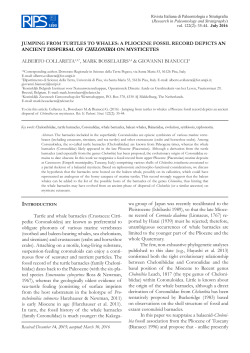
DUODENAL PORTION OF THE HEPATO-PANCREATIC
DUCT OF THE BOUTU, !NIA GEOFFRENSIS
KENJI KITO AND FUSAO YAMASAKI*
ABSTRACT
The duodenal portion of the hepato-pancreatic duct of the Boutu,
Inia geojfrensis (body length 204 cm, female), was examined macroscopically
and light microscopically. Passing through the muscle layer of the
duodenum, the hepato-pancreatic duct ran intramurally, and formed the
duodenal pouch, and opened into the lumen of the duodenum proper.
The intramural cystic gland, mucous in nature, was located in the relatively
thick submucosa of the duodenum, in which scattered bundles of muscle
fibers were arranged in a layered appearance. A tentative comparison for
some features of the duodenal portion of the hepato-pancreatic duct
suggests that the fresh water dolphins may be a phylogenetically peculiar
group in toothed whales.
INTRODUCTION
The hepato-pancreatic duct of the cetacean bile-passage generally dilates and
forms a well-developed glandular structure in the duodenal wall (Kamiya,
1962; Yablokov, Bel'kovich and Borisov, 1972). This glandular structure,
termed as the intramural cystic gland, supposedly plays a role in the storage
and excretion of the gall, compensating to some extent for the absence of the
gallbladder (Kamiya, 1962). According to the above two works, the intramural
cystic gland is located in the submucosa of the duodenum in whalebone
whales and the sperm whale, and in the muscle layer of the duodenum in
toothed whales except in the case of the sperm whale. It is, therefore,
suggested that the location of the intramural cystic gland in the duodenal wall
is an important character to consider the phylogeny of the cetaceans.
Some studies on the fresh water dolphins also have mentioned the
hepato-pancreatic duct passing through the duodenal wall: Takahashi and
Yamasaki (1972) and Yamasaki, Takahashi and Kamiya (1972) on Platanista
gangetica; Yamasaki, Takahashi and Kamiya (1975) and Takahashi, Yamasaki
and Kamiya (1976) on Pontoporia blainvillei; Zhou and Li (1981) on Lipotes
vexillifer; Zhou, Li and Pilleri (1982) on Inia boliviensis; and Yamasaki and Kito
(1984) on Inia geoffrensis. In order to discuss the fresh water dolphins, regarding the features of the bile-passage, it is necessary that these features of the
*Department of Biology, Sapporo Medical College, Sapporo 060, Japan
Sci. Rep. Whales Res. Inst.,
No. 36, 1985, 89-95
90
KITOANDYAMASAKI
I
n
i
αandL
i
p
o
t
e
sbedes
cr
i
b
e
da
tl
e
a
s
ta
sd
e
t
a
i
l
e
dl
ya
st
h
a
toft
h
eP
l
α
t
αn
i
s
t
aand
P
o
n
t
o
p
o
r
i
a
.Thep
r
e
s
e
n
tp
a
p
e
r
,a
st
het
h
i
r
dr
e
p
o
r
tont
h
emorphologicals
t
u
d
y
oft
h
ed
i
g
e
s
t
i
v
et
r
a
c
tofh臼 g
e
o
f
f
r
e
n
s
i
s
,d
e
s
c
r
i
b
e
sde
t
a
il
e
d
lyt
h
ehepato
・p
anc
r
e
a
t
i
cducti
nt
h
eduodenumandp
r
o
v
i
d
e
ssomefundamentalknowledget
o
con
s
i
d
e
rt
h
ephyl
o
g
e
n
e
t
i
cr
e
l
a
t
i
o
ns
hi
poft
h
ef
r
e
s
hwaterdolphinsi
nt
h
ec
e
t
a
c
e
a
n
s
.
句
MATERIALANDMETHODS
As
pec
imenoft
h
eBoutu(Amazoniand
o
l
p
h
i
n
)
,I
ηi
ag
e
o
f
f
r
e
ηs
i
s(bodyl
ength
204cm,femal
e
)wasprovidedf
o
rt
h
i
ss
t
u
d
ybyKamogawaSeaWorld,C
h
i
b
a
,
Japan,a
f
t
erdeath from anunide
n
t
i
f
i
e
dd
i
s
e
a
s
e
. Theduodenumwi
t
ht
h
e
hepat
o
・p
a
n
c
r
e
a
t
i
cductt
a
k
e
nfrom t
h
eabdominalc
a
v
i
t
y wasp
r
e
s
e
r
v
e
di
n
1
0% formal
ins
ol
u
t
i
o
nandt
r
a
n
s
p
o
r
t
e
dt
oourl
abora
t
o
ry
.Theb
il
e
p
a
s
s
a
g
e
o
u
t
s
i
d
eoft
h
eduodenumha
sbeenexcludedfromt
h
em
a
t
e
r
i
a
lt
r
a
n
s
p
o
r
t
e
d
,
s
ot
h
a
tt
h
ep
r
e
s
e
n
to
b
s
e
r
v
a
t
i
o
ni
sr
e
s
t
r
ic
t
e
dt
oonl
yt
h
ehepato-pan
c
r
e
a
t
i
cduct
l
o
c
a
t
e
di
nt
h
eduodenum.A
f
t
e
rm
a
c
r
o
s
c
o
p
i
c
a
lo
b
s
e
r
v
a
t
i
o
n
,t
h
em
a
t
e
r
i
a
lwas
embedde
di
nc
e
l
l
o
i
d
in,s
e
c
t
i
o
n
e
d,and s
t
a
i
n
e
dw
i
t
h hematox
yl
i
n
e
o
s
i
nf
o
r
l
i
ghtm
i
c
r
o
s
c
o
p
y
.
F
i
g
sI
aand I
b.Photomicrogiaphso
ft
h
el
ong
i
tud
i
n
a
ls
e
c
t
i
onoft
h
eduodenal
p
o
r
t
i
o
noft
heh
e
p
a
t
o・p
a
n
c
r
e
a
t
i
cduc
twhic
hi
sl
o
c
a
t
e
di
nt
h
eduodena
l
w
a
l
loft
h
e
I
n
i
ag
e
o
j
f
r
e
n
s
i
s
.Them
i
d
p
a
r
toft
hedu
ctwass
e
ct
i
o
n
e
dt
r
a
n
s
v
e
r
s
e
l
yandshowni
n
F
i
g
.2
.F
i
g
sl
aand l
bs
howt
h
ef
i
n
a
lo
n
e
f
o
u
r
t
handt
h
ei
n
i
t
i
a
lp
a
r
toft
he
duodenalpouch,r
e
s
p
e
c
t
i
v
e
l
y
.Thepouche
x
si
s
t
ss
u
b
m
u
c
o
s
a
l
l
yi
nt
h
eduodena
l
wal
l
.D
I
d
u
o
d
e
n
a
ll
u
m
e
n
;Op
トlumeno
fduodenalpouch;Tm-tun
i
c
amu
s
c
u
l
a
r
i
s
oft
heduodenum;Hp-hepato
・p
anc
r
e
a
t
i
cd
u
c
t;P
l
d
p
l
i
c
al
o
n
g
i
t
u
d
i
n
a
l
i
sd
u
o
d
e
n
i
;
anarrowshowst
h
eor
i
f
i
c
eoft
heduodenalp
o
u
c
h
.H-Es
t
a
i
n
.x 8
.
S
c
i
.Re
p
. Wha
l
e
sRe
s
.I
n
s
t
.
,
No
.3
6
,198
5
DUODENAL POUCH OFTHEBOUT U
91
OBSERVATION
ta
enumpropera
eduod
h
loft
l
a
ew
h
dt
e
r
e
t
cducten
i
t
a
e
r
c
n
o・pa
Thehepat
al
t
is
.Thed
b)
aandl
sl
g
i
F
tramuraly(
n
wardsi
ananal
eandr
l
epang
e
t
hers
t
a
r
slumenandformed
t
largedi
heduodenalwalen
nt
educti
h
soft
h
t
r
u
o
f
ee
r
h
t
d
l
u
o
,whichc
i
isduoden
nal
i
d
u
t
ongi
al
c
i
l
ep
h
h;t
aledduodenalpouc
c
o
hes
t
e
c
a
f
r
u
s
r
e
n
n
i
e
h
t
n
o
,
e
d
i
w
cm
6
.
0
d
n
a
g
n
o
l
cm
5
.
1
t
u
o
b
a
s
i
,
n
e
e
s
y
l
t
c
n
i
t
s
i
d
be
e
h
ot
t
n
c duct opened i
i
t
a
e
r
c
o・pan
. The hepat
he duodenum proper
of t
he
he midpan of t
ion of t
t
c
e
es
s
er
v
s
e uan
h
aph of t
r
g
o
r
c
i
m
o
t
. 2.A pho
ig
F
ner
n
ki
c
hi
het
soft
t
s
i
ons
numc
heduode
roft
e
y
a
el
l
c
emus
.Th
h
c
u
o
enalp
duod
a
s
o
ubmuc
as
el
kt
ic
h
het
nt
di
e
t
a
c
o
sl
hi
.Theduodenalpouc
s
rone
e
t
u
ino
h
andt
he
aoft
s
o
c
.Themu
)
w
o
r
r
a
en(
e
anbes
sc
r
e
b
i
lef
c
moothmus
hsomes
c
nwhi
i
e
erar
rm
o
hef
andsoft
enandgl
llum
na
ode
edu
h
toft
a
h
hant
rt
e
k
ic
h
st
hi
uc
po
enof
lum
l
en;Dp
llum
dena
duo
.DI
e
r
u
t
nna
usi
o
r
e
es
sar
t’
er
t
t
a
el
h
sandt
mucou
ubmucosa;
as
el
t
eduodenum;Tsh
soft
ri
a
ul
c
amus
c
ni
u
t
lpouch;T ma
n
e
duod
.
6
.X 1
n
i
a
t
Es
.He
r
u
t
c
u
r
t
ls
a
i
c
i
f
i
t
ar
*
.,
t
s
n
sR目 . I
e
. Whal
J
/
.Re
i
c
S
,1985
6
.3
o
N
92
KITO AND YAMASAKI
duodenal lumen through the slit-like orifice with 2 mm in length (Fig. la),
which was located about 9 cm away from the commencement of the
duodenum proper. The duodenal papilla was indistinct and no peculiar structure was found around the orifice.
Microscopically, the hepato-pancreatic duct ran in the submucosa of the
duodenum after passing through the muscle layer of the duodenum (Fig. lb).
There were no longitudinal or transverse folds and also no villi on the inner
surface of the duodenal pouch; luminal epithelium was seen to be worn-off.
As shown in Figs 1and2, the lamina propria mucosae of the hepato-pancreatic
duct was 0.3-1.2 mm thick, and was occupied with well-developed tubuloalveolar glands. This glandular structure, the intramural cystic gland, was
mucous in nature and apparently differed from that of the duodenum
proper, mostly consisting of serous glands. Goblet cells were not found
among the epithelial cells of the intramural cystic glands and lymphatic
nodules were also absent in the lamina propria mucosae of the hepato-pancreatic duct. No sphincteric smooth muscles could be seen either at the
proximal or distal portion of the duodenal pouch, though the outer longitudinal muscles of the duodenal wall irregularly and intermittently extended
to the submucosa around the proximal portion of the intramural cystic gland.
Scattered bundles of smooth muscle fibers gave a layered appearance near
the middle of the submucosa between both the mucosa of the duodenum and
the hepato-pancreatic duct.
DISCUSSION
Table 1 summarizes the macro- and microscopical features on the duodenal
portion of the hepato-pancreatic duct of the fresh water dolphins, based upon
the following studies, Takahashi and Yamasaki (1972) and Yamasaki et al.
(1972) on Platanista gangetica; Yamasaki et. al. (1975) and Takahashi et al.
(1976) on Pontoporia blainvillei; Zhou and Li (1981) on Lipotes vexillifer; and
Zhou et al. (1982) on Inia boliviensis; Yamasaki and Kito (1984) and the present study on Inia geoffrensis. This table is unfortunately incomplete because
all information on the features are not given from the above studies. Therefore, it is presently possible to compare the fresh water dolphins in only a few
characters which are clearly described.
The hepato-pancreatic duct of Inia is similar to that of Pontoporia and
Platanista in running analwards within the submucosa of the duodenum and
in entering the lumen of the duodenum proper through the orifice not surrounded by any peculiar prominences. While, the hepato-pancreatic duct of
Lipotes runs within the circular muscle layer of the duodenum and enters the
lumen of the duodenal ampulla through the orifice surrounded by a lip-like
prominence (Zhou and Li, 1981). The intramural cystic gland of I. geoffrensis
is mucous in nature as that of Pontoporia, and it also clearly forms the
duodenal pouch as that of Platanista. The duodenal pouch of Platanista is
Sci. Rep. Whales Res. Inst.,
No. 36, 1985
~
<: ;w.
i<'
~[
~
::>J
~
~~
\Jt"'
-°'~
"'::>J
mucous
0.3-1.2 mm
not found
scattered bundles
of muscle fibers
duodenum proper
indistinct
1.5 x 0.6 cm
Nature of the intramural
cystic gland
Thickness of the mucosa
Lymphatic nodules
Musculature around the
hepato-pancreatic duct
Location of opening of the
hepato-pancreatic duct
Duodenal papilla
Size of plica longitudinalis
duodeni**
circular muscle layer
of the duodenal wall
-
indistinct
2.0 x 1.0 cm
lip-like prominence
around the orifice
duodenal ampulla
abundant
-
duodenum proper
l.3mm
mucous*
absent*
muscle layer
Lipotes
vexillifer3!
0.4-0.6 mm
mucous*
absent*
submucosa*
Inia
boliviensis2!
indistinct
1.5 x 0.7 cm
1.5 x 0.9 cm
duodenum proper
thin circular layer,
many muscle fibers
scattered
abundant
0.5-2.4 mm
serous,
partially mucous
present
submucosa
Platanista
gangetica5!
indistinct
duodenum proper
well-developed longitudinal muscle layer
present
0.6-1.3 mm
mucous
absent
submucosa
Pontoporia
blainvillei 4J
References: 1) Yamasaki and Kito (1984) and the present study, 2) Zhou et al. (1982), 3) Zhou and Li (1981), 4) Yamasaki et al. (1975)
and Takahashi et al. (1976), 5) Takahashi and Yamasaki (1972) and Yamasaki et al. (1972).
- Data not given. * Data suggested from photomicrographs of the hepato-pancreatic duct. ** Length x width.
present
Duodenal pouch
submucosa
Inia
geojfrensis 1!
COMPARISON OF THE DUODENAL PORTION OF THE HEPATO-PANCREATIC DUCT IN THE FRESH WATER DOLPHINS
Location of the hepatopancreatic duct in the
duodenal wall
Character
TABLE 1.
t:I
(j()
<.O
c...,
c
0
I:;:)
M
::r:
...,
::r:
0
..,,
Cl
c
0
"':)
>
t""'
z
M
t:I
0
c
94
KITO AND YAMASAKI
more developed than that of I. geoffrensis and is especially characterized by the
presence of many circular folds on its inner surface and the presence of villi
of the mucosa; Pontoporia has no pouch in the duodenal wall*. There is no
mention of the nature of the intramural cystic gland in Lipotes and J. boliviensis,
though the photomicrographs of the hepato-pancreatic duct in the duodenal
wall in Lipotes (Fig. 2-2, Zhou and Li, 1981) and I. boliviensis (Plate 4A, Zhou et
al., 1982) suggest that both their glandular structures are mucous in nature
and they have no distinct duodenal pouch. If the duodenal pouch is really
missing in the bile-passage of I. boliviensis but present in that of I. geoffrensis,
the feature of the duodenal pouch appears to be one of the important
characters in which to classify these two closely related dolphins of the
genus Inia.
According to Kamiya (1962), the intramural cystic gland is located in the
submucosa of the duodenum in whalebone whales and it is located in the circular muscle layer of the duodenum in toothed whales except in the case of
the sperm whale, whose gland is located in the submucosa, the same as in
whalebone whales. Yablokov et al. (1972) added to Kamiya's view, based upon
their investigation on the duodenum of the white whale, Delphinapterus, that
the intramural cystic gland has its own musculature in toothed whales (except
the sperm whale) and is not found in whalebone whales and the sperm whale.
Among the fresh water dolphins, Lipotes has the intramural cystic gland in the
muscle layer of the duodenum as do most toothed whales, though it is not
mentioned whether the gland of Lipotes has its own musculature or not. The
Pontoporia, Platanista, and Inia have the intramural cystic gland in the submucosa as do whalebone whales and the sperm whale. The three differ, however,
from whalebone whales and the sperm whale in regards to the distributional
feature of the musculature in the submucosa. The intramural cystic gland of
Pontoporia is surrounded by a very thin submucosa and a remarkably developed muscle layer, mainly consisting of longitudinal muscles, of the
hepato-pancreatic duct; so that it appears to be located in the muscle layer of
the duodenum. The intramural cystic gland of Platanista is also surrounded
by a thin submucosa and a relatively thin muscle layer, and many muscle
fibers are distributed within the submucosa and lamina propria of the duct.
The intramural cystic gland of Inia is characterized by the scattered bundles
of muscle fibers intermittently running near the middle of the thick submucosa between both the mucosa of the duodenum and the hepato-pancreatic
duct. Thus, in toothed whales, the fresh water dolphins seem to be a peculiar
group consisting of dolphins having the intramural cystic gland with its own
musculature in the submucosa of the duodenum and those dolphins having
the intramural cystic gland in the muscle layer of the duodenum.
The duodenal portion of the hepato-pancreatic duct of the fresh water
* The
glandular structure of the hepato-pancreatic duct having no distinct duodenal pouch is thought
to be a kind of the intramural cystic gland.
Sci. Rep. Whales Res. Inst.,
No. 36, 1985
DUODENAL POUCH OF THE BOUTU
95
dolphins have some important and useful characters as do other structures,
such as the skelton, brain, the other digestive and respiratory organs, in order
to clarify the phylogenetic relationship among the fresh water dolphins and
cetaceans. For example, the location of the opening of the hepato-pancreatic
duct into the duodenal lumen is already considered as a plesiomorphous
character (located at duodenum proper) or an apomorphous character
(located at duodenal ampulla) in discussing the phylogeny of the fresh water
dolphins (Zhou, 1982). We will continue the discussion about the fresh water
dolphins when more details and new findings are gathered in the future
which will be sufficient to consider their phylogenetic relationship.
ACKNOWLEDGEMENTS
We wish to thank Dr T. Tobayama, Kamogawa Sea World, Chiba, for his
kindly supplying the specimen for this study, and Professor Dr K. Takahashi,
Department of Anatomy, Sapporo Medical College, Sapporo, for his valuable
advice.
REFERENCES
KAMIYA, T., 1962. On the "intramural cystic gland" of the cetacea. Acta Anat. Nippon, 37: 339-351. (in
Japanese with English summary).
TAKAHASHI, K. and F. YAMASAKI, 1972. Digestive tract of Ganges dolphin, Platanista gangetica. 11. Small and
large intestines. Okajimas Fol. anat. jap., 48: 427-452.
TAKAHASHI, K., F. YAMASAKI and T. KAMIYA, 1976. Extrahepatic bile-passage of Franciscana (La Plata
Dolphin), Pontoporia blainvillei. Okajimas Fol. anat. jap., 53: 115-126.
YABLOKOV, A.V., V.M. BEL'KOVICH and V.l. BoR1sov, 1972. Whales and dolphins. Acad. Sci. USSR, Moscow.
472 pp. (in Russian). (English translations were also referred; Joint Publications Research Service,
Virginia, 1974-Part I and 11).
YAMASAKI, F., K. TAKAHASHI and T. KAMIYA, 1972. Liver and bile-passage of Ganges dolphin, Platanista
gangetica. Okajimas Fol. anat. jap., 49: 365-390.
YAMASAKI, F., K. TAKAHASHI and T. KAMIYA, 1975. Digestive tract of La Plata dolphin, Pontoporia blainvillei.
11. Small and large intestines. Okajimas Fol. anat. jap., 52: 1-26.
YAMASAKI, F. and K. KITo, 1984. A morphological note on the intestine of the Boutu, with emphasis on its
length and ileo-colic transition compared with other platanistids. Sci. Rep. Whales Res. Inst., 35:
165-172.
ZHOU, K. and Y. L1, 1981. On the intestine of the Baiji, Lipotes vexillifer. Acta Zoo!. Sinica, 27: 248-253. (in
Chinese with English abstract).
ZHOU, K., Y. LI and G. PILLER!, 1982. The digestive tract of Inia boliviensis (D'ORBIGNY, 1934). Investigations
on cetacea, 13: 125-133.
ZHou, K., 1982. Classification and phylogeny of the superfamily Platanistoidea, with notes on evidence of
the morphology of the cetacea. Sci. Rep. Whales Res. Inst., 34: 93-103.
Sci. Rep. Whales Res. Inst.,
No. 36, 1985
© Copyright 2026 Paperzz