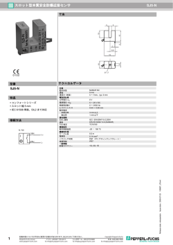
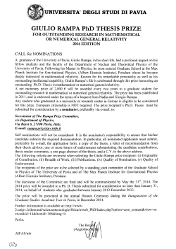
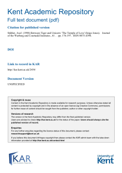
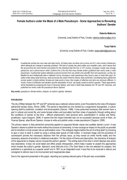
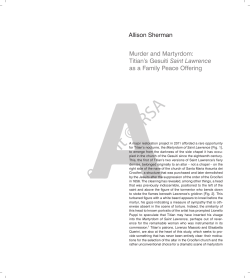
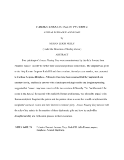
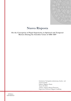
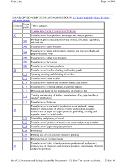
FEBS Letters 583 (2009) 3732–3737
journal homepage: www.FEBSLetters.org
Review
How Camillo Golgi became ‘‘the Golgi”
Paolo Mazzarello *, Carla Garbarino, Alberto Calligaro
Museum for the History of the University of Pavia, Department of Experimental Medicine, University of Pavia, Italy
a r t i c l e
i n f o
Article history:
Received 2 September 2009
Accepted 4 October 2009
Available online 13 October 2009
Edited by Daniela Corda
Keywords:
Camillo Golgi
Black reaction
Cell organelle
Golgi apparatus
Golgi complex
‘‘The Golgi”
Artefact
Electron microscopy
a b s t r a c t
On April 1898 Camillo Golgi communicated to the Medical–Surgical Society of Pavia, the discovery of
the ‘‘internal reticular apparatus”, a novel intracellular organelle which he observed in nerve cells
with the silver impregnation he had introduced for the staining of the nervous system. Soon after
the discovery it became evident that this cellular component, which was also named the ‘‘Golgi
apparatus”, was a ubiquitous structure in eukaryotic cells. However the reality of the organelle
was questioned for years and many cytologists considered the internal reticular apparatus as an
artefact due to the fixation and/or metallic impregnation procedure. The controversy was finally
solved in the mid-1950s by electron microscopy when the Golgi apparatus definitely acquired its
dignity of being a genuine cell organelle. The designation of ‘‘Golgi complex” entered officially in
the literature in 1956. Both the terms Golgi apparatus and Golgi complex are currently interchangeable. However a quick ‘‘the Golgi” and the introduction of Golgi in adjectival form are now prevalent
in the blooming scientific literature on the organelle. Thus Camillo Golgi underwent his final transformation and, becoming the eponym of the organelle he had discovered, he found a way to
immortality.
! 2009 Federation of European Biochemical Societies. Published by Elsevier B.V. All rights reserved.
1. Black silhouettes
At the end of the nineteenth century Camillo Golgi, professor of
General Pathology and Histology at the University of Pavia, was already internationally famous for his discoveries on the structure of
the central nervous system and for his descriptions of the human
malaria parasites development in the human blood [1].
Graduated in Pavia in 1865, he was trained in microscopy by
Giulio Bizzozero, a Rudolf Virchow’s pupil and one of the pioneer
of cell biology in Italy [2]. In 1873, while he was chief physician
in a hospital for chronic patients at Abbiategrasso not far from Milan, Golgi developed a method, the ‘‘black reaction” (known nowadays as the Golgi stain or the Golgi impregnation), which allowed
for the first time a full view of a single nerve cell. This histological
method, which is widely still in use, was based on hardening
blocks of nervous tissue in potassium dichromate, followed by
impregnation with silver nitrate. The final result was a precipitation of silver chromate, filling the nerve cell body and its prolongations, which could be followed and analyzed even at a great
distance from the cellular soma. Thus the black silhouette of the
nerve cell appears in all its morphological complexity with all its
ramifications (see Fig. 1).
* Corresponding author. Address: Sistema Museale di Ateneo, Strada Nuova 65,
27100 Pavia, Italy.
E-mail addresses: [email protected], [email protected] (P. Mazzarello).
The great advantage of this technique is that, for reasons that
are still unknown, the precipitate of silver chromate randomly
but selectively stains in black only a small proportion of nerve cells
(usually from 1% to 5%), and completely spares the others allowing
the individual elements to emerge from the nervous puzzle.
The discovery of the ‘‘black reaction” provided the spark to a
truly scientific revolution which allowed the morphology and the
basic architecture of the cerebral tissue to be evidenced in all its
complexity, thus contributing to the foundation of the modern
neurosciences.
Aided by his black reaction method, Golgi described the complex arborisation of the dendrites, discovered the branching of
the axons, analyzed several regions of the nervous system in detail
and provided beautiful illustrations of them. Among the remarkable Golgi’s descriptions of different brain regions, particularly
important were those of the cerebellum (where he described the
so called ‘‘Golgi cells”), the olfactory bulb, the hippocampus, and
the cerebral cortex. Taken together these investigations provided
a remarkable contribution to the advancement of the knowledge
on the structural organization of the nervous system.
Meanwhile he began to elaborate a general anatomic-physiological theory of brain organization, the ‘‘diffuse nervous net”,
according to which the ramified axons were connected (through
direct fusion or intimate contact) in a diffuse network along which
the nervous impulse propagated. Actually, it dealt with an illusory
network which resulted from a mistaken microscopic image created by superimposition and by interlocking of the axons of the diverse cells. Towards the end of the nineteenth century this concept
0014-5793/$36.00 ! 2009 Federation of European Biochemical Societies. Published by Elsevier B.V. All rights reserved.
doi:10.1016/j.febslet.2009.10.018
P. Mazzarello et al. / FEBS Letters 583 (2009) 3732–3737
3733
Fig. 1. Nerve cell stained with the black reaction. Drawing preserved with Golgi’s
papers, Museum for the History of the University of Pavia.
developed in polemical opposition to the neuron theory championed by the Spanish histologist Santiago Ramón y Cajal which affirms that the nervous system is anatomically and functionally
composed of individual cells, like any other tissue, and that nerve
cells act to each other through points of contact subsequently
named synapses by the British neurophysiologist Charles Scott
Sherrington.
Since the development of the black reaction in 1873, Golgi continued to modify the chemical and physical parameters of the reagents in order to improve the selectivity and the reproducibility
of the method under standard condition. The goal was however
never completely reached since an element of randomness remained, and still remains, in the Golgi stain. However by 1885,
Golgi was able to describe in his book Sulla fina anatomia degli organi centrali del sistema nervoso (On the fine anatomy of the central
organ of the nervous system) some modifications and variants of
the black reaction [3]. Particularly important was the introduction
of a osmium-dichromate solution as the first immersion step of the
reaction which allowed a fast staining of the nerve cells (the so
called rapid variant of the method).
Very interested in infectious diseases and concerned about public health, between 1886 and 1892 Golgi temporarily abandoned
neurobiological research and concentrated on studying human malaria (see Fig. 2). Not only he did worked out the cycle of development of the malaria parasite Plasmodium in the red blood cells, but
he also discovered the temporal coincidence between the recurrent
fever of the infection, and the multiplication of the parasite in human blood. Between 1893 and 1896 Golgi was mainly involved in
academic and administrative commitments as elected rector of the
University of Pavia. However he never lost his interest for the nervous system. In 1897 he returned in the laboratory to perform original research and started an investigation on the structure of the
spinal ganglia. The topic was at the centre of Golgi’s attention since
the T shape of the spinal ganglion cells seems to contradict the
‘‘law of dynamic polarization” of Santiago Ramón y Cajal and
Arthur van Gehuchten, one of the theoretical basis of the neuron
theory.
2. A fine and elegant network hidden within the cell body
During this research Golgi noticed the occurrence of a strange
reticular structure, hidden in the cytoplasm of the nerve cell body,
Fig. 2. Camillo Golgi around 1890. Museal System – University of Pavia.
Fig. 3. First published illustration of the Golgi Apparatus. From Ref. [5].
which was clearly detached from the membrane and from the nucleus. Unfortunately this bizarre new formation was not constantly
stainable. But Emilio Veratti (who in 1902 went on to describe the
sarcoplasmic reticulum [4]), working in Golgi’s laboratory, was
able to replicate and confirm the findings in the cell body of the
fourth cranial nerve. Golgi then persevered in his neurocytological
research and was able to obtain very good and reproducible
impregnation of the intracellular apparatus in the cell body of
the Purkinje cerebellar cells of the owl Tyto alba (see Fig. 3).
3734
P. Mazzarello et al. / FEBS Letters 583 (2009) 3732–3737
The apparatus was also detected in the cells of the sympathetic
system by Veratti who added 0.1 platinum chloride to the fixative
to facilitate the reaction [7].
Golgi continued to pursue with energy this line of research and
on 15 July 1898 he described at the Medical–Surgical Society of
Pavia the apparatus in cells of the spinal ganglia [8,9] and on 20
January 1899 he presented clear data on the age related variations
of this structure [10]. He noticed that in adult and older animals
the impregnation was only partial (see Fig. 5).
While he continued to perform research on the structure he had
discovered [11–14] he stimulated the interest of his co-workers to
search the structure in non-neural cell types (see Fig. 6).
3. Not only in the nervous system
Fig. 4. Golgi Apparatus, nerve cells of the cerebral cortex in the mouse. Drawing
preserved with Golgi’s papers, Museum for the History of the University of Pavia.
Fig. 5. Demonstration of the Golgi apparatus in ganglion cells partially stained with
the black reaction. Original Camillo Golgi’s drawing, Museum for the History of the
University of Pavia.
On a meeting of the Medical–Surgical Society of Pavia, in April
1898,1 Golgi felt thus confident to describe ‘‘a fine and elegant network within the cell body . . . completely internal in the nerve
cells . . . The distinctive appearance of this internal reticular apparatus is attributable to the prevalence of ribbon-like threads, their
manner of dividing, their anastomoses, and the pathways formed
by them . . . and to the presence in this apparatus of thin plates or
small discs, rounded and transparent in the centre, which appear
as nodal points of the net . . . the most distinctive feature of this
apparatus lies in its overall appearance, whereas it is clearly limited
towards the exterior . . . Towards the interior, in contrast, the filaments of the network penetrate to different planes” [5,6]. For his
shape and intracellular location Golgi named this structure ‘‘apparato reticolare interno” (internal reticular apparatus). He specified that
he had detected the apparatus in the Purkinje cells but he also stated
that ‘‘I am inclined to believe that this is most probably a standard
structure, at least among the major classes of nerve cells” (see Fig. 4).
1
Golgi wrote on many occasions that he presented his communication on April 19,
1898, but according to the minutes published in the Bollettino della Società MedicoChirurgica di Pavia, 13, III–IV, the communication occurred on April 22.
At the turn of the century Golgi had organized in Pavia a very
active laboratory of General Pathology and Histology. He was an
effective mentor and supervisor for his co-workers and disciples
as documented by the remarkable number of discoveries that they
made under his direction. After the first observations of the internal reticular apparatus in the nerve cells, Antonio Pensa, in Golgi’s
laboratory, succeeded in detecting this structure in the medullary
cells of the adrenal glands and he presented his findings to the
Medical–Surgical Society of Pavia in 1899 [15]. However, this first
indication that the apparatus could not only be present in nerve
cells was not considered conclusive given the common embryonic
origin of neurons and adrenal medullary cells. While the paper was
in press Pensa added a note to announce that ‘‘In our laboratory
Mr. Negri . . . in these days . . . could detect in pancreatic gland cells
and in the cells of some serous glands an endocellular apparatus
with some analogies with what I have described in the medullary
cells of the adrenal glands”. Negri, who was at the time a fifth year
medical student, quickly succeeded in demonstrating the network
in several non-neural cells, besides the pancreatic cell glands, i.e.
thyroid, epididymis, salivary glands and ovary [16]. Meanwhile
Edoardo Gemelli, another pupil of Golgi, described the apparatus
in the pituitary gland cells [17]. It was becoming evident that this
structure did not belong only to neurocytology, but was probably
ubiquitous in various eukaryotic cell types.
4. Before Golgi
When the Spanish histologist Ramón y Cajal read of the new
Golgi’s discovery, probably remembered a fact occurred some
years before which caused one of his ‘‘most deplorable disappointments”; as he reported in his recollections [18]:
‘‘In the years 1891 and 1892, I happened to immerse pieces of
the cerebrum of a young rabbit in a certain mixture of equal parts
of 3% potassium dichromate and 1% gold chloride solutions. A few
days later, sections of the pieces showed a splendid selective
reduction of the gold salt in the Golgi apparatus (then unknown)
of the cerebral pyramids. Enraptured with the wonderful results,
I devoted myself ardently to reiterated trials to determine the conditions of success. Well, the confounded reaction never appeared
again! I was guilty on that occasion of excessive scrupulousness
and timidity, since I did not dare to publish my rare discovery; it
seemed to me that it would be an abuse to announce a fact of
which confirmation was, at the time impossible. Had it not been
for such considerations, the so called reticular apparatus of Golgi,
which the neurologist of Pavia discovered in 1898 (by means of a
formula, indeed, which is notably uncertain), it would figure today
among my assets and under my name”.
In reality Golgi published his discovery only when, with the aids
of his pupil Veratti, was able to constantly reproduce the results.
But even before Ramón y Cajal (assuming that his vague and late
P. Mazzarello et al. / FEBS Letters 583 (2009) 3732–3737
3735
Fig. 6. Early protagonists of Golgi Apparatus research. We can recognise Golgi (fourth from right, first row), Pensa (second from left, first row), Veratti (second from right, first
row), Negri and Gemelli (first and third from left, second row, respectively) and a young Perroncito (first from right, second row).
others, the name of Adolphe von La Valette St. George (1867), Moritz Nussbaum (1882), Masanori Ogata (1883), Gustav Platner
(1886) and Auguste Prenant (1887) are sometime mentioned in
relation to early observations of the reticular apparatus. However
it is uncertain that they had observed this structure or something
else [19–21].
In any case it was Golgi and subsequently his school [22,23]
who defined the apparatus morphologically as an organelle, a stable component of the eukaryotic cell, and clearly differentiated it
from other cytological elements. Moreover the histologist of Pavia
must be credited to have extended to general cytology the application of the metallic impregnation, previously used mainly in
neurobiology.
5. Further contributions by the Golgi’s school
Fig. 7. Golgi Apparatus in the gastric mucosa cells of the frog. Original drawing
probably of Emilio Veratti, Museum for the History of the University of Pavia.
recollection refers to the same structure characterized by Golgi)
other histologists had observed cytoplasmic features that perhaps
could be identified as the internal reticular apparatus. Among the
After the first demonstrations of its presence in non-neural
cells, a number of students and researchers in Golgi’s laboratory
investigated the presence and the morphology of the reticular
apparatus in different conditions and in various cellular types.
Among the relevant studies performed in the fifteen years after
its discovery, important were those of Giuseppe Moriani, Ferruccio
Marcora and Emilio Veratti who described it in various pathological processes [24–26], Antonio Pensa in cartilage cells [27] and in
vegetal cells [28] Luigi Stropeni in hepatic cells [29], Giuseppe Carlo Riquier in the corpus luteum [30], Francesco Maccabruni in
megakaryocytes [31] Ernesto Brugnatelli in renal tubules [32], Carlo Barinetti in various tissues [33] (see Fig. 7).
Golgi himself continued to pursue his investigations on the
reticular apparatus and described a ‘‘photographic” method [34]
derived by a technique introduced in 1903 by Ramón y Cajal. He
was thus able to observe its variation in the shape and intracellular
disposition during the secretory process in the mucous glands of
the frog stomach. He found that the nucleus was located at the
base of the cell and the apparatus projected towards the secretory
3736
P. Mazzarello et al. / FEBS Letters 583 (2009) 3732–3737
surface [35]. One of his collaborators, Giuseppe D’Agata, extended
the investigation to the reticular apparatus in the epithelium of the
gastric mucosa [36]. One of the most important researches performed in Golgi’s laboratory at that time, was that of Aldo Perroncito who described the division of the apparatus, the so called
‘‘dictyokynesis”, in living spermatocytes of Paludina vivipera
[37,38]. According to Trautmann [19], this phenomenon had been
already observed by Gustav Platner, Friedrich Hermann and Arthur
Bolles-Lee but without understanding the meaning of the structure
they describe (see also Ref. [21]).
6. Artefact or reality?
Golgi’s discovery spurred a wealth of investigations at the international level. However the difficulties in handling the original silver Golgi’s method, and its capriciousness, made the reproduction
very difficult for non-expert histologists. So it was common to
speak ironically of the ‘‘water of Pavia”, the miraculous water that
made possible the reaction for the apparatus in Pavia while failed
elsewhere.
There were however also important and precocious confirmations. One of the first was provided by Friedrich Kopsch in 1902
by using an osmium technique [39] followed by Ramón y Cajal,
eternal rival of Golgi, but always along his tracks, who was among
the first to study the reticular apparatus outside Pavia. He was able
to demonstrate this structure in the nerve cells of the invertebrates
[40] and then to extend his investigations to some other types of
nerve and non-nerve cells [41–43] concluding in 1914 that ‘‘the
reticular Golgi apparatus is an anatomical feature constant in the
protoplasm of all living cells, both embryonic and adult” [43]. In
fact it had been observed also in vegetal cells [28,44,45] while
the demonstration of a structure equivalent to the reticular apparatus in the cardiac cells by Emerico Luna in 1911 allowed the differentiation of this organelle from the sarcoplasmic reticulum [4].
Thus, from the zoological laboratory of the Magdalen College, Oxford, came the important James Brontë Gatenby’s generalization
according to which ‘‘every sort of metazoan cell carefully examined
has been found to possess the typical apparatus of Golgi” [46]. The
histological demonstrations and conceptualizations were however
controversial. Beginning in 1899 the Swedish histologist Emil
Holmgren described a system of ramifying ”canals” in the cytoplasm of some types of cells, which he named ‘‘Trophospongium”,
that was in communication with ducts penetrating through the
external cell surface from neighbour cells (the ‘‘trophocytes”)
[47–50]. According to Holmgren this system of canals could play
a nutritive role and included, as an important component, the
reticulum described by Golgi. Also Ramón y Cajal agreed, at least
in part, with Holmgren’s hypothesis, even though he firmly denied
the communication of the Trophospongium with the surface and
with the structures outside the cell [43]. In the following years
the problem continued to be debated [51]; however Holmgren’s
system did not receive decisive confirmations and the term now
designates only the axon of invertebrates and the invagination of
glial processes around the cell body. Ramón y Cajal frequently referred in his publications on the subject to a ‘‘Golgi-Holmgren
reticular apparatus”. Other researchers indicated the structure as
‘‘Golgi-Kopsch apparatus”, ‘‘Netzapparat von Golgi” or ‘‘réseau
endocellulaire de Golgi”. In 1910 Carlo Besta designated this structure ‘‘Golgi apparatus” in the title of an article [52] and the term
became increasingly more frequent in the subsequent years, especially after the publication of a seminal work by Jòsef Nusbaum in
1913 [53] and its penetration in the English literature [51]. However, the supporters of the existence of the Golgi apparatus had
to confront the heated opposition of a large part of cell biologists.
The existence of the organelle was debated for years and many be-
gan to consider the Golgi apparatus as an artefact due to the fixation and/or metallic impregnation procedure [54–57]. Even the
first studies with electron microscopy did not confirm the existence of the Golgi apparatus supporting the view that this structure was in reality a composition of ‘‘myelin figure” artificially
induced in the cytoplasm during the preparation of the specimens
[58,59]. At the end, however, this new revolutionary instrument
was able to produce definite proof of the real existence of the Golgi
apparatus, unravelling the reality and the various components of
this organelle [60,61]. It is ironic that the electron microscope,
the very instruments that had provided the existence of synapses
between neurons, against Golgi’s concept of a continuous diffuse
neural network, also supplied the final proof that his ‘‘internal
reticular apparatus” was real [20,23,62–64]. A number of terms
were introduced when the electron microscope began to reveal
the complexity of this organelle, for example: Golgi bodies, Golgi
zone, Golgi area. The designation of ‘‘Golgi complex” entered officially in the literature in 1956 with the seminal work by Dalton
and Felix [65]. Both the terms Golgi apparatus and Golgi complex
are currently interchangeable. However a quick ‘‘the Golgi” and
the introduction of Golgi in adjectival form are now prevalent in
the blooming scientific literature on the organelle [23,66]. Thus Camillo Golgi underwent his final transformation and, becoming the
eponym of the organelle he had discovered, he found a way to
immortality.
References
[1] Mazzarello, P. (2009). Golgi (A. Badiani and H. Buchtel trans.), Oxford
University Press, New York.
[2] Mazzarello, P., Calligaro, A.L. and Calligaro, A. (2001) Giulio Bizzozero: a
pioneer of cell biology. Nature Rev. Mol. Cell. Biol. 2, 776–781.
[3] Golgi C. (1885) Sulla fina anatomia degli organi centrali del sistema nervoso.
Tip. S. Calderini e Figlio, Reggio Emilia.
[4] Mazzarello, P., Calligaro, A., Vannini, V. and Muscatello, U. (2003) The
sarcoplasmic reticulum. Its discovery and rediscovery. Nature Rev. Mol. Cell
Biol. 4, 69–74.
[5] Golgi, C. (1898) Intorno alla struttura delle cellule nervose. Bollettino della
Società Medico-Chirurgica di Pavia 13, 1–14. Partially translated by N. Geller
Lipsky (1989) with the title on the structure of nerve cells. J. Microsc. 155, 3–7.
[6] Golgi, C. (1898) Sur la structure des cellules nerveuses. Arch. Ital. Biol. 30, 60–71.
[7] Veratti, E. (1899) Über die feinere Struktur der Ganglienzellen des
Sympathicus. Anat. Anz. 15, 190–195.
[8] Golgi, C. (1898) Sulla struttura delle cellule nervose dei gangli spinali.
Bollettino della Società Medico-Chirurgica di Pavia 13, 53–63.
[9] Golgi, C. (1898) Sur la structure des cellules nerveuses des ganglions spinaux.
Arch. Ital. Biol. 30, 278–286.
[10] Golgi, C. (1899) Di nuovo sulla struttura delle cellule nervose dei gangli
spinali. Bollettino della Società Medico-Chirurgica di Pavia 14, 1–12.
[11] Golgi C. (1899) Sur la structure des cellules nerveuses de la moelle épinière, in
Volume jubilaire publié par la Société de Biologie. Masson & C., Paris, pp. 507–
530.
[12] Golgi, C. (1900) Sulla struttura delle cellule nervose del midollo spinale.
Bollettino della Società Medico-Chirurgica di Pavia 15, 1–32.
[13] Golgi, C. (1900) Intorno alla struttura delle cellule nervose della corteccia
cerebrale. Verh. Anat. Ges. 14, 164–176.
[14] Golgi C. (1901) Le reticulum intracellulaire et la structure fibrillaire
peripherique de la cellule nerveuse. Comptes rendus du XIII Congrès
Internationelle de Médecine. Section de Neurologie, Imprimerie Nationale,
Paris, pp. 582–586.
[15] Pensa, A. (1899) Sopra una fina particolarità di struttura di alcune cellule delle
capsule soprarenali. Bollettino della Società Medico-Chirurgica di Pavia 14,
76–85.
[16] Negri, A. (1900) Di una fina particolarità di struttura delle cellule di alcune
ghiandole dei mammiferi. Bollettino della Società Medico-Chirurgica di Pavia
15, 61–70.
[17] Gemelli, E. (1900) Contributo alla conoscenza sulla struttura della ghiandola
pituitaria nei mammiferi. Bollettino della Società Medico-Chirurgica di Pavia
15, 231–240.
[18] Ramón y Cajal S. (1989) Recollections of my life (Horne Craigie E. and Cano J.
trans.). The MIT Press, Cambridge MA, pp. 571-572.
[19] Trautmann J.C. (1988) Camillo Golgi (1843–1926) und die Entdeckung des
‘‘apparato reticolare interno” (Golgi-Apparat). Dissertation, Lübeck.
[20] Bentivoglio, M. and Mazzarello, P. (1998) The pathway to the cell and its
organelles: one hundred years of the Golgi apparatus. Endeavour 22, 101–105.
[21] Dröscher, A. (1998) Camillo Golgi and the discovery of the Golgi apparatus.
Histochem. Cell. Biol. 109, 425–430.
P. Mazzarello et al. / FEBS Letters 583 (2009) 3732–3737
[22] Mazzarello, P. (2003) La scuola scientifica di Camillo Golgi. Histochem. Ann.
Storia Univ. Ital. 7, 161–176.
[23] Bentivoglio, M. and Mazzarello, P. (1998) One hundred years of the Golgi
apparatus: history of a disputed cell organelle. Ital. J. Neurol. Sci. 19, 241–247.
[24] Moriani G. (1901) Di un apparato reticolare entro ad alcune cellule
cancerigene. Atti della Reale Accademia dei Fisiocritici in Siena 13 (Serie 4),
186–188.
[25] Marcora, F. (1901) Di una fine alterazione delle cellule nervose del nucleo
d’origine del grande ipoglosso consecutive allo strappamento e al taglio del
nervo. Bollettino della Società Medico-Chirurgica di Pavia 23, 134–137.
[26] Veratti, E. (1909) Sulla fine struttura delle cellule di alcuni tumori. Bollettino
della Società Medico-Chirurgica di Pavia 24, 34–44.
[27] Pensa, A. (1901) Osservazioni sulla struttura delle cellule cartilaginee.
Bollettino della Società Medico-Chirurgica di Pavia 16, 197–205.
[28] Pensa, A. (1910) Alcune formazioni endocellulari dei vegetali. Anat. Anz. 37,
325–333.
[29] Stropeni, L. (1908) Sopra una fine particolarità di struttura delle cellule
epatiche. Bollettino della Società Medico-Chirurgica di Pavia 23, 146–150.
[30] Riquier, G.C. (1909) L’apparato reticolare interno nelle cellule del corpo luteo.
Bollettino della Società Medico-Chirurgica di Pavia 24, 177–180.
[31] Maccabruni, F. (1909) Sulla fine struttura dei megacariociti. Bollettino della
Società Medico-Chirurgica di Pavia 24, 57–65.
[32] Brugnatelli, E. (1908) Di una fine particolarità di struttura degli epiteli dei
tubuli renali. Bollettino della Società Medico-Chirurgica di Pavia 23, 86–88.
[33] Barinetti, C. (1912) L’apparato reticolare interno e la centro-sfera nelle cellule di
alcuni tessuti. Bollettino della Società Medico-Chirurgica di Pavia 27, 273–296.
[34] Golgi C. (1908) Di un metodo per la facile e pronta dimostrazione
dell’apparato reticolare interno delle cellule nervose. Bollettino della Società
Medico-Chirurgica di Pavia 23, 81–87.
[35] Golgi, C. (1909) Di una minuta particolarità di struttura dell’epitelio della
mucosa gastrica ed intestinale di alcuni vertebrati. Bollettino della Società
Medico-Chirurgica di Pavia 24, 1–22.
[36] D’Agata, G. (1910) Sulle modificazioni dell’apparato reticolare interno
nell’epitelio della mucosa gastrica. Bollettino della Società MedicoChirurgica di Pavia 25, 517–522.
[37] Perroncito, A. (1910) Contributo allo studio della biologia cellulare.
Mitocondri, cromidii e apparato reticolare interno nelle cellule spermatiche.
Memorie della Reale Accademia dei Lincei. Classe de Scienze Fisiche,
Matematiche e Naturali 38, 224–261.
[38] Perroncito, A. (1911) Beiträge zur Biologie der Zellen (Mitochondrien,
Chromidien, Golgisches Binnennetz in den Samenzellen). Arch. Mikrosk.
Anat. 77, 311–321.
[39] Kopsch, F. (1902) Die Darstellung des Binnennetzes in spinalen Ganglienzellen
und anderen Körperzellen mittels Osmiumsäure. Sitzungsberichte der K.
Preussischen Akademie der Wissenschaften 40 (2), 929–932.
[40] Ramón y Cajal, S. (1903) Sobre la existencia de un aparato tubuliforme en el
protoplasma de las células nerviosas y epiteliales de la lombriz de tierra.
Boletín de la Sociedad Española de Historia Natural 3, 395–398.
[41] Ramón y Cajal, S. (1908) Les conduits de Golgi-Holmgren du protoplasma
nerveux et le réseau péricellulaire de la membrane. Travaux du Laboratoire de
Recherches Biologiques de l’Université de Madrid 5, 123–135.
[42] Ramón y Cajal, S. (1912) Fòrmula de fijaciòn para la demostraciòn fàcil del
aparato reticular de Golgi y apuntes sobre la disposiciòn de dicho aparato en la
retina, en los nervios y en algunos estrados patològicos. Trabajos del
Laboratorio de Investigaciones Biológicas de la Universidad de Madrid 10,
209–220.
3737
[43] Ramón y Cajal, S. (1914) Algunas variaciones fisiológicas y patológicas del
aparato reticular de Golgi. Trabajos del Laboratorio de Investigaciones
Biológicas de la Universidad de Madrid 12, 127–227.
[44] Smirnow, A.E. (1907) Ueber die Mitochondrien und den Golgischen Bildungen
analoge Strukturen in einigen Zellen von Hyacinthus orientalis. Anatomische
Hefte 3, 143–153.
[45] Bensley, R.R. (1910) On the nature of the canalicular apparatus of animal cells.
Biol. Bull. 19, 179–194.
[46] Gatenby, J.B. (1919) The cytoplasmic inclusions of the germ cells. Part V. The
gametogenesis and early development of the Limnaea stagnalis (L.), with
special reference to the Golgi apparatus and the mitochondria. Quart. J.
Microsc. Sci. 63, 445–493.
[47] Holmgren, E. (1899) Zur Kenntnis der Spinalganglienzellen von Lophius
piscatorius Lin. Anat. Hefte 12, 69–152.
[48] Holmgren, E. (1899) Zur Kenntnis der Spinalganglienzellen des Kaninchens
und des Frosches. Anat. Anz. 16, 161–171.
[49] Holmgren, E. (1900) Noch weitere Mitteilungen über den Bau der
Nervenzellen verschiedener Tiere. Anat. Anz. 17, 113–129.
[50] Holmgren, E. (1900) Weitere Mitteilungen über die ‘Saftkanälchen’ der
Nervenzellen. Anat. Anz. 18, 290–297.
[51] Penfield, W.G. (1921) The Golgi apparatus and its relationship to Holmgren’s
Trophospongium in nerve cells. Comparison during retispersion. Anat. Rec. 22,
57–77.
[52] Besta, C. (1910) Sull’apparato reticolare interno (apparato di Golgi) della
cellula nervosa. Anat. Anz. 36, 476–486.
[53] Nusbaum, J. (1913) Über den sogenannten inneren Golgischen Netzapparat
und sein Verhältnis zu den Mitochondrien, Chromidien und anderen
Zellstrukturen im Tierreich. Arch. Zellforsch. 10, 359–367.
[54] Baker, J. (1944) The structure and chemical composition of the Golgi element.
Quart. J. Microsc. Sci. 85, 1–71.
[55] Beker, J. (1949) Further remarks on the Golgi element. Quart. J. Microsc. Sci.
90, 293–307.
[56] Beker, J. (1954) What is the ‘‘Golgi controversy?”. J. R. Microsc. Soc. 73, 217–
221.
[57] Covell, W.P. and Scott, G.H. (1928) The relation between the granules stainable
with neutral red and the Golgi apparatus in nerve cells. Anat. Rec. 38, 377.
[58] Palade, G.E. and Claude, A. (1949) The nature of the Golgi apparatus I, II. J.
Morphol. 85, 35–112.
[59] Pease, D.C. and Baker, R.F. (1951) Electron microscopy of nervous tissue. Anat.
Rec. 110, 505–529.
[60] Dalton, A.J. and Felix, M.D. (1954) Cytologic and cytochemical characteristics
of the Golgi substance of epithelial cells of the epididymis – in situ, in
homogenates and after isolation. Am. J. Anat. 94, 171–208.
[61] Sjöstrand, F.S. and Hanzon, V. (1954) Ultrastructure of Golgi apparatus of
exocrine cells of mouse pancreas. Exp. Cell Res. 7, 415–429.
[62] Mazzarello P. and Bentivoglio M. (1998) The centenarian Golgi apparatus.
Nature 392, 543–544.
[63] Dröscher, A. (1998) The history of Golgi apparatus in neurons from its
discovery in 1998 to electron microscopy. Brain Res. Bull. 47, 199–203.
[64] Dröscher, A. (1998) From the ‘‘apparato reticolare interno” to ‘‘the Golgi”: 100
years of Golgi apparatus research. Virchows Arch. 434, 103–107.
[65] Dalton, A.J. and Felix, M.D. (1956) A comparative study of the Golgi complex. J.
Biophys. Biochem. Cytol. 2 (Suppl), 79–83.
[66] Bentivoglio, M. (1998) 1898: The Golgi apparatus emerges from nerve cells.
Trends Neurosci. 21, 195–200.
© Copyright 2026 Paperzz