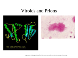
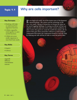
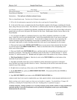
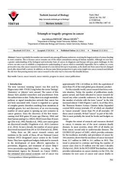
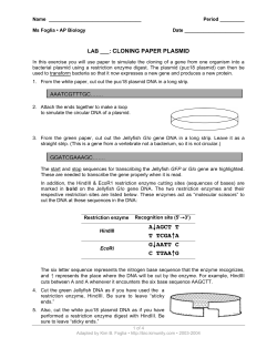
Perspectives Why have organelles retained genomes? Why have organelles retained genomes? The observation that chloroplasts and mitochondria have retained relics of eubacterial genomes and a proteinsynthesizing machinery has long puzzled biologists. If most genes have been transferred from organelles to the nucleus during evolution, why not all? What selective pressure maintains genomes in organelles? Electron transport through the photosynthetic and respiratory membranes is a powerful – but dangerous – source of energy. Recent evidence suggests that organelle genomes have persisted because structural proteins that maintain redox balance within bioenergetic membranes must be synthesized when and where they are needed, to counteract the potentially deadly side effects of ATP-generating electron transport. lastids and mitochondria descend from free-living eubacteria that, at the onset of symbiosis, must have possessed their own complete genomes. However, contemporary organelle genomes are greatly reduced, encoding only a handful of the proteins that their free-living eubacterial relatives possess1–3. During the process of organelle genome reduction, three possible fates awaited genes encoded therein: (1) some genes encoding proteins required to support existence as a free-living cell were forever lost during the course of reductive evolution; (2) many genes were transferred to what is now the nuclear genome; or (3) a small subset were retained within organelles – 50–200 protein-coding genes in the case of plastids4, 5–60 in the case of mitochondria5 and none in the case of most hydrogenosomes6,7 (for a glossary, see Box 1). Over evolutionary time, concentration of genetic material in the nucleus is strongly favoured by a population genetics principle known as Muller’s ratchet8, which predicts that deleterious, but sublethal, mutations accumulate more rapidly in asexually propagated genomes than in sexually propagated ones, ultimately dooming the former to extinction because the mutations cannot then be removed through recombination. In the case of organelles, which are predominantly asexual3, this means that genetic load should build up rapidly, providing a very strong selective pressure for genes to be transferred to, and fixed in, the nucleus, a predominantly sexual genome8–12. In addition, the chemistry of photosynthetic and respiratory electron transport in plastids and mitochondria generates high concentrations of various highly mutagenic reactive oxygen species that render extranuclear DNA especially prone to high mutation frequency1,13, exacerbating Muller’s ratchet8–12. This suggests that rates of nucleotide substitution (mutation plus fixation) in organelle genomes should be higher than in the nucleus, which is indeed the case for most animal mitochondria10. However, in plant mitochondria and chloroplasts, organelle substitution rates are not higher than in the nucleus, as Muller’s ratchet P Helen L. Race [email protected] ‡ Reinhold G. Herrmann herrmann@botanik. biologie. uni-muenchen.de § William Martin [email protected] 42 Iris Close, Aylesbury, Buckinghamshire, UK HP21 8YZ. ‡ Botanisches Institut der Ludwig-MaximiliansUniversitat, Menzinger, Strasse 67, D-8000 Munchen, Germany §Institut fur Genetik, Technische Universitat Braunschweig, Spielmannstrasse 7, D-38023 Braunschweig, Germany. 364 TIG September 1999, volume 15, No. 9 predicts; instead they are lower14,15, suggesting that compensatory mechanisms such as efficient DNA repair, high ploidy levels and occasional genetic recombination between organelles might counteract the long-term effects of asexuality in plants2. From the perspective of gene content, it is nonetheless clear that selective pressures have successfully chiseled away at organelle genomes for one or two billion years. Why are organelles unwilling to relinquish the final few percent of their genomes? What kind of darwinian selection? Previous conjecture on the evolutionary rationale behind organelle genomes has produced a number of possible explanations. One of the most widely held notions is that all genes have the potential to be expressed in the nucleus but some of the resulting proteins are too hydrophobic to be imported across the double membranes surrounding plastids and mitochondria16,17. Another proposal is that idiosyncratic base or codon usage might prevent nuclear expression of some organellar genes, locking them in organelles18. However, one particular example is at odds with both of these proposals, namely the large subunit of Rubisco, which with rare exceptions in predatory protists19, is always encoded in plastid DNA (Refs 4, 11). Its gene, rbcL, can be relocated from the tobacco chloroplast to the nucleus and the chloroplast-targeted precursor can restore autotrophic growth, albeit with severely reduced levels of Rubisco activity20. The RbcL protein is a highly hydrophilic, soluble stromal enzyme, such that the hydrophobicity argument16,17 fails to account for the plastid-localization of its gene. Furthermore, its codons have been recognized effectively by the cytosolic translational apparatus, such that the genetic code argument18 also does not account for retention of this gene in plastids. Another way of looking at the problem is to consider that organelle-encoded proteins might, in some way, be toxic to cells when passing through the cytosol as precursors2. In this argument, the genes remain caged not because the products cannot get back into the organelle, but because 0168-9525/99/$ – see front matter © 1999 Elsevier Science Ltd. All rights reserved. PII: S0168-9525(99)01766-7 Perspectives Why have organelles retained genomes? BOX 1. Glossary Autotroph Autotrophs are organisms that satisfy their carbon needs solely through the fixation of inorganic carbon. There are currently four biochemical pathways of CO2 fixation known: the Calvin cycle, the acetyl-CoA (Wood–Ljungdahl) pathway, the reverse citric acid cycle and the 3-hydroxypropionate pathway23. The latter three pathways occur in archaebacteria and eubacteria. The Calvin cycle is very widespread among eubacteria and is the only pathway of CO2 fixation known in eukaryotes. Hydrogenosome Hydrogenosomes are compartments found in many anaerobic unicellular eukaryotes that lack mitochondria6. Hydrogenosomes are surrounded by two membranes and produce ATP by anaerobic fermentations, that is, with the help of substrate-level phosphorylation without membrane-associated electron transport6. As a rule, they import pyruvate from the cytosol and produce, per mole of pyruvate, one mole each of CO2, H2 and acetate, which are excreted as waste products, and one mole of ATP, which is exported to the cytosol6. Recent evidence indicates that hydrogenosomes and mitochondria descend from the same eubacterial endosymbiont24. Photo-oxidation Photo-oxidation is light-dependent, electron transfer-mediated damage of the photosynthetic membrane. The electrochemical (redox) potentials generated by photosystems are considerable. If the physiological electron donors and acceptors of PS II and PS I are not available in sufficient quantities, or if the flow of electrons from H2O to CO2 is impaired, irradiated photosystems will still tend to perform electron transfer, but will necessarily resort to nonphysiological donors and acceptors, resulting in damaged or toxic molecules in and surrounding the photosynthetic membrane. A related term is photoinhibition, a light-dependent drop in the quantum yield of photosynthesis. Photoinhibition usually occurs when light-dependent excitation exceeds the rate of electron removal from PS II. A certain degree of photo-oxidation of the PS II reaction centre occurs during normal photosynthesis; for this reason, the turnover rate of the D1 protein (PsbA) is quite high3,25. Photosystem A photosystem is a discrete structural unit consisting of proteins and pigments in photosynthetic membranes that utilizes the energy from photons absorbed by the pigments to drive the light-dependent electron transfer (redox) reactions of photosynthesis. Cyanobacteria and plastids have two photosystems, designated as PS II and PS I. Other photosynthetic eubacteria only have one photosystem; proteobacteria possess a homologue of PS II, some Gram positive bacteria such as Heliobacillus possess a homologue of PS I (Ref. 26). Photosystems can be viewed as light-dependent drivers of redox reactions between electron donors and acceptors that would not otherwise react. For example, if water and oxidized plastoquinone (PQ) are mixed in a test tube, no reaction will occur, but in the presence of active PS II and light, electrons are extracted from water (producing O2) and transferred to PQ, yielding PQH2. Photosystems are not catalysts in the sense that enzymes are, because energy (in the form of absorbed light) is added to reactants during the redox reaction. Phototroph A phototroph can, by virtue of photosynthesis, use light to help to satisfy its energy (ATP) needs. Light-dependent synthesis of ATP is a two-step process. During photosynthesis, a proton gradient is generated across the photosynthetic membrane. The protons flow back across the membrane through an ATPase that utilizes this chemisomotic potential to synthesize ATP. Much of the ATP produced by photosynthesis in plastids is consumed during CO2 fixation. Plastid Plastids are compartments found in many eukaryotic cells. They are separated from the cytosol by two (in some organisms three, in others four) membranes and, in all cases studied to date, possess a remnant of a cyanobacterial genome. Photosynthesis in eukaryotes always occurs in plastids, but not all plastids perform photosynthesis. In potatoes tubers, for example, plastids are the site of starch accumulation (storage). In another example, the malaria parasite Plasmodium, the biochemical function(s) of the highly degenerate, non-photosynthetic plastid in this organism is not yet known27. Reactive oxygen species Components of electron transport chains can (and do) inadvertantly transfer electrons to O2 to yield the highly reactive superoxide radical •O2Ϫ. This reactive oxygen species is usually detoxified by superoxide dismutase, which produces H2O2, and subsequent catalases and peroxidases that break down H2O2 to nontoxic products. H2O2 that escapes detoxification can react with Fe2ϩ or Cuϩ to form the highly toxic hydroxyl radical •OH. These reactive oxygen species react readily with DNA and are thus extremely mutagenic1,28; they can also react readily with proteins, pigments or lipids, directly damaging cell constituents. Redox balance In general terms, redox balance refers to a state of cellular homeostasis in which the oxidized and reduced components of a given set of redox reactions exist at concentrations that would readily permit either accumulation of more of the oxidized compound or accumulation of more of the reduced compound – much like a buffer solution. In the case of photosynthetic membranes, if redox balance is not maintained, the physiological electron donors or acceptors become exhausted at which point photo-oxidation and/or the production of reactive oxygen species sets in. Rubisco Ribulose-1,5-bisphosphate carboxylase/oxygenase is the enzyme in the Calvin cycle that catalyzes the incorporation of CO2 into ribulose-1,5-bisphosphate, yielding two molecules of 3-phosphoglycerate (3-PGA). 3-PGA is phosphorylated by 3-phosphoglycerate kinase to 1,3-bisphosphoglycerate, which is then reduced by NADPHdependent glyceraldehyde-3-phosphate dehydrogenase, yielding glyceraldehyde-3-phosphate. The latter reaction is the NADPH-consuming reductive step of the Calvin cycle. In higher plants, Rubisco is an L8S8 heterohexadecamer, whereby the small (S) subunit is nuclear encoded and the large (L) subunit is plastid-encoded. The catalytic activity is localized in the large subunit. In prokaryotes, L2 forms of Rubisco are quite common. Because the CO2 concentration of air is, under most field conditions, rate-limiting for photosynthesis, Rubisco constitutes about 50% of the soluble protein in a typical leaf. the proteins might be harmful outside, as in the case of cytochrome c, which is exported from mitochondria to initiate apoptosis21. Yet another proposal is that it is just a matter of time before eventually everything will end up in the nucleus3. However, an intriguing yet often overlooked explanation for the persistence of organelle genomes was put forward in 1993 by Allen22. In a nutshell, he proposed that the expression of organelle genes encoding proteins with key roles in electron transport and energy coupling is regulated tightly through redox potentials generated by that same electron transfer. The immediate selective pressure demanding this tight regulation, he argued, was that electron transport chains, although very useful when operat- ing efficiently, are extremely harmful for the organelle (and cell and individual) when occasional and inevitable short circuits occur in these electron pathways. So harmful in fact, that cells unable to respond rapidly to photooxidation in plastids1 and enhanced production of reactive oxygen species in mitochondria21,22 are strongly counterselected. In the long run, only cells able to maintain redox balance in their bioenergetic membranes would survive. Thus, he surmised that organelle genomes have persisted for the simple reason that structural proteins of bioenergetic membranes must be synthesized when and where they are needed, to minimize the unavoidable side effects of electron transport and their deadly consequences for the cell. TIG September 1999, volume 15, No. 9 365 Perspectives Why have organelles retained genomes? FIGURE 1. Types of genes in organelles and their relatives Functional category No. of genes per genome 0 100 160 Photosynthesis and respiration Translation Predictions and observations Cofactor biosynthesis Amino acid biosynthesis Cellular processes Transporters and permeases Lipid metabolism Transcription Regulatory functions Energy metabolism Nucleotide biosynthesis DNA replication and repair Intermediate metabolism Synechocystis Plastids Rickettsia Mitochondria trends in genetics The functional categories refer to those used by Kaneko et al.29 Gene content in genomes of plastids (data summarized from Ref. 4) and mitochondria (data summarized from Ref. 5) is compared with that of free-living eubacterial relatives, the cyanobacterium Synechocystis sp.29 and the a-proteobacterium Rickettsia prowazekii 12. For plastids and mitochondria, bars indicate the number of different proteins encoded across all sequenced organelle genomes, irrespective of the number of different proteins encoded in any individual organelle genome. For example, 138 genes in the Synchechocystis genome fall into the functional category �photosynthesis and respiration’, whereas across 12 sequenced chloroplast genomes4, only 70 different genes can be found in at least one genome that fall into that category, suggesting that the difference (68 genes) has either been lost altogether or transferred to the nucleus. The plastid genomes surveyed (with GenBank accession numbers given in parentheses) were Zea mays (X86563), Oryza sativa (X15901), Nicotiana tabacum (S54304), Pinus thunbergii (D17510), Marchantia polymorpha (X04465), Euglena gracilis (Z11874), Porphyra purpurea (U38804), Odontella sinensis (Z67753), Chlorella vulgaris (AB001684), Cyanophora paradoxa (U30821) and the non-photosynthetic plastids of Epifagus virginiana (M81884) and Plasmodium falciparum (X95275, X95276). This logic led to three explicit predictions22: (1) organelle genomes should primarily encode structural proteins involved in maintaining redox balance across bioenergetic membranes, in addition to the ribosomal machin366 TIG September 1999, volume 15, No. 9 ery necessary for their rapid synthesis in the organelle; (2) organelle genes for structural proteins involved in redox balance should be transcriptionally regulated in response to redox state; and (3) eubacterial redox-sensing systems that perceive the redox status of membrane components and transduce that signal into a control mechanism at the transcriptional level, should have existed in the free-living ancestors of organelles and be present (but not necessarily encoded) in the organelle. How do those predictions fare from the standpoint of today’s data? The first prediction involves the functions of genes expected to be encoded in organelles. We have compared the number of protein-coding genes per functional category that are present in the genomes of organelles and their free-living relatives, a cyanobacterium and an ␣proteobacterium (Fig. 1). For both plastids and mitochondria, there are clearly two main functional categories of genes remaining in their genomes: proteins of the bioenergetic membrane (photosynthesis and respiration) and proteins involved in gene expression. Specifically, of the 46 proteins common to nine functional chloroplast genomes recently studied11, 24 are constituents of the photosynthetic membrane, 16 are ribosomal proteins, three are subunits of the RNA polymerase, one is Rubisco, and the remaining two are of unknown function (Table 1). Obviously, ribosomal RNAs are also essential to the translation process and are therefore ubiquitous in organelle genomes2,3,5; yet many organelles import some of their transfer RNAs from the cytosol30. However, organelle genomes have retained almost no trace of other functional categories4. In particular, there are only few candidates for the job of conveying signals from the bioenergetic membranes to the DNA (regulatory functions3; Fig. 1). This is in line with Allen’s predictions because as long as the machinery is in place to transduce the signal the location of the genes for that machinery is unimportant. If we have a closer look at the composition of protein complexes within the thylakoid membrane from the standpoint of the location of their genes (Fig. 2), it is immediately apparent that those proteins whose genes are most resistant to transfer to the nucleus are located at or close to the functional physical core of the photosynthetic reaction centres (Fig. 2b). For mitochondria, the picture is similar – only two proteins are encoded in all mitochondrial genomes studied to date3,5: cytochrome b and cytochrome oxidase subunit II, both integral components of the respiratory chain. Of the 3168 protein-coding genes in the Synechocystis genome, only 46 have been retained in all sequenced genomes of photosynthetic plastids and 44 of those 46 are directly involved in electron transportrelated processes or in the gene expression apparatus necessary to synthesize these structural proteins in the organelle (see Table 1). Thus, the types of genes retained by organelles are in line with the first prediction. Moreover, the locations of their products, with respect to the �dangerous’ part of electron transport, the photosystem reaction centres of thylakoids in particular, fit the predictions splendidly. As outlined above (and explained in slightly more detail below) electron transport is indeed �dangerous’ because if a structural component of the electron transport chain is not available in sufficient quantity, electrons are taken from or are transferred to inappropriate acceptors and donors. Perspectives Why have organelles retained genomes? TABLE 1. The functional core of proteins encoded in plastid genomes (numbers of genes) Genome Synechocystis Anyb plastid genome Allb plastid genomes Electron transport and related processes Gene expression PS II Cyt b6/f PS I ATPase CO2 fixation RNA polymerasea Ribosome 25 16 13 8 7 2 12 12 4 9 8 5 24 2 1 12 4 3 53 48 16c Other Total 3025 159 2d 3168 256 46 a Includes sigma factors. b Refers to sequenced genomes of ten photosynthetically functional plastids (i.e. Plasmodium and Epifagus were excluded from analysis). Data are from Refs 4, 11 and 29; categories are from Ref. 29. c Sixteen in Ref. 11, but only 15 if Chlorella is counted (see Ref. 4). d Open reading frames of unknown function: ycf4 (homologous to Synechocystis protein sll0226) and ycf9 (homologous to Synechocystis protein sll1281; see http://www.kazusa.or.jp:8080/cyano/). This can result in photo-oxidation (see glossary) in the case of plastids1,22 or the production of reactive oxygen species such as the extremely toxic superoxide radical via uncontrolled reduction of O2 (Refs 1, 21, 22). Even plastid coding for Rubisco makes sense from the standpoint of redox balance, because if electrons from the thylakoid membrane cannot be transferred rapidly enough to CO2, photo-oxidation of the membrane and cell death are the consequence22, providing selection for rapid regulation of this plastid protein in strict photoautotrophs. Furthermore, the loss of the vast majority of genes from plastid DNA occurs independently in many different lineages11. This finding fits well with the first prediction because it means that the set of proteins encoded in plastid genomes has been retained in many different lineages independently, suggesting that it is selection rather than chance that underlies the pattern of plastid proteins (Fig. 2). Indeed, the most highly degenerate plastid genomes, from the standpoint of protein-coding gene content4, are found in parasites that are no longer dependent upon photosynthetic electron transport, such as Epifagus (a parasite of beech trees)14 or Plasmodium (a parasite of humans)27. In hydrogenosomes, which are organelles related to mitochondria that produce ATP but without electron transport across membranes6, DNA is usually lacking altogether7,24, fully consistent with the hypothesis22. In view of this, loss of electron transport in organelles should lead not only to a reduction in gene content, but also to a relaxation of functional constraints for the entire organelle genome, and hence an increased nucleotide substitution rate. This has been shown to be the case for Epifagus14. Although similar studies have not been reported for Plasmodium, alignments of proteins encoded in its plastid genome (available on ftp// 134.169.70.80/pub/incoming/Plasmodium) clearly reveal a relaxation of functional constraints. Thus, prediction number one is compatible with current data. plastid genes were not subject to sophisticated transcriptional regulation at all33. Now Allen and colleagues34 have presented evidence confirming prediction number two, and it is worthwhile refreshing our memory of photosynthetic electron transport to prime us for their findings. The two energy-converting photosystems operating in chloroplast photosynthesis are connected in an electrochemical series (Fig. 3). One of the electron carriers linking these multimeric protein complexes is plastoquinone (PQ), which resides within the lipid bilayer. PQ is reduced by photosystem II (PS II), which �pulls’ electrons from water into the PQ pool, yielding PQH2. PQH2 is oxidized back to PQ by the cytochrome b6/f complex if, and only if, FIGURE 2. Nuclear and plastid coding of proteins in the photosynthetic membrane (a) cpDNA-encoded in at least one sequenced plastid genome (b) cpDNA-encoded in all sequenced genomes of photosynthetic plastids Photosystem II Cytochrome b6/f Photosystem I trends in genetics Plastoquinone: a controller of plastid gene expression? The second prediction demands redox-modulated regulation of organelle gene transcription. Although specific redox effects can be seen on the pattern of de novo protein synthesis in isolated chloroplasts and mitchondria32, direct evidence for redox control of organelle transcription has been hard to come by. At one time, it was thought that Plastid-encoded gene products are designated in green, and nuclear-encoded gene products are in yellow. The schematic model of the organization of the higher plant photosynthetic membrane is modified from Ref. 23. For specific designations of individual proteins, see Fig. 3. In (a), a protein is designated green if it is encoded in at least one of the photosynthetically active genomes listed in the legend of Fig. 1. In (b), a protein is designated green only if it is encoded in all of those genomes. Note that the set of proteins in (b) encompasses primarily such proteins as are close to the photochemically active photosystem cores. TIG September 1999, volume 15, No. 9 367 Perspectives Why have organelles retained genomes? FIGURE 3. Functional phylogenetic model of the photosynthetic membrane C6H1206 Redox-modulated Redox-modulated Transcriptional regulation of chloroplast genes Thioredoxin regulation of chloroplast genes RuBisCO ADP + Pi a D tp Response regulator? NADP+ + H+ PetH Sensor kinase? PsbQ PsbP Mn4 DY PsbY H2O PsbY PsbO Lumen AtpB FtrB PsaC VII AtpA P700 Lhc a 19–23 kD Chl a/b 730 19–23 kD Chl a/b 680 d AtpE 3H+ AtpH III AtpH III AtpH III AtpH III AtpH III AtpH III A0 Lhc a ATPF I PsaJ IX PetG V 2H+ 2H+ AtpC g A1 PsaB PetC 2 PsaA PsaF III PsaN XII PetE PC – 3H+ 1 /2 O2 + 2H+ Photosystem II Plastid-encoded proteins PetA cyt f PsaG V PsaH VI PQ PsaM XIII PsaI VII ZY PQH2 PetB cyt b6 P680 PsaL XI Rieske PetL VI Pheo a b PetF Fd PsaD PsaE II IV 1a X 1b PsaA PsaB 2H+ PetM VII PetD IV B FNR 2H+ Psb Psb L K PsbB PsbD D2 PsbA D1 PsbC PsbJ cp47 QA cp43 Q PsbF cyt b559/1 PsbE cyt b559/2 PsbW PsbN PsbM PsbX CP29 CP26 CP24 Lhc b 25–28 kD LHC II Chl a/b (stationary) PsbS PsbH P-protein PsbT PsbI PsbR Trx ATP b b a FtrA PetF Fd 2 e– PsaK X Stroma Lhc b 25–28 kD LHC II Chl a/b (mobile) dA NADPH ATPG II AtpH IV Higher plant chloroplast Cytochrome b6/f Nuclear-encoded proteins Electron flux Photosystem I Proton flux Heme centers ATP synthase Iron sulfur centers Water-splitting center trends in genetics Electron and proton fluxes through the multi-subunit complexes in the higher plant thylakoid membrane are indicated, as are points where redox-controlled sensing pathways might originate: thioredoxin35 and a new route suggested for sensing redox state of the PQ pool34. A table of complete gene and gene product names for proteins designated here can be found in Ref. 4 (reprints available from W.M. upon request). The structural model is adapted and modified from the model of the spinach chloroplast membrane31, taking into account newer findings on the similar organization of the PS II and PS I core polypeptides36,37. Because the diagram refers to spinach plastids31, nuclear vs. plastid encoding of proteins differs from both Fig. 2a and b. The flux of electrons from water to CO2 is shown by heavy blue arrows. Thin blue arrows show the minor flux of electrons to the thioredoxin system that regulates the enzymatic activity of many chloroplast proteins38. Recent evidence for redox-regulated transcription of plastid genes34 is indicated, whereby the precise signal transduction pathway involved is as yet unknown (see text). electrons are pulled further through the chain by photosystem I (PS I). The ratio of PQH2 to PQ is a measure of the redox balance in the photosynthetic membrane. If PS II activity becomes limiting, the supply of PQH2 is rapidly exhausted. If PS I becomes limiting, PQH2 accumulates and continuing PS II activity results in the excess electrons being transferred to any available acceptors, in particular oxygen, producing highly toxic reactive oxygen species1,13,21,22. Obviously, redox balance and adequate supplies of both PQ and PQH2 are essential for safe (and efficient) energy conversion. Allen and co-workers have now provided evidence that the redox state of plastoquinone (the ratio of PQH2 to PQ) in the thylakoid membrane controls transcription of chloroplast genes encoding proteins in the reaction centres of both photosystems34. It has often been shown that selective excitation of PS I in plants, brought about by growth under specific light wavelengths, produces an increase in the stoichiometry of PS II relative to PS I as the organism endeavours to maximize photosynthetic efficiency39 (or protect itself from oxidative stress, depending on how one views the process). The reverse is also true: specific excitation of PS II results in the accumulation of additional PS I complexes, so that the rate-limiting component in the electron transport chain is replenished and redox balance restored. By controlling the illumination of mustard seedlings (Sinapis alba), Pfannschmidt et al. found that the rate of transcription of the plastid-encoded psaAB genes, encoding the two major reaction centre subunits of PS I, increased as the electrochemical activity of this photosystem progressed from being light-saturated to lightlimited34. In other words, under conditions in which more 368 TIG September 1999, volume 15, No. 9 PS I was needed to establish equal rates of electron flow through PS II and PS I (redox balance), more PS I-specific transcript (psaAB) was produced. Conversely, the rate of transcription of the psbA gene, encoding the D1 reaction centre protein of PS II, increased as the activity of this photosystem became light-limited. The changes in transcription rate of psaAB and psbA, which could be distinguished within minutes of changing the illumination conditions, were mirrored by complementary changes in the mRNA abundance and, in the longer term, in the quantities of functional photosystems within the membranes. To try to pinpoint the signal, they went a step further by assaying the effect of redox state on transcription in isolated chloroplasts34. Chloroplasts were purified and treated with subsaturating concentrations of site-specific electron transport inhibitors in order to chemically control the redox state of PQ (in addition to using specific intensities and wavelengths of light). 3-(3Ј,4Ј-dichlorophenyl)1,1Ј-dimethyl urea (DCMU) and 2,5-dibromo-3-methyl-6isopropyl-p-benzoquinone (DBMIB) prevent electron flow into and out of the membrane pool of PQ, respectively. When plastoquinone was reduced, in this instance by treatment with DBMIB, transcription of psaAB was increased within minutes. Conversely, when PQ was oxidized by treatment with DCMU, psaAB transcription was quickly retarded34. The consistency of these findings, obtained by two independent approaches – wavelength of light and chemical inhibition – indicate that the signal might indeed be redox state, not light itself. Because they were obtained in isolated plastids, all of the machinery necessary to Perspectives Why have organelles retained genomes? transduce the signal and regulate transcription must reside in the plastid. Thus, prediction number two22 has withstood its test: organelle genes for structural proteins critical to redox balance do appear to be transcriptionally regulated in the organelle in response to redox state. These findings uncover various new questions. For example, psbA transcription was light-regulated in illuminated seedlings, but it did not respond to the redox state of PQ in the isolated system. This does not rule out the possibility of regulation of psbA by redox state of PQ in vivo; rather, it might point to an as yet unidentified regulatory circuit that over-rides the PQ response34. Furthermore, the new findings do not rule out the existence of additional, post-transcriptional regulatory mechanisms. Molecular biologists will be eager to get to the root of such issues in plastids, where redox-controlled gene expression was previously inaccessible, and in their free-living cyanobacterial cousins, where evidence for redox regulation of gene expression has been found40,41. FIGURE 4. Bacterial twocomponent systems Physiological signal Sensor kinase Signal input domain systems are indeed redoxcontrolled44, a good example of which is the arc system in Escherichia coli, which senses altered redox potentials resulting from changes in oxygen concentration48. Conclusions Redox control of organelle gene expression would permit a rapid Membrane response, via a short, simple signalling pathway, to changes in the Phosphotransfer ATP His domain physical environment, allowing the ADP cell (and the organelle) to protect His-P itself from harm stemming from its own electron transport chain. Catalytic Phosphoryl Further attempts at the reduction of domain transfer organelle genomes might be onResponse regulator going in various eukaryotic linAsp eages, but this process appears to Asp-P be held in check by the constant need to synthesize structural proRegulatory teins that maintain redox balance domain Output in bioenergetic membranes when domain and where they are needed. This straightforward and potentially Physiological response profound selective pressure could For example be sufficient to maintain genomes transcriptional activation trends in genetics and ribosomes in organelles over evolutionary time22. It would account directly for the curious Redox signalling pathways In a typical two-component pathway an environmental change affecting the signal input finding that although organelle The concept of redox signalling in phodomain induces phosphorylation of a histidine genome reduction proceeds in partosynthetic cells is not new. Previous side chain on the phosphotransfer domain of the allel in independent lineages, reducwork has focused on gene expression histidine sensor kinase, resulting in a high-energy tion leaves the same set of genes in whole algal cells where ferredoxin phosphoramidate bond45. The phosphoryl group is residing in the organelle11. Furtherand thioredoxin have been implicated then transferred to an aspartate side chain in the in transducing signals from the accepmore, it would account for the regulatory domain of the response regulator, which tor side of PS I to regulate the psbA finding that organelles clearly do changes the properties of the protein, causing a gene at the post-transcriptional level31. not retain a random sample of response. In many two-component systems, this retheir ancestral eubacterial genomes. PQ has also been implicated in regulatsponse is activation of transcription through DNAIndeed, organelle gene content ing expression of the nuclear-encoded binding of the response regulator. The Porphyra plastid genome encodes a member of the histidine boils down to a very limited subset genes for light-harvesting complex II sensor kinase family (accession number P51392). thereof, encompassing specific funcproteins in a green alga42. (Pathways A nuclear-encoded chloroplast protein that reacts tional classes of genes: those for these and the current observations with antibodies against histidine kinases is known involved in maintaining redox balare summarized in Fig. 3.) (Y14198). The ycf27 and ycf29 genes in the ance in bioenergetic membranes Although not directly addressed by Porphyra (P51358 and P51343) and Cyanophora and components the gene expression the recent work of Pfannschmidt (P48259 and P48359) plastid genomes encode apparatus necessary to synthesize et al., the mechanism responsible for members of the response regulator family. the former. communicating the redox status of Predictions one and two are membrane-associated electron carriers to the chloroplast or nuclear DNA might be based upon a supported by available data, prediction number three is bacterial two-component regulatory system22,34. In brief, that the ancestors of plastids and mitochondria contained this consists of a sensor protein, commonly membrane- not only the components of photosynthesis and respiassociated, that transfers the ␥-phosphate of ATP to one ration, but also regulatory systems that utilize changes in of its own histidine residues (autophosphorylation) in redox potential to control synthesis of key proteins in response to an environmental signal43,44. The phosphate these electron transport chains22. Numerous two-compogroup is subsequently transferred to an aspartate residue nent systems have been identified in the Synechocystis of the cognate substrate, the response regulator, which is genome29 and homologues of both sensor kinases and commonly a DNA-binding protein able to specifically response regulators are still encoded in some plastid interact with an RNA polymerase (Fig. 4). Such systems genomes. The dfr gene4 in the Porphyra plastid genome have been well-studied in eubacteria43–47, where they are encodes a member of the histidine sensor kinase family, heavily involved in maintaining cellular homeostasis whereas the ycf27 and ycf29 genes in the Porphyra and and regulate such critical functions as nitrogen assimi- Cyanophora plastid genomes encode members of the lation, osmotic pressure, virulence and chemotaxis to response regulator family. Nuclear-encoded sensor kinases name but a few. Numerous prokaryotic two-component and response regulators that possess remarkable similarity TIG September 1999, volume 15, No. 9 369 Perspectives Why have organelles retained genomes? to cyanobacterial homologues have been characterized from plants49,50, but none has yet been shown to be imported into chloroplasts or to respond to redox state. Time will tell whether one or more of the Synechocystis two-component systems might turn out to have nuclearencoded homologues in plants that sense the redox state of PQ in plastids and regulate PS I and PS II gene transcription. References 1 Allen, J.F. and Raven, J.A. (1996) Free-radical-induced mutation vs. redox regulation: costs and benefits of genes in organelles. J. Mol. Evol. 42, 482–492 2 Martin, W. and Herrmann, R.G. (1998) Gene transfer from organelles to the nucleus: how much, what happens and why? Plant Physiol. 118, 9–17 3 Herrmann, R.G. (1997) in Eukaryotism and Symbiosis (Schenk, H.E.A. et al., eds), pp. 73–118, Springer-Verlag 4 Stoebe, B. et al. (1998) Distribution and nomenclature of protein-coding genes in 12 sequenced chloroplast genomes. Plant Mol. Biol. Rep. 16, 243–255 5 Gray, M.W. et al. (1998) Genome structure and gene content in protists mitochondrial DNAs. Nucleic Acids Res. 26, 865–878 6 Müller, M. (1993) The hydrogenosome. J. Gen. Microbiol. 139, 2879–2889 7 Akhmanova, A. et al. (1998) A hydrogenosome with a genome. Nature 396, 527–528 8 Kurland, C.G. (1992) Evolution of mitochondrial genomes and the genetic code. BioEssays 14, 709–714 9 Moran, N.A. (1996) Accelerated evolution and Muller’s ratchet in endosymbiotic bacteria. Proc. Natl. Acad. Sci. U. S. A. 93, 2873–2878 10 Lynch, M. (1996) Mutation accumulation in transfer RNAs: molecular evidence for Muller’s ratchet in mitochondrial genomes. Mol. Biol. Evol. 13, 209–220 11 Martin, W. et al. (1998) Gene transfer to the nucleus and the evolution of chloroplasts. Nature 393, 162–165 12 Andersson, S.G.E. et al. (1998) The genome sequence of Rickettsia prowazekii and the origin of mitochondria. Nature 396, 133–140 13 Blackstone, N.W. (1995) A units-of-evolution perspective on the endosymbiont theory of the origin of the mitochondrion. Evolution 49, 785–796 14 Wolfe, K.H. et al. (1992) Rapid evolution of the plastid translational apparatus in a nonphotosynthetic plant: loss or accelerated sequence evolution of tRNA and ribosomal protein genes. J. Mol. Evol. 35, 304–317 15 Lynch, M. (1997) Mutation accumulation in nuclear, organelle and prokaryotic transfer RNA genes. Mol. Biol. Evol. 14, 914–925 16 Palmer, J.D. (1997) Organelle genomes: going, going, gone! Science 275, 790–791 17 von Heijne, G. (1986) Why mitochondria need a genome. FEBS Lett. 198, 1–4 18 Doolittle, W.F. (1998) You are what you eat: a gene transfer ratchet could account for bacterial genes in eukaryotic nuclear genomes. Trends Genet. 14, 307–311 19 Morse, D. et al. (1995) A nuclear-encoded form II RuBisCO in dinoflagellates. Science 268, 1622–1624 Acknowledgements We thank John F. Allen for valuable discussions and for making data available prior to publication, and Rainer Figge for critical reading of the text. H.L.R. thanks the Gray Laboratory Cancer Research Trust for their hospitality during the preparation of this article; W.M. and R.G.H. thank the Deutsche Forschungsgemeinschaft for financial support. 20 Kanevski, I. and Maliga, P. (1994) Relocation of the plastid rbcL gene to the nucleus yields functional ribulose-1,5bisphosphate carboxylase in tobacco chloroplasts. Proc. Natl. Acad. Sci. U. S. A. 91, 1969–1973 21 Blackstone, N.W. and Green, D.R. (1999) The evolution of a mechanism of cell suicide. BioEssays 21, 84–88 22 Allen, J.F. (1993) Control of gene expression by redox potential and the requirement for chloroplast and mitochondrial genomes. J. Theor. Biol. 165, 609–631 23 Strauss, G. and Fuchs, G. (1993) Enzymes of a novel autotrophic CO2 fixation pathway in the phototrophic bacterium Chloroflexus aurantiacus, the 3-hydroxypropionate cycle. Eur. J. Biochem. 215, 633–643 24 Müller, M. et al. (1999) The genome of Rickettsia prowazekii and some thoughts on the origins of mitochondria and hydrogenosomes. BioEssays 21, 377–381 25 Campbell, D. et al. (1998) The cyanobacterium Synechococcus resists UV-B by exchanging photosystem II reaction-center D1 proteins. Proc. Natl. Acad. Sci. U. S. A. 95, 364–369 26 Xiong, J. et al. (1998) Tracking molecular evolution of photosynthesis by characterization of a major photosynthesis gene cluster from Heliobacillus mobilis. Proc. Natl. Acad. Sci. U. S. A. 95, 14851–14856 27 Wilson, R.J. and Williamson, D.H. (1997) Extrachromosomal DNA in the Apicomplexa. Microbiol. Mol. Biol. Rev. 61, 1–16 28 Demple, B. and Harrison, L. (1994) Repair of oxidative damage to DNA: enzymology and biology. Annu. Rev. Biochem. 63, 915–948 29 Kaneko, T. et al. (1996) Sequence analysis of the genome of the unicellular cyanobacterium Synechocystis sp. strain PCC6803. II. Sequence determination of the entire genome and assignment of potential protein-coding regions. DNA Res. 3, 109–136 30 Entelis, N.S. et al. (1998) Structural requirements of tRNALys for its import into yeast mitochondria. Proc. Natl. Acad. Sci. U. S. A. 95, 2838–2843 31 Herrmann, R.G. (1996) in Molecular Genetics in Photosynthesis (Andersson, B. et al., eds), pp. 1–44, Oxford University Press 32 Allen, C.A. et al. (1995) Redox conditions specify the proteins synthesised by isolated chloroplasts and mitochondria. Redox Rep. 1, 119–123 33 Gruissem, W. et al. (1988) Transcriptional and posttranscriptional control of plastid mRNA levels in higher plants. Trends Genet. 4, 258–263 34 Pfannschmidt, T. et al. (1999) Photosynthetic control of chloroplast gene expression. Nature 397, 625–628 35 Danon, A. and Mayfield, S.P. (1994) Light-regulated translation of chloroplast messenger RNAs through redox potential. Science 266, 1717–1719 36 Schubert, W.D. et al. (1998) A common ancestor for oxygenic and anoxygenic photosynthetic systems: a comparison based on the structural model of photosystem I. J. Mol. Biol. 280, 297–314 37 Rhee, K.H. et al. (1998) Three-dimensional structure of the plant photosystem II reaction centre at 8 Å resolution. Nature 396, 283–286 38 Buchanan, B.B. (1991) Regulation of CO2 assimilation in oxygenic photosynthesis: the ferredoxin/thioredoxin system. Arch. Biochem. Biophys. 288, 1–9 39 Anderson, J.M. (1986) Photoregulation of the composition, function, and structure of thylakoid membranes. Annu. Rev. Plant Physiol. 37, 93–136 40 Reyes, J.C. and Florencio, F.J. (1995) Electron transport controls transcription of the glutamine synthetase gene (glnA) from the cyanobacterium Synechocystis sp. PCC 6803. Plant Mol. Biol. 27, 789–799 41 Jiang, F. et al. (1997) Evidence for redox regulation of the transcription factor NtcA, acting both as an activator and a repressor, in the cyanobacterium Anabaena PCC 7120. Biochem. J. 327, 513–517 42 Escoubas, J-M. et al. (1995) Light intensity regulation of cab gene transcription is signaled by the redox state of the plastoquinone pool. Proc. Natl. Acad. Sci. U. S. A. 92, 10237–10241 43 Stock, J.B. et al. (1989) Protein phosphorylation and regulation of adaptive responses in bacteria. Microbiol. Rev. 53, 450–490 44 Allen, J.F. (1993) Redox control of transcription: sensors, response regulators, activators and repressors. FEBS Lett. 332, 203–207 45 Robinson, V.L. and Stock, A.M. (1999) High energy exchange: proteins that make or break phosphoramidate bonds. Structure 7, R47–R53 46 Bauer, C. et al. (1993) Control of photosystem genes in Rhodobacter capsulatus. Trends Genet. 9, 56–60 47 Joshi, H.M. and Tabita, F.R. (1996) A global two component signal transduction system that integrates the control of photosynthesis, carbon dioxide assimilation, and nitrogen fixation. Proc. Natl. Acad. Sci. U. S. A. 93, 14515–14520 48 Guest, J.R. (1992) Oxygen-regulated gene expression in Escherichia coli. J. Gen. Microbiol. 138, 2253–2263 49 Yeh, K-C. and Lagarias, J.C. (1998) Eukaryotic phytochromes: light regulated serine/threonine protein kinases with histidine kinase activity. Proc. Natl. Acad. Sci. U. S. A. 95, 13976–13981 50 Imamura, A. et al. (1998) Response regulators implicated in His-to-Asp phosphotransfer signalling in Arabidopsis. Proc. Natl. Acad. Sci. U. S. A. 95, 2691–2696 Pharmainformatics: A Trends Guide This excellent supplement from Elsevier Trends Journals is included with this issue of Trends in Genetics and provides essential information about bioinformatics for the pharmaceutical industry. Extra copies are available at a cost of £10 sterling (US $16.50) each, with a minimum order of ten copies. All orders received by mid-September will be shipped in time for classes starting in the new academic year. To find out more, including special discounts for bulk orders, please contact Thelma Reid ([email protected]), Special Project Sales Manager, Elsevier Trends Journals, 68 Hills Road, Cambridge, UK CB2 1LA. Tel: +44 1223 311114; Fax: +44 1223 321410. 370 TIG September 1999, volume 15, No. 9
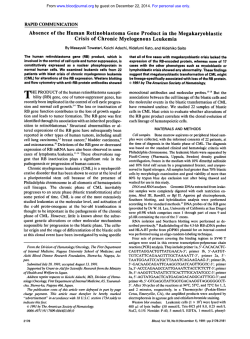
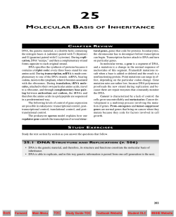
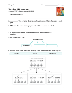
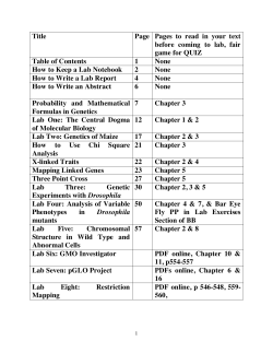
© Copyright 2026 Paperzz