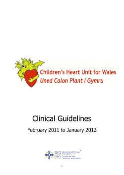
EXPERIMENTAL STUDY
Analysis of cardiac and pulmonary injuries resulting
from an experimental penetrating thoracic injury
Mahmut Tokur, M.D.,1 Mehmet Ergin, M.D.,2 Mehmet Okumuş, M.D.3
1
Department of Thoracic Surgery, Sutcu Imam University Faculty of Medicine, Kahramanmaras;
2
Department of Emergency Medicine, Konya Necmettin Erbakan University Faculty of Medicine, Konya;
3
Department of Emergency Medicine, Sutcu Imam University Faculty of Medicine, Kahramanmaras
ABSTRACT
BACKGROUND: This study was planned to analyze the internal and external anatomical findings of cardiac injuries and the presence
of accompanying pulmonary injuries in intentionally inflicted thoracic injuries to swine models.
METHODS: We inflicted a penetrating heart injury in six suis domesticus female swine models. Two cardiac injuries, one on the left
paratracheal of fourth intercostal space (ICS) and the other on the right side were inflicted on each model by the same researcher
using a 20-cm long scalpel. All animals were then sacrificed for morphological evaluation.
RESULTS: After strikes to the left fourth ICS, external evaluation showed that 50% of the subjects suffered a single laceration and
that 33% suffered multiple lacerations. Internal evaluation showed additional intracardiac injuries in all five subjects. However, the
subject that suffered a single laceration on the outer surface of the heart had multiple internal injuries while another subject that had
multiple outer lacerations had only one intracardiac injury. Only three subjects suffered cardiac injuries and only two out of those three
with pulmonary injuries after right fourth intercostal intrusions.
CONCLUSION: This experiment has shown that external evaluation of the heart tissue may not alone be sufficient to determine
the extent of cardiac injuries and accompanying pulmonary injuries caused by penetrating thoracic injuries.
Key words: Myocardial injury; penetrating; trauma.
INTRODUCTION
MATERIALS AND METHODS
Although rarely seen, penetrating heart injuries are important thoracic pathologies because they carry a high-risk for
mortality and require rapid diagnosis and treatment.[1] We
conducted this study to analyze the internal and external
anatomical findings of cardiac injuries and the presence of accompanying pulmonary injuries in intentionally inflicted thoracic injuries to swine models and explained the mechanisms
that contribute to high mortality rates in related injuries.
Overview of the Experimental Animal Groups
Address for correspondence: Mehmet Ergin, M.D.
Sille Parsana Mahallesi, Barış Caddesi, Lale Park Evlerino: 111/4,
Selçuklu, Konya, Turkey
Tel: +90 332 - 223 65 00 E-mail: [email protected]
Qucik Response Code
Ulus Travma Acil Cerrahi Derg
2014;20(4):227-230
doi: 10.5505/tjtes.2014.58538
Copyright 2014
TJTES
Ulus Travma Acil Cerrahi Derg, July 2014, Vol. 20, No. 4
Six suis domesticus type female swine models, all of which
were 1-year-old and weighed between 50 kg and 55 kg,
were used in this experiment. All animals were kept under
12 hour/12 hour light/dark cycle at a constant temperature
(22°C) and allowed free access to food and tap water. All experimental procedures were approved by the Cukurova University School of Medicine Ethical and Experimental Committee. All investigators complied with the 1996 “Guide for the
Care and Use of Laboratory Animals.” All efforts were undertaken to reduce the total number of animals required for the
experiment and to minimize their discomfort. Feeding of the
animals was stopped 24 hours prior to experiment, with water being available until 2 hours prior. Intravenous cannulations
were performed in animals through V. Auricularis lateralis and
infusion of 0.9% isotonic saline was started at the beginning
of the experiment. Atropine sulfate at 0.2 mg/kg was given to
reduce secretions prior to anesthesia. Xylazine (Rompun), at
a dose of 2 mg/kg, and ketamine, at a dose of 13 mg/kg, were
started to induce anesthesia, and this combination continued
227
Tokur et al. Analysis of cardiac and pulmonary injuries resulting from an experimental penetrating thoracic injury
the third subject (Table 1). All injured subjects had lacerations
on the frontal surface of the left ventricle, with additional
damage to coronary arteries in two of these subjects. External evaluation showed that 50% of the subjects suffered a
single laceration and that 33% suffered multiple lacerations.
Intracardiac evaluation showed additional injuries in all five
subjects. We found a partial interventricular septum injury
and a full-layer mitral valve papillary muscle injury in one
subject; partial interventricular septum injury in one subject;
partial mitral valve papillary muscle injury in one subject; and
partial left ventricle posterior wall injuries in two subjects.
Based on these observations, the subject that suffered a single laceration on the outer surface of the heart had multiple
internal injuries, while subjects that had multiple external lacerations had only one intracardiac injury. All five subjects who
had cardiac injuries also had pulmonary injuries.
to be infused throughout the experiment. Supplemental oxygen was provided to the subjects during the experiment.
Induction of Penetrating Thorax Injury and
Insertion of Catheters and Thoracotomy
Operation
Thoraces of the animals covering the sternum and ribs were
shaved and then properly disinfected using povidone iodine
(Batikon®) solution. Two cardiac injuries, one on the left
paratracheal of fourth intercostal space (ICS) and the other
on the right paratracheal of fourth ICS were inflicted on each
model by the same researcher using a 20-cm long scalpel (Fig.
1a). All animals were then sacrificed with high dose ketamine
hydrochloride. The hearts of the animals were removed for
morphological evaluation. The outer surfaces of the hearts
were opened first, followed by the left ventricles and then
the right ventricles and injury tracings were evaluated and
classified (Fig. 1b-d). Accompanying pulmonary injuries were
also detected and recorded. The data is given as a number or
percentages in the text.
Only three subjects suffered cardiac injuries resulting from
right 4th intercostal incursion, while the other three had no
injuries (Table 2). External morphological evaluation showed
that all of these injuries were to the right ventricles. One
subject had further damage to the apex cordis. On internal
evaluation, two subjects had partial interventricular septum
injuries whereas one subject had a full-layer interventricular
RESULTS
Strikes to the left fourth ICS caused cardiac injuries in all, but
(a)
(b)
Y
X
(c)
(d)
Figure 1. (a) Localization of entrance point for scalpel on both hemithorax. (b-d) The process for
morphological evaluation of cardiac findings after penetrating thorax injury.
228
Ulus Travma Acil Cerrahi Derg, July 2014, Vol. 20, No. 4
Tokur et al. Analysis of cardiac and pulmonary injuries resulting from an experimental penetrating thoracic injury
Table 1. Morphological signs explored following the injuries inflicted through the left 4th intercostal space paratracheal region
Test subject
1
234
5
6
External cardiac injuries
Left ventricle
+
+–+
+
+
Coronary artery
–
+–+
–
–
Internal cardiac injuries
Interventricular septum
+ (partial)
–
–
–
–
+ (partial)
Papillary muscle of mitral valve
+ (full layer)
–
–
–
+ (partial)
–
Posterior wall of left ventricle
–
+ (partial)
–
+ (partial)
–
–
Lung injuries
Left middle lobe
+
–
–
–
–
–
Left lower lobe
–
+
+
–
+
+
+: Injury was present; –: Injury wasn’t present.
Table 2. Morphological signs explored following the injuries inflicted through the right 4th intercostal space paratracheal region
Test subject
1
2
3
4
5
6
External cardiac injuries
Right ventricle
–
+
+
–
–
+
Apex cordis
–
+
–
–
–
–
Internal cardiac injuries
Interventricular septum
Lung injuries
–
+ (full layer)
+ (partial)
–
–
Right middle lobe
–
+
–
–
–
Right lower lobe
–
–
–
–
–
+ (partial)
+
+: Injury was present; –: Injury wasn’t present.
septum injury. Only two out of those three models had accompanying pulmonary injuries.
Injury tracing in the second subject was further investigated,
and it was found that the scalpel penetrated the right fourth
ICS and went through the right ventricle and interventricular
septum, passed the left ventricle and exited on the other side
of the heart after penetrating the apex cordis. Injuries to the
left ventricle and coronary artery were concluded to result
from left fourth ICS penetration while the injury to the apex
cordis was a result of right fourth ICS penetration.
DISCUSSION
Cardiac damage should be considered whenever injuries to
the anterior thoracic region, medial to both nipples, and between the sternal jugulum and the upper abdomen are present, unless proven otherwise.[2] In our experimental model,
we were able to observe eight cardiac injuries in 12 inflicted
penetrating injuries to the thorax.
There are intracardiac injuries reported in the literature folUlus Travma Acil Cerrahi Derg, July 2014, Vol. 20, No. 4
lowing penetrating and even blunt thoracic trauma. Tokur et
al. presented a case series of 11 penetrating cardiac injuries
that were surgically intervened.[3] In their series, they detected a valve laceration in one patient in post-surgery echocardiography, a lacerated coronary artery in another patient
during the surgery, and a left ventricle posterior wall injury in
another patient. Cha et al. reported that among 71 penetrating cardiac injury patients observed over a period of 10 years,
48 survived their injuries and 11 had post-surgery sequales.
Among their sequales were ventricular septal defects, aortic valvular damage, atrial septal defects, conduction defects,
and tricuspid valvular lesions.[4] Jenson et al.[5] reported a case
with atrial septal defects and mitral valve anterior leaflet perforation, which were detected and surgically repaired 7 years
after sustaining a penetrating cardiac injury. In another study,
Stahl et al. reported a case with tricuspid and mitral valve
papillary muscle rupture and intraventricular septal rupture
following a blunt trauma, and concluded that indirect forces
that increase intraventricular hydrostatic pressures could
cause ruptures in the atria or valves.[6] In our experiment, in
which we evaluated the tracings of the injuries, we observed
intracardiac injuries accompanying injuries to the external
surface of the heart, with the possibility of multiple intracar229
Tokur et al. Analysis of cardiac and pulmonary injuries resulting from an experimental penetrating thoracic injury
diac injuries even when only one injury was present on the
external surface.
Tokur et al.[3] reported accompanying pulmonary injuries in six
patients out of 11 who suffered penetrating cardiac injuries.
Similarly, Kaplan et al.[7] reported that in a series of 63 penetrating cardiac injury cases, 34% had accompanying pulmonary injuries. In another study, Yavuz et al.[8] reported that 54
(57.5%) out of 94 patients had isolated cardiac injuries, whereas 40 (42.5%) sustained additional injuries along with cardiac
injuries, with the most common accompanying injuries being
pulmonary injuries. Furthermore, they stated that the highest
rate of mortality was observed in groups with accompanying
pulmonary injuries. Our findings of accompanying pulmonary
injuries are in line with the findings in the literature.
Evidence in the literature shows that echocardiography performed in the emergency department to detect hemopericardium in penetrating and blunt cardiac injuries is an important
diagnostic intervention and that the use of ultrasonography reduces the time between a patient’s arrival to the hospital and
surgical intervention being performed.[9] On the other hand,
detailed examinations such as computed tomography can be
performed in only a few hemodynamically stable patients.
Echocardiography evaluation performed prior to surgery provides valuable information in valvular and atrial/ventricular
injuries. Performing transesophageal echocardiography during
the surgery, on the other hand, can be useful in evaluating intraventricular injuries and valve functions in patients for whom
pre-surgery echocardiography cannot be performed.[10]
The results of our experiment showed that evaluation of the
external surface of the heart is not sufficient to determine
the severity of cardiac injury and/or the presence of accompanying pulmonary injury in injuries sustained following penetrating thoracic injuries. Physicians should therefore consider
injuries to the external surface of the heart as the “tip of
the iceberg” and explore for additional damage. Undetected
intracardiac and pulmonary injuries may have complications
and adverse effects during the post-surgery period. All these
indicate that thorough pre-surgery preparation and surgical
exploration for damage should be performed in patients with
penetrating cardiac injuries. However, it is known that there
is not enough time, equipment, or personnel available for the
application of heart-lung machines in all cases with penetrating cardiac injuries.
Conflict of interest: The authors have no commercial associations or sources of support that might pose a conflict of
interest. All authors have made substantive contributions to
the study, and all authors endorse the data and conclusions.
There is no conflict of interest between authors.
REFERENCES
1.Campbell NC, Thomson SR, Muckart DJ, Meumann CM, Van Middelkoop I, Botha JB. Review of 1198 cases of penetrating cardiac trauma. Br
J Surg 1997;84:1737-40. CrossRef
2. Çakır Ö, Eren Ş, Balcı AE, Özçelik C, Eren N. Penetrating cardiac injuries.
[Article in Turkish] Türk Göğüs Kalp Damar Cer Derg 1999;7:112-6.
3. Tokur M, Ergin M, Kurkcuoglu C. Penetrating heart injuries and common difficulties encountered during emergency surgery. J Curr Surg
2012;2:89-95.
4. Cha EK, Mittal V, Allaben RD. Delayed sequelae of penetrating cardiac
injury. Arch Surg 1993;128:836-41. CrossRef
5. Jenson B, Kessler RM, Follis F, Wernly JA. Repair of atrial septal defect
due to penetrating trauma. Tex Heart Inst J 1993;20:241-3.
6. Stahl RD, Liu JC, Walsh JF. Blunt cardiac trauma: atrioventricular valve
disruption and ventricular septal defect. Ann Thorac Surg 1997;64:1466-8.
7. Kaplan M, Demirtaş M, Alhan C, Aka SA, Dağsalı S, Eren E, et al. Cardiac injuries: experience with 63 cases. [Article in Turkish] Türk Göğüs
Kalp Damar Cer Derg 1999;7:287-90.
8. Yavuz C, Çil H, Başyiğit İ, Demirtaş S, İslamoğlu Y, Tekbaş G, et al.
Factors affecting mortality in penetrating cardiac injuries: our 10-year.
Turkish J Thorac Cardiovasc Surg 2011;19:337-43. CrossRef
9. Rozycki GS, Feliciano DV, Ochsner MG, Knudson MM, Hoyt DB, Davis
F, et al. The role of ultrasound in patients with possible penetrating cardiac
wounds: a prospective multicenter study. J Trauma 1999;46:543-52. CrossRef
10. Meyer DM, Jessen ME, Grayburn PA. Use of echocardiography to detect occult cardiac injury after penetrating thoracic trauma: a prospective
study. J Trauma 1995;39:902-9. CrossRef
DENEYSEL ÇALIŞMA - ÖZET
OLGU SUNUMU
Deneysel penetran toraks yaralanmasına bağlı kardiyak ve pulmoner yaralanmaların analizi
Dr. Mahmut Tokur,1 Mehmet Ergin,2 Mehmet Okumuş3
1
2
3
Sütçü İmam Üniversitesi Tıp Fakültesi, Göğüs Cerrahisi Anabilim Dalı, Kahramanmaraş;
Konya Necmettin Erbakan Üniversitesi Tıp Fakültesi, Acil Tıp Anabilim Dalı, Konya;
Sütçü İmam Üniversitesi Tıp Fakültesi, Acil Tıp Anabilim Dalı, Kahramanmaraş
AMAÇ: Bu çalışmada, domuz deneklerde kasten yapılan torasik yaralanma sonucu meydana gelen kardiyak yaralanmaların iç ve dış anatomik bulguları ile eşlik eden pulmoner yaralanmaların incelenmesi amaçlandı.
GEREÇ VE YÖNTEM: Biz altı adet suis domesticus cinsi dişi domuz denekte penetran kardiyak yaralanma oluşturduk. Aynı araştırmacı tarafından
20 cm uzunluğundaki bistüri ile biri sağ paratrakeal 4. interkostal aralıktan (İKA) ve diğeri sol paratrakeal 4. İKA’dan olmak üzere iki adet penetran
kardiyak yaralanma gerçekleştirildi. Tüm hayvanlar morfolojik inceleme için sakrifiye edildi.
BULGULAR: Sol 4. İKA’dan uygulanan darbe neticesinde dış yüzey incelemesi %50’sinde tek ve %33.3’ünde birden fazla kesi oluştuğunu gösterdi. İç
yüzey incelemesi ise beş denekte ek intrakardiyak yaralanma olduğunu gösterdi. Ancak dış yüzeyde tek yaralanması olan denekte birden çok iç yüzey
yaralanması, dış yüzeyde birden çok yaralanması olan denekte ise bir adet intrakardiyak yaralanma tespit edildi. Sağ 4. İKA’dan uygulanan darbe
sonucunda ise üç denekte yaralanma oluştuğu ve üç domuzdan iki tanesinde akciğer yaralanmasının mevcut olduğu görüldü.
TARTIŞMA: Bu deney, penetran toraks yaralanmaları sonucu oluşan kardiyak hasarın şiddetini ve eşlik eden akciğer yaralanmasını ortaya koymada,
sadece kalp dış yüzey bulgularının yeterli olamayacağını işaret etmektedir.
Anahtar sözcükler: Miyokardiyal hasar; penetran; travma.
Ulus Travma Acil Cerrahi Derg 2014;20(4):227-230
230
doi: 10.5505/tjtes.2014.58538
Ulus Travma Acil Cerrahi Derg, July 2014, Vol. 20, No. 4
© Copyright 2026 Paperzz