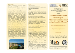
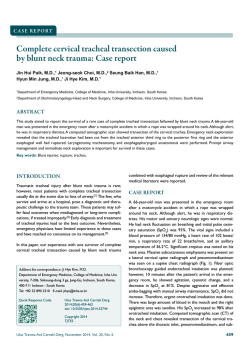
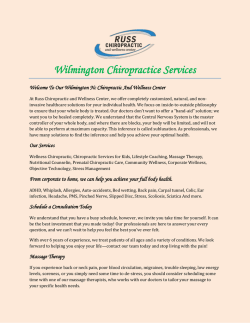
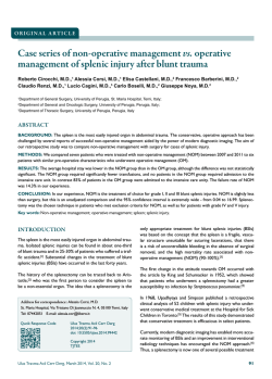
Case Report / Olgu Sunumu
Metatarsal Neck Fractures Are not Always Isolated
Injuries: Case Report of a Lisfranc Injury
Metatarsal Boyun Kırıkları Her Zaman İzole Yaralanmalar Değildir: Lisfranc Yaralanması Olgu Sunumu
Baran Kömür1, Serhat Mutlu1, Barış Yılmaz2, Harun Mutlu3, Nurettin Heybeli1
Clinic of Orthopedics and Traumatology, Kanuni Sultan Süleyman Training and Research Hospital, İstanbul, Turkey
Clinic of Orthopedics and Traumatology, Fatih Sultan Mehmet Training and Research Hospital, İstanbul, Turkey
3
Clinic of Orthopedics and Traumatology, G.O.P Taksim Training and Research Hospital, İstanbul, Turkey
1
2
ABSTRACT
A 40-year-old male presented to the emergency room with tenderness, swelling, and ecchymosis in the middle/medial and dorsal aspects of his right
foot after falling from a height of about 1 meter at work. He had a medial cuneiform bone fracture-dislocation, first metatarsal base luxation, displaced
second metatarsal neck fracture, and nondisplaced third metatarsal neck fracture. The Lisfranc fracture-dislocation was treated with two cannulated
screws using a dorsomedial approach. The displaced second metatarsal fracture was treated by open reduction and internal fixation using a Kirschner
wire. The nondisplaced third metatarsal fracture was treated conservatively. (JAREM 2014)
Key Words: Luxation, Lisfranc injury, metatarsal neck fracture, medial cuneiform fracture
ÖZET
Kırk yaşında erkek hasta 1 metre yüksekten düşme sonrası sağ ayak ortasında medial ve dorsal bölgede hassasiyet, şişlik ve ekimoz ile acil servise
başvurdu. Medial kuneiform kırıklı çıkığı, birinci metatars bazis luksasyonu, deplase ikinci metatars boyun kırığı ve non-deplase üçüncü metatars boyun
kırığı mevcuttu. Lisfranc kırıklı çıkığı dorsomedial yaklaşımla 2 adet kanüllü vida ile tespit edildi. Deplase ikinci metatars boyun kırığı açık redüksiyon ve
1 adet Kirschner teli ile fikse edildi. Nondeplase üçüncü metatars boyun kırığının tedavisine ise konservatif kalındı. (JAREM 2014)
Anahtar Sözcükler: Luksasyon, Lisfranc yaralanması, metatarsa boyun kırığı, medial kuneiform kırığı
INTRODUCTION
Tarsometatarsal injuries that lead to separation between the midand forefoot are called Lisfranc injuries, named after the French
surgeon Jacques Lisfranc de St. Martin who described tarsometatarsal amputations in the Napoleonic Era (1). A Lisfranc injury is a
subtle problem that can easily escape notice. Almost half of all tarsometatarsal joint injuries are diagnosed and treated late because
they are rare and have various forms. Although the injuries to these
joints are usually ligamentous, they can involve bone tissue. The
injuries that involve bone tissue are called fracture-dislocations
and they usually result from axial and rotational forces in highenergy trauma, such as falls from height and motorcycle accidents
(2). Their incidence is 1 per 60.000 annually (3). This paper reports a
Lisfranc injury accompanying metatarsal neck fractures. In Lisfranc
injuries occurring with metatarsal base fractures, co-occurring
metatarsal neck fractures have never been reported.
CASE PRESENTATION
A 40-year-old male presented to the emergency room after injuring his right foot at work after falling 1 m on a plantar flexed foot.
His vital signs were stable and he had an isolated injury of his
right foot. The physical examination revealed pain, swelling, and
ecchymosis at the proximal metatarsophalangeal joint, with tenderness and swelling broadly on the dorsal aspect of the foot and
locally over the second and third metatarsals. The neurovascular
examination showed no open wounds and no distal neurovascuAddress for Correspondence / Yazışma Adresi: Dr. Harun Mutlu, Clinic
of Orthopedics and Traumatology, G.O.P Taksim Training and Research
Hospital, İstanbul, Turkey
Phone: +90 555 623 26 23 E-mail: [email protected]
lar pathology. The radiological evaluation showed second and
third metatarsal neck fractures (Figure 1). Comparative radiography and computed tomography (CT) showed a nondisplaced
medial cuneiform fracture and Lisfranc injury of the proximal
metatarsophalangeal joint (Figure 2). A short-leg cast was applied because of the swelling, and the patient was admitted for
elevation and ice application. To reduce the chance of compartment syndrome developing, the surgery was delayed for 2 days,
until the edema had regressed. At surgery, the periosteum on
the cuneiform bone was scraped off via a dorsomedial approach.
The cuneiform fracture and first metatarsal dislocation were reduced. Using a Kirschner wire (K-wire), two 4.0 mm cannulated
screws were inserted via fluoroscopic guidance at a 45° angle
through the medial cuneiform fragments, fixing the Lisfranc dislocation. The second metatarsal fracture was approached via a
dorsal incision in the web between the second and third metatarsals. The extensor tendon was dissected and the fracture was
reduced with a K-wire (Figure 3). The third metatarsal was not
fixed because it appeared stable. A short-leg plaster was applied
after checking the reduction using fluoroscopy. Three weeks later, the K-wires were removed after observing metatarsal bone
union. Once union was seen radiographically, the patient was allowed gradually increasing weight-bearing. After the sixth week,
the plaster cast was removed to let the foot contact the ground
for full weight-bearing with full range of motion. The patient returned to work and at the 1-year follow-up his daily life activities
were as they were before the injury.
Received / Geliş Tarihi: 16.02.2014
Accepted / Kabul Tarihi: 11.03.201
Çevrimiçi Yayın Tarihi / Available Online Date: 17.06.2014
© Telif Hakkı 2014 AVES Yayıncılık Ltd. Şti. Makale metnine www.jarem.org web sayfasından ulaşılabilir.
© Copyright 2014 by AVES Yayıncılık Ltd. Available online at www.jarem.org
DOI: 10.5152/jarem.2014.483
Kömür et al.
Case Report of a Lisfranc Injury
a
a
Figure 1. a, b. (a) Anteroposterior (AP) and (b) oblique X-rays showing the Lisfranc injury and metatarsal neck fracture
DISCUSSION
The Lisfranc joint complex comprises three columns in the foot:
the medial column is formed by the first metatarsal and medial
cuneiform; the central column is formed by the second and third
metatarsals and the intermediate and lateral cuneiforms; and the
lateral column consists of the cuboid and its relationship with the
fourth and fifth metatarsals. In the coronal plain, the transverse
arc of the midfoot is stable. Although many ligaments contribute
to this stability, the most important contribution is that of the Lisfranc ligament between the medial cuneiform and second metatarsal; this is the strongest of these ligaments (4).
Diagnosis of a Lisfranc injury begins with the patient’s history and
physical examination. The time of the injury and time elapsed
should be determined carefully to choose among treatment alternatives. A Lisfranc injury should be considered when plantar
ecchymosis is seen. The patient should be monitored closely
for compartment syndrome. A radiological and CT (if necessary)
study must be performed (5).
Lisfranc injuries usually result from one of three mechanisms: lateral translocation of a metatarsal caused by forced abduction of
the forefoot; axial weight-bearing when the toes are dorsiflexed
and ankle is in the equinus position; and crush injury trauma involving plantar translocation caused by force acting on the dorsal metatarsal (6). Our patient described a fall from height with
the toes dorsiflexed and ankle plantar flexed (the second type
described above). A fracture dislocation of the base of the first
metatarsal usually occurs after such trauma. In this case, there
was a fracture dislocation between the first metatarsal and me-
Figure 2. Computed tomography of the medial cuneiform fracture
dial cuneiform and neck fractures of the second and third metatarsals. In our case, the metatarsal neck fractures were probably
caused by redirection of the axial force due to falling by the lifts
in the front of his work shoes, which could have resulted in a rotational axial force to the forefoot and metatarsals.
Plaster casting for 6~12 weeks is recommended for non-displaced
Lisfranc injuries. Surgery should be considered for displaced injuries, because of the chance of developing post-traumatic midfoot
arthrosis or progressive dislocation and displacement. Closed reduction may be used for displaced dislocations and a K-wire may
be used when this fails. The presence of bone fragments and entrapped soft tissues might prevent the reduction (7). Surgery should
wait until any edema regresses, to reduce the risk of developing
compartment syndrome. In our case, we delayed surgery for 2 days.
There are various surgical treatments for Lisfranc injuries. The
current treatment is to reduce the medial and central column
injuries using K-wires and to ensure osteosynthesis using cannu-
Kömür et al.
Case Report of a Lisfranc Injury
a
a
Figure 3. a, b. Fixation using two cannulated screws and a K-wire: (a) AP and (b) oblique radiographs
lated screws (2, 4, 7). It is crucial to restore the Lisfranc ligament
between the middle cuneiform and second metatarsal. When the
lateral column is injured, fixation of the fourth and fifth metatarsals to the cuboid bone is advised (8). In our case, the Lisfranc injury was reduced using K-wires and osteosynthesis was promoted
using two cannulated screws. The second metatarsal fracture was
also fixed using a K-wire. In some Lisfranc injuries, the metatarsal
joint is luxated both proximally and distally, producing a “swimming metatarsal” (6). A Lisfranc injury accompanied by multiple
metatarsal neck fracture-dislocations has also been reported (9).
However, ours is the first reported Lisfranc injury accompanied by
metatarsal neck fractures without luxation.
CONCLUSION
After diagnosing common isolated injuries, such as metatarsal
neck fractures, problems that have a high probability of going
unnoticed, such as a Lisfranc injury, should be considered. No
consensus has been reached on whether to fix these using screws
or K-wires. In our opinion, using a compression screw better promotes full recovery.
Informed Consent: Written informed consent was obtained
from the patient who participated in this case.
Peer-review: Externally peer-reviewed.
Author Contributions: Concept - B.K., N.H.; Design - H.M.; Supervision - S.M., B.Y.; Funding - S.M., H.M., B.K.; Materials - N.H.,
B.K.; Data Collection and/or Processing - H.M., S.M.; Analysis
and/or Interpretation - B.Y., N.H.; Literature Review - H.M.; Writing - B.K., N.H.; Critical Review - S.M., B.Y., N.H.
Conflict of Interest: No conflict of interest was declared by the
authors.
Financial Disclosure: The authors declared that this study has
received no financial support.
Hasta Onamı: Yazılı hasta onamı bu olguya katılan hastadan
alınmıştır.
Hakem değerlendirmesi: Dış bağımsız.
Yazar Katkıları: Fikir - B.K., N.H.; Tasarım - H.M.; Denetleme S.M., B.Y.; Kaynaklar - S.M., H.M., B.K.; Malzemeler - N.H., B.K.;
Veri toplanması ve/veya Processing - H.M., S.M.; Analiz ve/veya
yorum- B.Y., N.H.; Literatür taraması - H.M.; Yazıyı yazan - B.K.,
N.H.; Eleştirel İnceleme - S.M., B.Y., N.H.
Çıkar Çatışması: Yazarlar çıkar çatışması bildirmemişlerdir.
Finansal Destek: Yazarlar bu çalışma için finansal destek
almadıklarını beyan etmişlerdir.
REFERENCES
1. Görgeç M, Esenkaya İ, Nalbantoğlu U, Türkmen M. Lisfranc eklem
yaralanmaları ve tedavisi Acta Orthop Traumatol Turc 1994; 28: 240-4.
2. Scolaro J, Ahn J, Mehta S. Lisfranc fracture dislocations Clin Orthop
Relat Res 2011; 469: 2078-80. [CrossRef]
3. Hardcastle PH, Reschauer R, Kutscha-Lissberg E, Schoffmann W. Injuries to the tarsometatarsal joint: incidence, classification and treatment. J Bone Joint Surg Br 1982; 64: 349-56.
4. Miswan MFM, Singh VA, Yasin NF. Outcome of surgically treated
Lisfranc injury: A review of 34 cases. Ulus Travma Acil Cerrahi Derg
2011; 17: 504-8. [CrossRef]
Kömür et al.
Case Report of a Lisfranc Injury
5.
Ross G, Cronin R, Hauzenblas J, Juliano P. Plantar ecchymosis sign: a
clinical aid to diagnosis of occult Lisfranc tarsometatarsal injuries. J
Orthop Trauma 1996; 10: 119-22. [CrossRef]
6. Singh PA, Singh PA, Chadha M. Lisfranc kırıklı çıkık ile birlikte ters
yüzen birinci metatars ve yüzen üçüncü metatars: Nadir bir yaralanma. Acta Orthop Traumatol Turc 2010; 44: 169-71. [CrossRef]
7. Stavlas P, Roberts CS, Xypnitos FN, Giannoudis PV. The role of reduction and internal fixation of Lisfranc fracture-dislocations: A sys-
tematic review of the literature. International Orthopaedics (SICOT)
2010; 34: 1083-91. [CrossRef]
8. Sangeorzan BJ, Benirschke SK, Gould MT. Tarsometatarsal Lisfranc
injuries: evaluation and management. In: Wiss D, ed. Master Techniques of Orthopaedic Surgery: Fractures. 2nd edition. Philadelphia,
PA: Lippincott-Williams&Wilkins, 2006: 605-17.
9. Drosos GI, Gavalas NS, Papoutsidakis AN. Concomitant Lisfranc
fracture-dislocation and displaced neck fractures of the lesser metatarsals: A case report. J Foot Ankle Surg 2000; 6: 49-54. [CrossRef]
© Copyright 2026 Paperzz