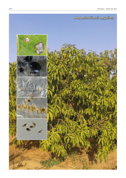
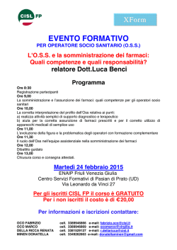
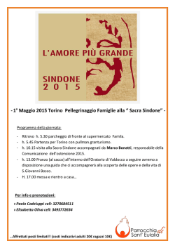
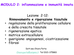
I Nematol.
medit. (1977), 5: 305-311.
I
Laboratorio di N ematologia agraria del C.N.R.
and
Istituto di Botanica dell'Universita - 70126 Bari
CELL WALL BREAKDOWN AND CELLULAR RESPONSE
IN DEVELOPING GALLS INDUCED BY LONGIDORUS APULUS
by
TERESA BLEVE-ZACHEO, G. ZACHEO, F. LAMBERTI and O. ARRIGONI
Some aspects of the histological and histochemical modifications
induced by Longidorus apulus Lamberti et Bleve-Zacheo in the roots
of celery (Apium graveolens L.) have been reported previously (BleveZacheo et al., 1977). Further studies, reported here, have extended
the information on cell response to L. apulus feeding particularly
with reference to morphological and ultrastructural changes.
MATERIALS AND METHODS
Celery seedlings, one month old, were transplanted into 100 ml
plastic pots containing steam sterilized sand. One hundred female
L. apulus, from a population reared on celery in the glasshouse,
were added in a small volume of water to the root zone of the plants
when transplanted. The pots were then placed in a controlled-temperature chamber at 22 + 1" C. Seven and 15 days after inoculation
the damaged root apices were excised, fixed for 2 h in 3% glutaraldehyde in 0.1 M phosphate buffer at pH 7.2, washed overnight in
buffer and postfixed for 2 h in 1 % osmium tetroxide, at 4° C. Alternatively, the apices were fixed for 2 h in 2% potassium permanganate in
phosphate buffer at pH 7. Both types of fixed roots were dehydrated
in a graded ethanol-propylene oxide series and embedded in Araldite.
Sections 2 [Lm thick were stained in an aqueous solution of
toluidine blue for light microscopy. Ultrathin sections were stained
11
305
1 h in uranyl acetate and post-stained 10' in lead citrate (Reynolds,
1963) and examined at 60 kV in a JEM 100B electron microscope.
RESULTS
The feeding site of L. apulus on celery roots was limited to the
apical meristematic area. The odontostyle was inserted perpendicular
to the root axis and usually penetrated six to seven cell layers. Cells
in the vicinity of the feeding site were swollen and the cytoplasm
became granular and stained intensely with toluidine blue. The cells
around the feeding site became prematurely hypertrophic with the
nuclei appressed to the cell walls (Fig. 1, a and b). In the area of
the root, opposite the site of odontostyle insertion, cell division was
active but disordered, resulting in disorganization of the tissues.
This abnormal tissue proliferation resulted in the formation of a gall.
Some slight disorganization of the root tissue also occurred for a
distance of a few millimetres immediately above the gall.
The hypertrophic cells formed near the point of odontostyle
insertion ceased division; the cells had little cytoplasm and the walls
became convoluted. On the opposite side of the root the cell walls
appeared normal but the cytoplasm was dense with many organelles,
abundant vacuoles and some were binucleate (Fig. 1, c).
The procambium may be modified in two ways. When the penetration of the odontostyle is limited to the cortical meristem, as
observed in cross section, the procambial cells were polygonal and
small near the site of insertion and larger on the opposite side;
later, when cellular differentiation occurred, as a result of nematode
feeding, the pericycle cells underwent a periclynal division and later
became hypertrophic, causing compression of the endodermis (Fig.
1 d).
When the odontostyle of the nematode reached the procambial
cells, these first became hypertrophic, then a process of lysis of the
cell wall started which led to the formation of a syncytia like structure.
Some of the cell walls were intact, but many were disintegrated and
associated with other cultural material that has been lysed. This
syncytium was multinucleate; it appeared to progressively absorb
adjacent hypertrophic cells with vescicolous nuclei (Fig. 2, a, b, c).
A similar process has also been observed in the cortical meristem,
where whole layers of cells, with dotted walls were present, from
-
306-
Fig. 1 - a: cross section of celery roots with cells damaged by the feeding
action of Longidorus apulus (fs); b: feeding site (fs ); c: feeding site of the nema~
tode with hyp ertrophic cells (ipe) compared with normal meristematic cells (me);
d: abnormal growth of the pericycle with consequent compression of the endodermis; e: cell wall breakdown.
Fig. 2 - a, b (electron micrograph), and c: lysigen cavities (Ie) showin g partially dissolved cell wa lls and several nucl ei; cells surrounding the cavi ties have
thi ckened walls and dense cytoplasm w ith filling bodies (fb); d and e : hypertrophic multinucl ea te cells.
Fig. 3 - a, b, and c: e lectron micrographs showin g breakdown (b) and di ssolution of the cell walls (arrows) 6500 x.
the epidermis to the central cylinder (Fig. 1, e). The breakdown of
the cell walls resulted in the formation of cavities along the cortical
meristem (Fig. 2, d, e). Electron microscopy observations indicated
that there were no preferential sites from which the dissolution of
the cell wall started.
The cell walls delimiting the syncytium were always thickened
with a rather irregular inner profile. Electron dense material was
present near the sites of lysis and this presumably originated from
the wall dissolution (Fig. 3, a, b, c).
In roots which were severely damaged after 15 days exposure
to the nematode feeding hypertrophic and sometimes multinucleate
cells of the cortical meristem were adjacent to the syncytia; 6 to 8
nuclei have been counted (Weischer and Wyss, 1976). The cytoplasm
of these cells stained more intensively with toluidine blue than less
damaged cells.
DISCUSSION
The structural modifications observed in root cells of celery
attacked by the ectoparasitic L. apulus differ little from those described
for endo- and semi-endoparasitic nematodes (Bird, 1972; Endo, 1975;
Jones and Gunning, 1976; Rebois et aI., 1976). Although there is a
structural similarity in response to nematode feeding, we consider
that the physiological response is different. Feeding by endo - and
semi endoparasitic nematodes causes an intense flux of metabolites
through the modified cells, which can thus be considered as true
transfer cells. The syncytia caused by the trophic action of L. apulus,
however, appear to act more as lysigen cavities, which are an intermediate step to the complete destruction of the root apex.
ACKNOWLEDGMENTS
The cooperation of Mr. Emanuele Olivieri who has worked on
this matter preparing his thesis is gratefully appreciated.
-
310-
SUMMARY
Studies on the morphological and ultrastructural modifications caused by
Longidorus apulus Lamberti et Bleve-Zacheo, in celery (Apium graveolens L.)
root cells indicate that the nematode feeds upon the meristematic cells by inserting its odontostyle perpendicularly to the root axis. In response to feeding the
root apex develops asymmetrically because of an increase of cell division in the
area opposite the insertion of the odontostyle; on the side where the odontostyle
penetrates cell walls dissolve and syncytia are formed.
RIASSUNTO
Alterazioni cellulari indotte da Longidorus apulus in galle di radici di Sedano.
Studi sulIe alterazioni cellulari in galle, indotte da Longidorus apulus Lamberti et Bleve-Zacheo in radici di Sedano (Apium graveolens L.), indicano che il
nematode si nutre su cellule meristematiche introducendo l'odontostile perpendicolarmente all'asse della radice. In risposta, l'apice radicale si sviluppa asimmetricamente poiche, nell'area opposta a quella d'inserzione dell'odontostile,
si ha una divisione cellulare. Nel punto di penetrazione dell'odontostiIe Ie pareti
cellulari si dissolvono e si formano delle cellule sinciziali.
LITERATURE
CITED
BIRD A. F., 1972 - Cell wall breakdown during the formation of syncytia induced in
plants by root-knot nematodes. Int. 1. Parasit., 2: 431-432.
BLEVE-ZACHEO T., ZACHEO G. e LAMBERTI F., 1977 - Reazioni istologiche ed istochimiche indotte da Longidorus apulus in radici di Sedano e Cicoria. N ematol.
medit., 5: 85-92.
ENDO B. Y., 1975 - Pathogenesis of nematode-infected plants. An. Rev. Phytopath.,
13: 213-238.
JONES M. G. K. and GUNNING B. E. S., 1976 - Transfer cells and nematode induced
giant cells in Helianthemum. Protoplasma, 87: 275-279.
REBOIS R. V., MADDEN Ph. A. and ELDRIDGE B. J., 1975 - Some ultrastructure changes
induced in resistant and susceptible soybean roots following infection by
Rotylenchulus renitormis. 1. NematoI., 7: 122-139.
REYNOLDS E. S., 1963 - The use of lead citrate at high pH as an electron-opaque
stain in electron microscopy. 1. Cell BioI., 17: 208-212.
WEI SCHER B. and WySS U., 1976 - Feeding behaviour of Xiphinema on grape.
Nematologica, 22: 319-325.
Accepted for publication on 18 October 1977.
-
311-
© Copyright 2026 Paperzz