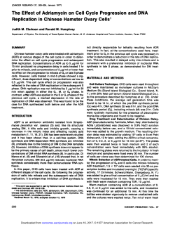
270 Persoonia – Volume 35, 2015 Neopestalotiopsis egyptiaca 271 Fungal Planet description sheets Fungal Planet 372 – 4 December 2015 Neopestalotiopsis egyptiaca A.M. Ismail, G. Perrone & Crous, sp. nov. Etymology. Name reflects the country Egypt where the fungus was collected. Classification — Sporocadaceae, Xylariales, Sordariomycetes. Conidiomata pycnidial, globose, formed on PDA within 7 d, mostly solitary, scattered, semi-immersed or erumpent, to 300 μm diam; releasing slimy, black conidial masses. Conidio phores septate, branched at base, sometimes reduced to conidiogenous cells, hyaline, smooth-walled, septate, up to 42 μm long. Conidiogenous cells discrete, cylindrical, hyaline, smooth, proliferating 2 – 3 times percurrently at apex, 15–25 × 3–5 μm. Conidia smooth, fusiform, straight and sometimes slightly curved, 4-septate consisting of three thick-walled pale to dark brown median cells, of which the upper two are darker brown than the lower cell (these cells also tend to be finely roughened), and two thin-walled pale to dark olivaceous apical and basal cells, (22.5 –)23 – 26(– 28) × 6 –7.5 μm. Apical cell giving rise to 2 – 3 unbranched, tubular, hyaline appendages, (15–)16.5 – 21(– 25) μm long; basal cell with a single, hyaline, centric, unbranched appendage, 4.5 –7.5 μm long. Culture characteristics — On PDA colonies reached up to 90 mm diam after 10 d at 25 °C with smooth edge, whitish, slightly raised, circular appearance, with sparse to moderate aerial mycelium on the surface with black, scattered conidio mata. On reverse, olivaceous, with distinct zonation. Notes — Based on a recent multi-locus phylogenetic study of Pestalotiopsis, Maharachchikumbura et al. (2014) divided the complex into three genera: Pestalotiopsis, Neopestalotiopsis and Pseudopestalotiopsis. Morphologically, Neopestalotiopsis can be easily distinguished from Pseudopestalotiopsis and Pestalotiopsis by its versicolorous median cells. Conidiophores in Neopestalotiopsis are indistinct and often reduced to coni diogenous cells. Based on the phylogenetic analysis of the sequence data of tub2, tef1 and ITS, N. egyptiaca is phylogenetically closely related to N. australis. However, the conidia of N. australis are wider and the median cells are darker than those of N. egyptiaca. Typus. Egypt, Ismailia, on leaves of Mangifera indica (Anacardiaceae), Apr. 2014, A.M. Ismail (holotype CBS H-22294, culture ex-type CBS 140162 = CPC 26132, CBS 140163 = CPC 26133; ITS sequence GenBank KP943747, LSU sequences GenBank KT950860, KT950861, tub2 sequence GenBank KP943746, tef1 sequence GenBank KP943748, MycoBank MB813837). Colour illustrations. Mangifera indica in Egypt; symptomatic leaf, colony on PDA, conidiophores giving rise to conidia and appendaged conidia. Scale bar = 10 µm. Ahmed M. Ismail, Plant Pathology Research Institute, Agricultural Research Center, 12619 Giza, Egypt; e-mail: [email protected] Giancarlo Perrone & Donato Magista, Istituto di Scienze delle Produzioni Alimentari, Via Amendola 122/O, 70126 Bari, Italy; e-mail: [email protected] & [email protected] Pedro W. Crous & Johannes Z. Groenewald, CBS-KNAW Fungal Biodiversity Centre, P.O. Box 85167, 3508 AD Utrecht, The Netherlands; e-mail: [email protected] & [email protected] © 2015 Naturalis Biodiversity Center & Centraalbureau voor Schimmelcultures
© Copyright 2026 Paperzz