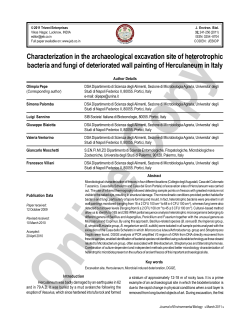
FEMS Microbiology Letters 233 (2004) 341–346
www.fems-microbiology.org
Cerato-platanin protein is located in the cell walls of ascospores,
conidia and hyphae of Ceratocystis fimbriata f. sp. platani
Silvia Boddi a, Cecilia Comparini b, Roberto Calamassi a, Luigia Pazzagli c,
Gianni Cappugi c, Aniello Scala b,*
b
a
Dipartimento di Biologia Vegetale, Universita di Firenze, Piazzale delle Cascine 28, 50144 Firenze, Italy
Dipartimento di Biotecnologie Agrarie, Sezione di Patologia Vegetale, Universita di Firenze, Via della Lastruccia 10, 50019 Sesto Fiorentino, Italy
c
Dipartimento di Scienze Biochimiche, Universita di Firenze, Viale Morgagni 50, 50134 Firenze, Italy
Received 23 December 2003; received in revised form 1 March 2004; accepted 2 March 2004
First published online 10 March 2004
Abstract
Cerato-platanin (CP), a protein of about 12.4 kDa from Ceratocystis fimbriata f. sp. platani (Cfp), accumulated in the mycelium
and was located in the cell walls of Cfp ascospores, hyphae and conidia suggesting that this protein had a role in forming the fungal
cell wall apart from the already known fact that it is secreted early in culture and elicits phytoalexin synthesis and/or plant cell death.
The finding was obtained with three immunological techniques: a quantitative ELISA which determines the amount of CP in the
mycelium, an immunofluorescence assay, and immunogold labelling to define the exact localization of CP in the Cfp cells.
Ó 2004 Federation of European Microbiological Societies. Published by Elsevier B.V. All rights reserved.
Keywords: ELISA; Immunofluorescence; Immunogold labelling; Plant–pathogen interaction
1. Introduction
Cerato-platanin (CP), a small, moderately hydrophobic protein consisting of 120 amino acids, is produced by the Ascomycete Ceratocystis fimbriata (Ell.
and Halst.) Davidson f. sp. platani Walter (Cfp) [1]. Cfp
is the causative agent of canker stain of plane, a severe
disease with a high incidence in European populations
of Platanus acerifolia (Ait.) Willd. [2,3]. CP is reported
to be released abundantly in shake culture at an early
stage, and to elicit defense-related responses, such as
phytoalexin synthesis and/or cell death, in both host and
non-host plants [1,4,5].
According to various databases (SwissProt, EMBL
and GenBankTM), CP is the reference protein of a small
protein family that includes three other proteins produced by other Ascomycota: the snodprot1 protein,
*
Corresponding author. Tel.: +39-055-4573227; fax: +39-0554573232.
E-mail address: aniello.scala@unifi.it (A. Scala).
from Phaeosphaeria nodorum (SwissProt Accession no.
O74238), the allergen Asp f 15 precursor (Asp f 13),
from Aspergillus fumigatus (SwissProt O60022), and the
heat-stable 19 kDa antigen (CS-Ag), from Coccidioides
immitis (SwissProt Q00398). In addition, Wilson et al.
[6] have recently characterized the gene sp1 from Leptosphaeria maculans; this gene encoded a secreted protein closely related to the CP protein family. All these
proteins are characterized by high sequence homology,
but not always by clear functional similarities. However,
they are all secreted, and in some cases seem to be involved in biological recognition phenomena. Snodprot1
is produced during infection of wheat leaves by
P. nodorum [7]. The Asp f 15 precursor (Asp f 13) has
been characterized together with many other allergens
from A. fumigatus in order to find a serological procedure to diagnose allergic bronchopulmonary aspergillosis, a severe disease of the lungs in humans caused by
A. fumigatus [8]. CS-Ag is one of a class of trypsin-like
serine proteinases produced by both the saprophytic and
the parasitic phases of C. immitis, causing San Joaquin
0378-1097/$22.00 Ó 2004 Federation of European Microbiological Societies. Published by Elsevier B.V. All rights reserved.
doi:10.1016/j.femsle.2004.03.001
342
S. Boddi et al. / FEMS Microbiology Letters 233 (2004) 341–346
Valley fever or coccidioidomycosis, a human respiratory
disease, and was proposed as a Coccidioides-specific
antigen for the diagnosis of this fungus [9]. The recombinant SP1 protein induced an autofluorescence
response on host leaves [6].
Cerato-platanin shares some structural and functional characteristics with the hydrophobin family: (i) a
low molecular mass; (ii) hydrophobicity; (iii) the sequence Cys–Ser–Asn is aligned with the signature sequence of the hydrophobins (Cys–Cys–Asn), except for
the conservative substitution Cys ! Ser; (iiii) the high
homology of the N-terminal region of CP with that of
cerato-ulmin, a hydrophobin from Ophiostoma spp. [10–
12]. However, CP differs from the hydrophobins in
having only four cysteines, all involved in two disulphide
bonds, and in having a distinctive hydropathicity profile,
unlike that of both class I and class II hydrophobins.
In the present paper we show that CP is located in the
mycelial cell walls of Cfp by three different methods, all
based on the immunorelationship between CP and a CPspecific rabbit antiserum.
2. Materials and methods
2.1. Fungal cultures
The origin of the reference Cfp strain Cf AF 100 has
been previously described [1]. Details of the other Cfp
strains (CF 3, CF 5, CF 6, CF 7, CF 8, CF 9, CF 11, CF
12, CF 15, CF 16, CF 17, CF 18, CF 22, CF 23, CF 24,
CF 25, CF 27, CF 42) are given in Santini and Capretti
[13]. The strains were routinely cultured on potato
dextrose agar (PDA) (Difco, Detroit, MI, USA). For
long-term storage, conidia and mycelial fragments collected from 4-day-old liquid shaken mini cultures (3 ml)
in potato dextrose broth (PDB) were resuspended in
10% (v/v) glycerol and stored at )70 °C.
In the experiments described below, mycelial fractions including aerial hyphae and hyaline and cylindrical
conidia were carefully collected from the surface of a 6day-old culture, centrifuged at 5000g for 10 min, washed
three times with 0.9% (w/v) NaCl and twice with distilled water, and then resuspended to obtain the appropriate concentrations of hyphae and conidia. The
ascospores were collected from mature perithecia produced on the surface of 2–3-week-old cultures on PDA,
and diluted in octane.
2.2. Purification of CP and antiserum preparation
Cerato-platanin was purified from culture filtrate of
Cfp strain Cf AF 100 according to the procedure in
Pazzagli et al. [1]. Rabbit CP-specific antiserum was
prepared according to Scala et al. [9], divided into 1 ml
aliquots and stored at )20 °C. Pre-immune serum was
collected from a rabbit before the first injection and
stored at )20 °C to be used as a negative control.
2.3. Quantitative ELISA
The amount of CP in Cfp mycelium was quantified by
ELISA. A freeze-dried aliquot (10 mg) of each sample
was extracted for 2 min with 500 ll of 60% (v/v) ethanol
and centrifuged for 5 min at 13000g. The supernatant
was dried under vacuum. The residue was solubilized in
500 ll phosphate buffered saline (PBS), 0.1 M, pH 7.2,
and used for coating the wells (50 ll per well) (Falcon
3911 Microtest flexible plates, Becton Dickinson Labware, Oxnar, CA, USA) in the ELISA. The culture filtrates were serially twofold diluted, from 1:1 to 1:8; 50 ll
samples of each dilution were added in triplicate to the
wells and maintained at 37 °C for 3 h. Serial dilutions of
CP and uninoculated PDB were used as positive and
negative controls respectively. The wells were washed
three times with cold PBS and any remaining binding
sites were blocked with 50 ll per well of 0.5% (w/v)
gelatin in PBS for 2 h at 37 °C. After saturation, 50 ll of
CP antiserum, diluted 1:1000 in PBS containing 0.25%
(w/v) gelatin and 0.2% (v/v) Tween 20, was added.
Rabbit pre-immune serum was used in the control wells.
After overnight incubation, the multiwell plates were
washed with cold PBS and incubated at 37 °C for 2 h in
the presence of 50 ll per well of goat anti-rabbit IgG/
peroxidase (Sigma–Aldrich, St. Louis, MO, USA) diluted 1:2000 in PBS containing 0.25% (w/v) gelatin and
0.2% (v/v) Tween 20. The wells were washed three times
with cold PBS, after which a 150 ll substrate solution
(0.4 mg ml1 o-phenylenediamine dihydrochloride,
0.012% (v/v) H2 O2 in 0.1 M citrate-phosphate buffer,
pH 5.0) was added; after 30 min of incubation at room
temperature in the dark, the optical density at 492 nm
(OD492 ) was measured with a Model 550 Microplate
reader (Bio-Rad, Hercules, CA, USA). The concentration of CP in the samples was determined using a
standard curve. The standard linear calibration curves
at OD492 vs. the log of the purified CP concentration
had a correlation coefficient >0.95 using purified CP
over concentrations ranging from 3 ng to 1 lg per well.
The concentration of 1 lg per well gave an OD492 of
about 0.700. Negative samples always yielded an
OD492 < 0:030.
2.4. Immunofluorescence assay
Aliquots (20 ll) of hyphal, conidial and ascospore
suspensions at a concentration of about 105 cells ml1
were dispensed to 12-well Multitest slides (ICN Biomedicals, Aurora, OH, USA) and dried at 60 °C. The
wells were washed with PBS, filled with 20 ll of CP
antiserum diluted 1:100 in PBS and incubated for 2 h at
room temperature. Control wells were filled with pre-
S. Boddi et al. / FEMS Microbiology Letters 233 (2004) 341–346
immune serum diluted 1:100. After washing three times
with PBS, the wells were filled with 20 ll each of a 1:40
dilution of goat antirabbit IG-fluorescein isothiocyanate
conjugate (Sigma–Aldrich) in PBS and incubated for 2 h
at room temperature. The slides were washed with distilled water and examined under a Leitz Phloemopack
2.1 microscope with an incidence light excitation system,
equipped with UV filters and a 75-W Leitz 100Z Xenon
lamp.
2.5. Immunogold labelling
Hyphae, conidia and ascospores were fixed in 2.5%
(v/v) glutaraldehyde in 0.1 M Na-phosphate buffer (pH
7.2) overnight at 4 °C. After washing in the same buffer
the material was postfixed in 1% OsO4 buffer for 1 h,
washed three times with buffer, and dehydrated in an
ethanol series of 30, 50, 70, 90 and 100% (v/v), at room
temperature. The samples were infiltrated in 2:1 (v/v)
ethanol:LR White (Sigma) for 1 h; 1:2 (v/v) ethanol:LR
White for 2 h; 100% LR White overnight at 4 °C and
finally embedded in LR White resin according to the
method of Tagu et al. [14] with modifications. Ultrathin
sections were cut with an LKB ultramicrotome, collected on formvar-coated gold grids and processed for
immunogold labelling to localize the CP. The sections,
collected on formvar-coated gold grids, were first incubated in PBS with 1% bovine serum albumin (BSA,
British BioCell International, Cardiff, UK) (pH 8.2) for
20 min. They were then incubated for 1–2 h in CP antiserum (primary antibody) diluted 1:100 in the first
buffer. The grids were washed five times for 5 min with
the buffer, then incubated for 30 min at room temper-
343
ature on a drop of goat anti-rabbit IgG antibodies
conjugated to 10 nm gold particles (Sigma) diluted 1:20
in the buffer [15] (modified). After rinsing with the buffer
and distilled water, the grids were stained with uranyl
acetate and lead citrate. Examination was in a Philips
EM 201C transmission electron microscope operating at
80 kV.
Control reactions were performed under the same
conditions using pre-immune serum instead of the CP
antiserum, or the gold-conjugated secondary antibody
(goat anti-rabbit IgG) alone.
2.6. Statistical analysis
ELISA data of the CP content of Cfp isolate mycelium were analysed by ANOVA, fixed model. Homogeneous groups were identified by means of the Tukey
HSD test.
3. Results and discussion
The cellular content of CP in Cfp mycelium, as
quantified by ELISA, was 16.61 0.42 ng mg1 fresh
weight mycelium for the reference isolate CF AF 100.
Results with the other isolates did not differ statistically
from that of the reference isolate.
The IF assay showed that CP occurred on the surface
of the conidia, hyphae and ascospores of Cfp isolate CF
AF 100 (Fig. 1). The fluorescence reaction remained
intense even after numerous washes with 80% ethanol,
suggesting that the CP was a stable component of the
Fig. 1. Immunofluorescence labelling of hyphae and conidia (A, scale bar ¼ 50 lm) and ascospores (B, scale bar ¼ 10 lm) with CP antiserum.
344
S. Boddi et al. / FEMS Microbiology Letters 233 (2004) 341–346
fungal surface. No fluorescence was observed in samples treated with rabbit pre-immune serum (data not
shown).
Immunoelectron microscopy showed unequivocally
that CP was a component of the Cfp cell walls. The
positive reaction was located on the walls of hyphae
(Fig. 2(A) and (B)), conidia (Fig. 2(C)) and ascospores
(Fig. 2(D) and (E)) of Cfp. CP was more abundant on
the ascospore and conidial wall than on the hyphal wall
(Fig. 2(C)–(E)). Labelling on the conidia was distributed
across the entire thickness of the wall, while on the ascospore wall the gold granules were located more densely on the electron-transparent layer near the
plasmamembrane than on the more outward layer.
There was labelling also on the fibrillar material at the
surface of ascospores (Fig. 2(D) and (E)). No labelling
was found on control sections where pre-immune serum
was used or where the first antibody was omitted (data
not shown).
All the experimental evidence demonstrated that CP
was a significant component of the cell walls of Cfp
conidia, hyphae and ascospores and of the fibrillar
material at the surface of Cfp ascospores. This finding
suggests that this protein may have some new structural
functions, apart from the already known fact that it is
secreted early in culture and elicits phytoalexin synthesis
and/or cell death in both host and non-host plants
[1,4,5]. CP resembles the hydrophobins, known to be
located on the surface of the conidial and hyphal walls,
where they form insoluble complexes that have a
structural role in forming aerial hyphae [16–19]. Hydrophobins are also often excreted abundantly in the
form of monomeric proteins into the culture medium by
submerged feeding hyphae, and/or they are involved in
Fig. 2. Transmission electron microscopy of immunogold labelling of Ceratocystis fimbriata f. sp. platani. A, B: Hyphae with gold granules on the
wall. Scale bar ¼ 0.3 lm. C: labelling distributed across the conidial wall. Scale bar ¼ 0.2 lm. D, E: Gold granules on the ascospore wall and on the
fibrillar material on the surface of ascospores (arrowhead). Scale bar ¼ 0.3 lm. Abbreviations: g, golgi body; m, mitochondrion; n, nucleus; pm,
plasmamembrane; w, wall.
S. Boddi et al. / FEMS Microbiology Letters 233 (2004) 341–346
ensuring contact and communication between the fungus and its environment. In the outermost layer of the
fungal cell walls the hydrophobins self-assemble into
polymeric and amphipatic monolayers with a higher
content of beta-sheets than the monomeric proteins, as
has been demonstrated in the case of amyloid fibrils
[20,21]. Other hydrophobic cell wall proteins make the
cell walls hydrophobic as well and have a role in
the formation of aerial hyphal structures, or in securing
the adhesion of the hyphae to the hydrophobic surface
of the host [22,23]. However, the capacity of being secreted into the environment and of being involved in
biological recognition phenomena with other organisms,
is a characteristic common only to some of the hydrophobins and CP family members. The phytopathogenic
fungi are reported to produce proteins a morphologic
and/or pathogenic role. In all these cases elucidation of
the protein functions was facilited through our obtaining fungal mutants with traditional mutagenesis methods, and was made more effective by targeted gene
disruption. Thus cerato-ulmin hydrophobin from the
Ophiostoma species was shown to be involved in causing
the hydrophobicity of the mycelium grown on solid
culture, its adhesion to vector insects, and elm wilting
symptom expression [24–27]. Disruption of the gene
codifing unsecreted MPG1 hydrophobin produced mutants of Magnaporthe grisea with lower levels of virulence, conidiation and appressorium formation [28,29],
while in the case of the SC3 gene of Schizophyllum
commune the composition of the fungal cell wall was
affected by its disruption [30]. The HCf-1 hydrophobin
of Cladosporium fulvum was demonstrated to be required for efficient water-mediated dispersal of conidia
[31]. In other cases the targeted deletion of genes did not
lead to differences in the morphology and hydrophobicity of spores, or in virulence on the host plant, as was
the case with the cpph1 hydrophobin gene of Claviceps
purpurea [32], and the sp1 CP family member protein
gene from Leptosphaeria maculans [6].
At the moment, the role of CP presence in the fungal
cell wall is unclear, but our research group, having sequenced the cp gene (NCBI accession number
AJ311644), is now focusing on targeted mutants unable
to produce CP. These will help to elucidate the role of
CP in the pathogenesis of plane canker stain and in the
structural integrity of cell walls and/or their morphogenesis. Studies are also under way to determine whether
CP self-assembles at the interface between the fungal
cells and the air, and whether this modifies the betastructure content.
Acknowledgements
This work was financially supported by grants from
MIUR (ex 40%) to A. Scala and G. Cappugi.
345
References
[1] Pazzagli, L., Cappugi, G., Manao, G., Camici, G., Santini, A. and
Scala, A. (1999) Purification, characterization, and amino acid
sequence of cerato-platanin, a new phytotoxic protein from
Ceratocystis fimbriata f. sp. platani. J. Biol. Chem. 274, 24959–
24964.
[2] Anselmi, N., Cardin, L. and Nicolotti, G. (1994) Plane decline in
European and Mediterranean countries: associated pests and their
interactions. OEPP/EPPO Bull. 24, 159–171.
[3] Panconesi, A. (1999) Canker stain of plane trees: a serious danger
to urban plantings in Europe. J. Plant Pathol. 81, 3–15.
[4] Pazzagli, L., Cappugi, G., Manao, G., Camici, G., Santini, A.,
Comparini, C., Scala, A. (2001). Cerato-platanin, a hydrophobinlike protein, is rapidly released in culture by Ceratocystis fimbriata
f. sp. platani. Proceedings of the of the Fifth Congress of the
European Foundation for Plant Pathology, Taormina-Giardini
Naxos, Italy, 18–22 September 2000, pp. 280–282.
[5] Scala, A., Pazzagli, L., Comparini, C., Santini, A., Tegli, S.,
Cappugi, G. (2004). Cerato-platanin, an early-produced
protein by Ceratocystis fimbriata f. sp. platani, elicits phytoalexin
synthesis in host and non-host plants. J. Plant Pathol. 86, in press.
[6] Wilson, L.M., Idnurm, A. and Howlett, B.J. (2002) Characterization of a gene (sp1) encoding a secreted protein from Leptosphaeria maculans, the blackleg pathogen of Brassica napus. Mol.
Plant Pathol. 3, 487–493.
[7] Sharen, A.L. and Krupinski, G. (1970) Cultural and inoculation
studies of Septoria nodorum, cause of Glume Blotch of wheat.
Phytopathology 60, 1480–1485.
[8] Kurup, V.P., Banerjee, B., Hemmann, S., Greenberger, P.A.,
Blaser, K. and Crateri, R. (2000) Selected recombinant Aspergillus
fumigatus allergens bind specifically to IgE in ABPA. Clin. Exp.
Allergy 30, 988–993.
[9] Pan, S. and Cole, G.T. (1995) Molecular and biochemical
characterization of a Coccidioides immitis-specific antigen. Infect.
Immun. 63, 3994–4002.
[10] Ebbole, D.J. (1997) Hydrophobins and fungal infection of plant
and animals. Trends Microbiol. 5, 405–408.
[11] Wessels, J.G.H. (1994) Development regulation of fungal cell wall
formation. Annu. Rev. Phytopathol. 32, 413–437.
[12] Wessels, J.G.H. (1997) Hydrophobins: proteins that change the
nature of the fungal surface. Adv. Microb. Physiol. 38,
1–45.
[13] Santini, A. and Capretti, P. (2000) Analysis of the Italian
population of Ceratocystis fimbriata f. sp. platani using RAPD
and minisatellite markers. Plant Pathol. 49, 461–467.
[14] Tagu, D., De Bellis, R., Balestrini, R., De Vries, O.M.H., Piccoli,
G., Bonfante, P. and Martin, F. (2001) Immunolocalization of
hydrophobin HYDPt-1 from the ectomycorrhizal basidiomycete
Pisolithus tinctorius during colonization of Eucalyptus globulus
roots. New Phytol. 149, 127–135.
[15] Trembley, M.L., Ringli, C. and Honneger, R. (2002) Differential
expression of hydrophobins DGH1, DGH2 and DGH3 and
immunolocalization of DGH1 in strata of the lichenized basidiocarp
of Dictyonema glabratum.
New
Phytol. 154,
185–195.
[16] Wessels, J.G.H. (1999) Fungi in their own right. Fungal Genet.
Biol. 27, 134–145.
[17] Whiteford, J.R. and Spanu, P.D. (2002) Hydrophobins and the
interactions between fungi and plants. Mol. Plant Pathol. 3, 391–
400.
[18] W€
osten, H.A.B. (2001) Hydrophobins: multipurpose proteins.
Annu. Rev. Microbiol. 55, 625–646.
[19] W€
osten, H.A.B. and Willey, J.M. (2000) Surface-active proteins
enable microbial aerial hyphae to grow into the air. Microbiology
146, 767–773.
346
S. Boddi et al. / FEMS Microbiology Letters 233 (2004) 341–346
[20] Ritva, S., Torkkeli, M., Paananen, A., Linder, M., Kisko, K.,
Knaapila, M., Ikkala, O., Vuorimaa, E., Lemmetyinen, H. and
Seeck, O. (2003) Self-assembled structures of hydrophobins HFBI
and HFBIIa. J. Appl. Crystall. 36, 499–502.
[21] Torkkeli, M., Ritva, S., Ikkala, O. and Linder, M. (2002)
Aggregation and self-assembly of hydrophobins from Trichoderma reesei: low-resolution stuctural models. Biophys. J. 83,
2240–2247.
[22] Claessen, D., Rink, R., de Jong, W., Siebring, J., de Vreugd, P.,
Hidde Boersma, F.G., Dijkhuizen, L. and W€
osten, H.A.B. (2003)
A novel class of secreted hydrophobic proteins is involved in aerial
hyphae formation in Streptomyces coelicolor by forming amyloidlike fibrils. Genes Dev. 17, 1714–1726.
[23] W€
osten, H.A.B., Bohlmann, R., Eckerskorn, C., Lottspeich, F.,
Bolker, M. and Kahmann, R. (1996) A novel class of small
amphipathic peptides affect aerial hyphal growth and surface
hydrophobicity in Ustilago maydis. EMBO J. 15, 4274–4281.
[24] Del Sorbo, G., Scala, F., Parrella, G., Lorito, M., Comparini, C.,
Ruocco, M. and Scala, A. (2000) Functional expression of the gene
cu, encoding the phytotoxic hydrophobin cerato-ulmin, enables
Ophiostoma quercus, a non-pathogen on elm, to cause symptoms of
Dutch elm disease. Mol. Plant Microbe Interact. 13, 43–53.
[25] Scala, A., Pattuelli, M., Coppola, L., Guastino, M., Tegli, S., Del
Sorbo, G., Mittempergher, L. and Scala, F. (1997) Dutch elm
disease progression and quantitative determination of cerato-ulmin
in leaves, stems and branches of elms inoculated with Ophiostoma
novo-ulmi and O. ulmi. Physiol. Mol. Plant Pathol. 50, 349–360.
[26] Tegli, S. and Scala, A. (1996) Isolation and characterization of
non cerato-ulmin producing laboratory induced mutants of
Ophiostoma novo-ulmi. Mycol. Res. 100, 661–668.
[27] Temple, B., Horgen, P.A., Bernier, L. and Hintz, W.E. (1997)
Cerato-ulmin, a hydrophobin secreted by the causal agents of
Dutch elm disease, is a parasitic fitness factor. Fungal Genet. Biol.
22, 39–53.
[28] Beckerman, J.L. and Ebbole, D.J. (1996) MPG1, a gene encoding
a fungal hydrophobin of Magnaporthe grisea, is involved in
surface recognition. Mol. Plant Microbe Interact. 9,
450–456.
[29] Talbot, N.J., Kersham, M.J., Wakley, G.E., de Vries, O.M.H.,
Wessels, J.G.H. and Hamer, J.E. (1996) MPG1 encodes a fungal
hydrophobin involved in surface interactions during infectionrelated development of Magnaporthe grisea. Plant Cell 8,
985–999.
[30] van Wetter, M.A., W€
osten, H.A.B., Sietsma, J.H. and Wessels,
J.G.H. (2000) Hydrophobin gene expression affects hyphal composition in Schizophyllum commune. Fungal Genet. Biol. 31, 99–
104.
[31] Whiteford, J.R. and Spanu, P.D. (2001) The hydrophobin HCf-1
of Cladosporium fulvum is required for efficient water-mediated
dispersal of conidia. Fungal Genet. Biol. 32, 159–168.
[32] Mey, G., Correia, T., Oeser, B., Kersham, M.J., Garre, V., Arntz,
C., Talbot, N.J. and Tudzynski, P. (2003) Structural and
functional analysis of an oligomeric hydrophobin gene from
Claviceps purpurea. Mol. Plant Pathol. 4, 31–41.
© Copyright 2026 Paperzz