CHAPTER 4
Tumours of the Oral Cavity and
Oropharynx
Squamous cell carcinomas amount to more than 90% of malignant tumours of the oral cavity and oropharynx. As in other
parts of the upper aerodigestive tract, there is a strong and
synergistic association with tobacco smoking and alcohol
abuse. In some regions, particularly the Indian subcontinent,
oral cancer is among the most frequent malignancies, largely
due to tobacco chewing.
The WHO Working Group has made an attempt to unify the terminology used to define the histological features of precursor
lesions throughout the head and neck region. Although there
has been considerable progress in the understanding of the
genetic and molecular events underlying the progression of
precancerous lesions to invasive carcinomas, this has yet to be
translated into novel therapeutic strategies.
WHO classification of tumours of the oral cavity and oropharynx
Malignant epithelial tumours
Squamous cell carcinoma
Verrucous carcinoma
Basaloid squamous cell carcinoma
Papillary squamous cell carcinoma
Spindle cell carcinoma
Acantholytic squamous cell carcinoma
Adenosquamous carcinoma
Carcinoma cuniculatum
Lymphoepithelial carcinoma
8070/3
8051/3
8083/3
8052/3
8074/3
8075/3
8560/3
8051/3
8082/3
Epithelial precursor lesions
Benign epithelial tumours
Papillomas
Squamous cell papilloma and verruca vulgaris
Condyloma acuminatum
Focal epithelial hyperplasia
Granular cell tumour
Keratoacanthoma
9580/0
8071/1
Salivary gland tumours
Salivary gland carcinomas
Acinic cell carcinoma
Mucoepidermoid carcinoma
Adenoid cystic carcinoma
Polymorphous low-grade adenocarcinoma
Basal cell adenocarcinoma
Epithelial-myoepithelial carcinoma
Clear cell carcinoma, not otherwise specified
Cystadenocarcinoma
Mucinous adenocarcinoma
Oncocytic carcinoma
Salivary duct carcinoma
8550/3
8430/3
8200/3
8525/3
8147/3
8562/3
8310/3
8450/3
8480/3
8290/3
8500/3
8050/0
Myoepithelial carcinoma
Carcinoma ex pleomorphic adenoma
Salivary gland adenomas
Pleomorphic adenoma
Myoepithelioma
Basal cell adenoma
Canalicular adenoma
Duct papilloma
Cystadenoma
Soft tissue tumours
Kaposi sarcoma
Lymphangioma
Ectomesenchymal chondromyxoid tumour
Focal oral mucinosis
Congenital granular cell epulis
8982/3
8941/3
8940/0
8982/0
8147/0
8149/0
8503/0
8440/0
9140/3
9170/0
Haematolymphoid tumours
Diffuse large B-cell lymphoma (DLBCL)
Mantle cell lymphoma
Follicular lymphoma
Extranodal marginal zone B-cell lymphoma of MALT type
Burkitt lymphoma
T-cell lymphoma (including anaplastic large cell lymphoma
Extramedullary plasmacytoma
Langerhans cell histiocytosis
Extramedullary myeloid sarcoma
Follicular dendritic cell sarcoma / tumour
9680/3
9673/3
9690/3
9699/3
9687/3
9714/3
9734/3
9751/1
9930/3
9758/3
Mucosal malignant melanoma
8720/3
Secondary tumours
__________
1
Morphology code of the International Classification of Diseases for Oncology (ICD-O) {821} and the Systematized Nomenclature of Medicine (http://snomed.org).
Behaviour is coded /0 for benign tumours, /3 for malignant tumours, and /1 for borderline or uncertain behaviour.
164 Tumours of the oral cavity and oropharynx
TNM classification of carcinomas of the oral cavity and oropharynx
TNM classification of carcinomas of the lip and oral cavity 1,2
TNM classification of carcinomas of the oropharynx 1,2
T – Primary tumour
TX
Primary tumour cannot be assessed
T0
No evidence of primary tumour
Tis Carcinoma in situ
T1
Tumour 2 cm or less in greatest dimension
T2
Tumour more than 2 cm but not more than 4 cm in greatest dimension
T3
Tumour more than 4 cm in greatest dimension
T4a (lip)
Tumour invades through cortical bone, inferior alveolar nerve, floor
of mouth, or skin (chin or nose)
T4a (oral cavity)
Tumour invades through cortical bone, into deep/extrinsic muscle
of tongue (genioglossus, hyoglossus, palatoglossus, and styloglossus), maxillary sinus, or skin of face
T4b (lip and oral cavity)
Tumour invades masticator space, pterygoid plates, or skull base;
or encases internal carotid artery
Note: Superficial erosion alone of bone/tooth socket by gingival primary is
not sufficient to classify a tumour as T4.
T – Primary tumour
TX
Primary tumour cannot be assessed
T0
No evidence of primary tumour
Tis Carcinoma in situ
T1
Tumour 2 cm or less in greatest dimension
T2
Tumour more than 2 cm but not more than 4 cm in greatest dimension
T3
Tumour more than 4 cm in greatest dimension
T4a Tumour invades any of the following: larynx, deep/extrinsic muscle
of tongue (genioglossus, hyoglossus, palatoglossus, and styloglossus), medial pterygoid, hard palate, and mandible
T4b Tumour invades any of the following: lateral pterygoid muscle,
pterygoid plates, lateral nasopharynx, skull base; or encases the
carotid artery
N – Regional lymph nodes##
NX Regional lymph nodes cannot be assessed
N0
No regional lymph node metastasis
N1
Metastasis in a single ipsilateral lymph node, 3 cm or less in great
est dimension
N2
Metastasis as specified in N2a, 2b, 2c below
N2a Metastasis in a single ipsilateral lymph node, more than 3 cm but
not more than 6 cm in greatest dimension
N2b Metastasis in multiple ipsilateral lymph nodes, none more than 6 cm
in greatest dimension
N2c Metastasis in bilateral or contralateral lymph nodes, none more
than 6 cm in greatest dimension
N3
Metastasis in a lymph node more than 6 cm in greatest dimension
Note: Midline nodes are considered ipsilateral nodes.
M – Distant metastasis
MX Distant metastasis cannot be assessed
M0 No distant metastasis
M1 Distant metastasis
Note: Midline nodes are considered ipsilateral nodes.
Stage grouping
Stage 0
Stage I
Stage II
Stage III
M – Distant metastasis
MX Distant metastasis cannot be assessed
M0 No distant metastasis
M1 Distant metastasis
Stage grouping
Stage 0
Stage I
Stage II
Stage III
Stage IVA
Stage IVB
Stage IVC
Stage IVA
Tis
T1
T2
T1, T2
T3
T1, T2, T3
T4a
Any T
T4b
Any T
N0
N0
N0
N1
N0, N1
N2
N0, N1, N2
N3
Any N
Any N
M0
M0
M0
M0
M0
M0
M0
M0
M0
M1
Stage IVB
Stage IVC
Tis
T1
T2
T1, T2
T3
T1,T2,T3
T4a
T4b
Any T
Any T
N0
N0
N0
N1
N0, N1
N2
N0, N1, N2
Any N
N3
Any N
M0
M0
M0
M0
M0
M0
M0
M0
M0
M1
## The regional lymph nodes are the cervical nodes.
## The regional lymph nodes are the cervical nodes.
1
2
{947,2418}.
A help desk for specific questions about the TNM classification is available at www.uicc.org/index.php?id=508 .
WHO and TNM classificaton 165
Tumours of the oral cavity and
oropharynx: Introduction
Tumours of the oral cavity and oropharynx may be either epithelial, mesenchymal, or haematolymphoid. The epithelial
tumours may be classified as those originating within the epithelium lining of the
oral cavity and oropharynx and those
derived from salivary gland tissue. Both
will be included in this chapter, including
precursor lesions where appropriate.
For the haematolymphoid diseases, the
reader is referred to the WHO Classification of Tumours of Haematopoietic and
Lymphoid Tissues {1197}, for mesenchymal ones to the WHO Classification of
Tumours of Soft Tissue and Bone {775}.
Oral Cavity
The oral cavity extends from the lips to the
palatoglossal folds. The outer vestibule is
enclosed by the cheeks and lips and
forms a slit-like space separating it from
the gingivae and teeth. It is limited above
and below by mucosal reflections from
the lips and cheeks.
The space bordered by the teeth and gingivae is the oral cavity proper. It is bounded inferiorly by the floor of the mouth and
tongue and superiorly by the hard palate.
The buccal mucosa extends from the
commissure of the lips anteriorly to the
palatoglossal fold posteriorly. It is lined by
thick, non-keratinized stratified squamous
epithelium and contains variable numbers
of sebaceous glands (Fordyce spots or
granules) and minor salivary glands. The
duct of the parotid gland (Stensen’s duct)
opens on a papilla or fold opposite the
upper second permanent molar tooth.
The mucous membrane related to the
teeth is the gingiva. The gingival mucosa
surrounds the necks of the teeth and the
alveolar mucosa overlies the alveolar
bone and extends to the vestibular reflections. The junction between these two
parts is marked by a faint scalloped line
called the mucogingival junction. The gingival mucosa is pink and firmly attached
to the underlying bone and necks of the
teeth (attached gingiva) except for a free
marginal area. It is usually non-keratinized
or parakeratinized. The alveolar mucosa
is reddish and covered by thin, non-kera-
tinized stratified squamous epithelium.
Minor salivary glands may be seen in the
alveolar mucosa and occasionally the
attached gingiva.
The hard palate is continuous anteriorly
with the maxillary alveolar arches and
posteriorly with the soft palate. A median
raphe extends anteriorly from this junction
to the incisive fossa into which the
nasopalatine foramen opens. Most of the
palatal mucosa is firmly bound to the
underlying bone forming a mucoperiosteum. It is covered by orthokeratinized
stratified squamous epithelium and posteriorly contains many minor mucous salivary glands.
The oral part of the tongue (anterior two
thirds) lies in front of the V-shaped sulcus
terminalis. It is mobile and attached to the
floor of the mouth anteriorly by a median
lingual fraenum. The dorsal part is covered by stratified squamous epithelium
and contains several types of papillae.
The most numerous are the hair-like filiform papillae which are heavily keratinized. There are less numerous and
evenly scattered fungiform papillae which
form pink nodules and contain taste buds.
Taste buds here and in other oral sites are
occasionally mistaken for junctional
melanocytic proliferation or Pagetoid infiltration. In front of the sulcus terminalis
there are 10-12 circumvallate papillae.
These contain many taste buds on the
surface and in a deep groove that surrounds each papilla. In addition, the ducts
of minor serous salivary glands
Fig. 4.1 Taste buds. Normal intraepithelial taste
buds are sometimes confused with melanocytic
lesions and pagetoid infiltration..
166 Tumours of the oral cavity and oropharynx
P.J. Slootweg
J.W. Eveson
(von Ebner’s glands) open into the base of
the groove. At the postero-lateral aspect
of the tongue where it meets the
palatoglossal fold there are the leaf
shaped foliate papillae. These also may
contain taste buds on the surface and the
core of the papillae often contains lymphoid aggregates similar to those in the
rest of the Waldeyer ring. In addition,
there are minor salivary glands in the
underlying lingual musculature. The ventrum of the tongue is covered by thin, nonkeratinized stratified squamous epithelium which is continuous with similar
mucosa in the floor of the mouth. Minor
salivary glands (glands of Blandin and
Nuhn) are present, predominantly
towards the midline and deep within the
lingual musculature. They can extend to
involve the tip of the tongue.
The floor of the mouth is a horseshoeshaped area between the ventrum of the
tongue medially and the gingivae of the
lower teeth anteriorly and laterally. It
extends to the palatoglossal folds distally
and is in continuity with the retromolar pad
behind the lower third molar tooth. The
mucosa covers the major sublingual
glands
and
the
submandibular
(Wharton’s) ducts which open anteriorly
onto the submandibular papillae on either
side of the median sublingual fraenum. It
is important to note that 75% of oral squamous cell carcinomas have been reported to arise in an area that comprises the
floor of the mouth and adjacent lingual
mucosa, sublingual sulcus and retromolar
region {1767}. This region forms only
about 20% of the total mucosal area. The
zone of increased susceptibility has been
called the �drainage area’ as it is thought
that any carcinogens present in the mouth
pool there before being swallowed. It is
obvious, therefore, that any precursor
lesions in these areas should be regarded
as highly suspicious.
Oropharynx
The oropharynx lies behind the oral cavity. It is bounded superiorly by the soft
palate and inferiorly by a hypothetical
horizontal line level with the tip of the
epiglottis. Anteriorly are the isthmus of
the fauces and the posterior third of the
tongue, and the lateral wall is formed by
the palatopharyngeal arches and the
palatine tonsils. The posterior wall contains the pharyngeal tonsils.
The palatine tonsils are two masses of
lymphoid tissue situated in the triangular
recess (tonsillar sulcus) between the
anterior and posterior faucial pillars.
They extend from the soft palate to the
dorsum of the tongue. The surface is
convoluted and deep clefts or crypts can
penetrate almost its full thickness. The
bulk of the tonsil consists of lymphoid tissue arranged in nodules or follicles.
There are no afferent lymphatics and no
subcapsular sinuses. Squamous cell
carcinomas at this site can invade
deeply into the underlying tissues, base
of tongue and lateral pharyngeal wall.
They also have a particular tendency to
extend upwards into the nasopharynx.
The soft palate is a mobile, muscular flap
attached to the posterior edge of the
hard palate and extending to a free margin posteriorly. The uvula forms a small,
conical, midline process. The oral surface of the soft palate is covered by nonkeratinized stratified squamous epithelium and contains many minor mucous
glands. The uvula contains mainly fat
and a few muscle fibres but minor salivary glands may also be seen and occasionally salivary gland tumours develop
at this site.
The pharyngeal part of the tongue is
immobile and has a bossellated surface
due to the presence of underlying lymphoid tissue forming the lingual tonsils.
Minor salivary glands are also present.
Lymphatic drainage of mouth and
oropharynx
The main sites of lymphatic drainage
from the mouth and oropharynx are the
jugulodigastric, submandibular and submental lymph nodes. Lymph vessels
from the gingiva usually drain to the submandibular lymph nodes but those in the
lower incisor region run to the submental
nodes. Most of the vessels from the
palate run to the jugulodigastric group
but some involve the retropharyngeal
nodes. There is a rich lymphatic plexus in
the tongue and the main vessels can be
subdivided into marginal and central.
The marginal vessels drain the lateral
third of the dorsum and contiguous later-
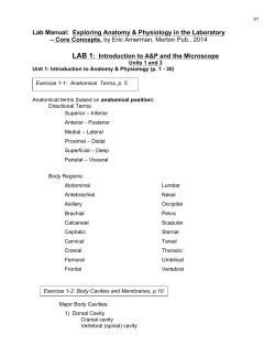
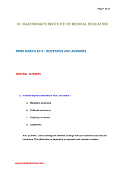
Fig. 4.2 Global incidence rates of tumours of the oral cavity and oropharynx (all ages) in males. Age-standardized rates (ASR, world standard population) per 100,000 population and year. From J. Ferlay et al.,
Globocan 2000 {730}.
Fig. 4.3 Incidence and mortality rates for tumours of the oral cavity and pharynx (excl. nasopharynx), all
ages, in males. Age-standardized rates (ASR, world standard population) per 100,000 population and year.
From J. Ferlay et al., Globocan 2000 {730}.
al border and part of the ventrum of the
tongue. They run to the ipsilateral submandibular nodes. Those towards the tip
of the tongue drain to the submental
nodes. Central lymph vessels drain to the
submandibular nodes on both sides.
Some marginal and central vessels run
directly to the jugulodigastric group but
some can pass direct to the jugulo-omohyoid nodes. Vessels from the area of the
circumvallate papillae and posterior third
of the tongue drain to the jugulodigastric,
jugulo-omohyoid or intermediate nodes,
either unilaterally or bilaterally. Most of
the lymphatics of the palatine tonsils
drain to the jugulodigastric nodes.
Introduction 167
Squamous cell carcinoma
Definition
An invasive epithelial neoplasm with varying degrees of squamous differentiation
and a propensity to early and extensive
lymph node metastases, occurring predominantly in alcohol and tobacco-using
adults in the 5th and 6th decades of life.
ICD-O code
8070/3
Epidemiology
More than 90% of malignant neoplasms
of the oral cavity and oropharynx are
squamous cell carcinomas of the lining
mucosae with relatively rare neoplasms
arising in minor salivary glands and soft
tissues. It is important to specify which
anatomical sites are included in epidemiological data. Separate assessment of
incidence rates for the oral cavity and
oropharynx is complicated by the difficulty of assigning a site of origin to
tumours that are often advanced.
Males are affected more often than
females because of heavier indulgence
in both tobacco and alcohol habits in
most countries: in India the highest rates
of intraoral cancer may be found in
women who chew tobacco heavily. The
male to female ratio is, however, globally
lower for cancer of the oral cavity than for
cancer of the oropharynx, perhaps suggesting that higher exposure to tobacco
smoking and alcohol drinking are
required to induce oropharyngeal than
oral cancer {796}.
Globally some 389,650 cases occurred
in the year 2000; 266,672 for the oral
cavity (ICD-9 140-5) and 122,978 for the
oropharynx (ICD-9 146,8-9) {1981}. This
represents 5% of all cancers for men and
2% for women.
In males, the country with the highest
rate in the western world is currently
France, with extremely elevated rates
also in French-speaking Switzerland,
Northern Italy, Central and Eastern
Europe (especially Hungary) and parts of
Latin America. Rates are elevated
amongst both men and women throughout South Asia. In the USA incidence
rates are two-fold higher in Black men
than White men {1981}. Very high rates in
the IARC database for Melanesia, presumably associated with areca nut and
tobacco habits, are based on small numbers and need confirmation {730,1981}.
The high incidence rates in Australasia
are explained by lip cancer in fairskinned races which has a comparatively low mortality rate.
Much of Europe and Japan is experiencing alarming rises in incidence, with a
strong cohort effect, those born from
approximately 1930 onwards showing
significantly increased incidence and
mortality. In North America there are statistically significant falls in Whites, but
Blacks continue to show worse outcomes. Globally, with the exception of
the most highly specialized treatment
centres, survival rates have not improved
for decades.
Significant increases in incidence in
younger subjects, particularly males,
have been reported from many western
countries in recent decades {1534,2259}.
N. Johnson
S. Franceschi
J. Ferlay
K. Ramadas
S. Schmid
D.G. MacDonald
J.E. Bouquot
P.J. Slootweg
Etiology
Tobacco smoking and alcohol
The dominant risk factors are tobacco use
and alcohol abuse, which are strongly
synergistic {228}. Alcohol and tobacco
account for 75% of the disease burden of
oral and oropharyngeal malignancies in
Europe, the Americas and Japan
{227,1862}. For the highest levels of consumption compared to the lowest ones
relative risks from 70 to over 100 have
been shown {287,1811}. Relative risks in
case-control studies showing a supermultiplicative effect in the oral cavity, between
additive and multiplicative in the oesophagus, and multiplicative in the larynx,
reflecting degree of contact with both
these agents at these sites {797}.
Most of the rise in western countries in
recent years has been attributed to rising
alcohol consumption in northern Europe
{1597 and rises in tobacco consumption
in parts of southern Europe. Significant
risk increases have also been reported
amongst non-drinking smokers and, to a
lesser extent, non-smoking heavy
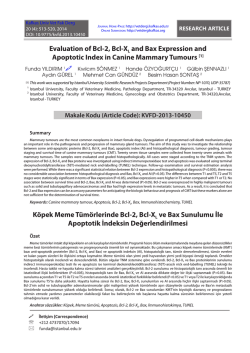
Fig. 4.4 Trends in mortality from cancer of the oral cavity and pharynx in some European countries. The
large differences observed (currrently 10-fold in between Hungary and Finland) largely reflect past success
and failure in tobacco and alcohol control. From F. Levi et al. {1483}.
168 Tumours of the oral cavity and oropharynx
drinkers {1406}. Studies that have
attempted to estimate a difference
between wine, beer and hard liquors generally indicate that heavy consumption of
all types of alcoholic beverage confers
risk, the differences in risk estimates
being largely due to socio-cultural correlates of drinking patterns in various populations {142,1404}.
Ultraviolet light and contact with smoking
appliances are important for lip vermillion.
Tobacco chewing
Oral smokeless tobacco is a major cause
of oral {969} and oropharyngeal {2908}
squamous cell carcinoma in the Indian
subcontinent, parts of South-East Asia,
China and Taiwan and in emigrant communities therefrom, especially when consumed in betel quids containing areca
nut and calcium hydroxide (lime). Areca
nut has been declared a known human
carcinogen by an IARC Expert Group
(2003). In India chewing accounts for
nearly 50% of cancers of the oral cavity
and oropharynx in men and over 90% in
women {108}. Traditional tobacco products used in Sudan and the Middle East,
which are powdered and fermented and
mixed with sodium bicarbonate, contain
very high levels of tobacco-specific
nitrosamines and are highly carcinogenic
{1171}. Those forms of non-flue cured
smokeless tobacco used as oral snuff in
Scandinavia and North America is less
carcinogenic {1230} – though they cause
nicotine addiction.
Human papillomavirus (HPV) infection
HPVs, especially those genotypes of
known high oncogenic potential in uterine
cervix and skin such as HPV 16 and 18,
are found in a variable but small proportion of oral, and up to 50% of tonsillar and
oropharyngeal SCCs, especially the tonsil. Recent studies suggest that HPV may
be responsible for a small fraction of oral,
and up to 40% of oropharyngeal, cancers
{888,1077}. This has lead to speculation
that HPV infection, perhaps arising from
oral/genital contact, might be important in
some cases {2284}. Of interest is the
observation that HPV-containing cancers
at these sites do not generally show TP53
mutations, contrary to HPV DNA-negative
cancers {660,1077}. It is well known that
HPV 16 E6 protein inactivates p53 protein, suggesting that HPV and smoking
might operate, in part, on the same criti-
A
B
C
D
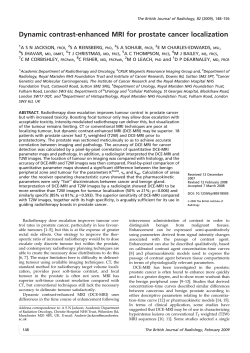
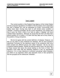
Fig. 4.5 Squamous cell carcinoma. A Exophytic growth involving the left buccal mucosa and overlying skin
in a 65 year old male who chewed betel quid and smoked tobacco. B An exophytic growth arising from the
left palate. C Squamous cell carcinoma of the tongue. D Early squamous cell carcinoma of lateral border
of the tongue.
cal step in the multistage process of carcinogenesis at these sites.
Prevention
Recent work on risk factors in younger
cases emphasises the importance of
early and heavy tobacco and alcohol
use, the protective effect of diets rich in
fresh fruits and vegetables, but with a
substantial minority without these established risk factors {1534}.
The protective effect of diets rich in trace
elements and antioxidant vitamins is well
demonstrated in many countries, especially in Italian studies {1628,2563}.
Though more controversial, a contribution
from poor oral hygiene is also suggested
{108,2548}.
Second primary tumours
It has been recognised for a long time that
patients with oral cancer are at risk of
second tumours in the upper aerodigestive tract. This has been reported to occur
in 10-35% of cases {2676}. These may be
synchronous with the index tumour or, if
occurring after an interval of longer than
six months are described as metachronous. Recurrence of the index tumour
after treatment can be diagnosed by the
pathologist where the tumour is in deeper
tissue and not associated with the epithelial surface. However, the most frequent
situation of second tumours is when they
arise from surface epithelium adjacent to
the treated index tumour. On morphological grounds these are diagnosed as second primary tumours. The increasing use
of molecular biological techniques has
allowed distinction to be made between
molecularly distinct second primary
tumours and second field tumours
derived from the same genetically altered
field as the index tumour {248}.
Localization
Tumours may arise in any part of the oral
cavity. The most common sites vary geographically reflecting different risk factors.
Lip SCC arise almost exclusively on
the lower lip. Within the oral cavity, the
subsites at which tumours may be located
include: buccal mucosa, upper and
lower gingiva, hard palate, anterior twothirds of the tongue, including dorsal,
ventral and lateral surfaces, and the floor
of mouth. Many tumours are large at presentation and the tumour site is then
recorded as essentially the centre of the
tumour. Analysis of small symptomless
tumours shows the highest frequency in
floor of mouth, ventrolateral tongue and
soft palate complex {1655}. This suggests
that tumours arise at these sites, but
spread preferentially to involve other sites
such as tongue, being then recorded
as lingual lesions. The clinical relevance
of this observation is to emphasise the
Squamous cell carcinoma 169
A
B
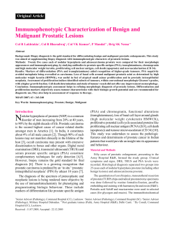
Fig. 4.6 A Well-differentiated squamous cell carcinoma (SCC), characterized by abundant formation of keratin pearls. B Moderately differentiated SCC. Cells form
large anastomosing areas in which keratin pearls are formed. They are not very numerous and the main component consists of cells with pronounced cytonuclear
atypia.
importance of close examination of highrisk sites. The oropharynx consists of the
base of the tongue (posterior third), vallecula, tonsil with tonsillar fossae and pillars, glossotonsillar sulci, posterior wall
and superior wall composed of the inferior surface of the soft palate and the uvula.
The most common oropharyngeal site of
involvement for SCC is the base of
tongue.
Clinical features
Signs and symptoms
Patients with small oral and oropharyngeal SCC are often asymptomatic or may
present with vague symptoms and minimal physical findings. Hence, a high
index of clinical suspicion is needed to
diagnose small lesions, especially if the
patients have tobacco and alcohol
habits. Patients may present with red
lesions, mixed red and white lesions, or
white plaques. Co-existing white plaques
(leukoplakia) may be observed adjacent
to carcinomas and this implies an origin
in a pre-existing white lesion though the
prevalence of this association varies
considerably in different populations.
However, most patients present with
signs and symptoms of locally advanced
disease. The clinical features may vary
according to the affected intraoral subsite. Mucosal growth and ulceration,
pain, referred pain to the ear, malodour
from the mouth, difficulty with speaking,
opening the mouth, chewing, difficulty
and pain with swallowing, bleeding,
weight loss, and neck swelling are the
common presenting symptoms of locally
advanced oral and oropharyngeal cancers. Occasionally, patients present with
enlarged neck nodes without any symp-
A
toms from oral or oropharyngeal lesions.
Extremely advanced cancers present as
ulceroproliferative growths with areas of
necrosis and extension to surrounding
structures, such as bone, muscle and
skin. In the terminal stages, patients may
present with orocutaneous fistula,
intractable bleeding, severe anaemia
and cachexia.
Cancer of the buccal mucosa may present as an ulcer with indurated raised
margin, exophytic or verrucous growth or
with the site of origin depending upon the
preferential side of chewing and placement of betel quid. In advanced stages,
these lesions infiltrate into the adjacent
bone and overlying skin. Cancer of the
tongue may appear as a red area interspersed with nodules or as an ulcer infiltrating deeply, leading to reduced mobility of the tongue. These tumours are
B
Fig. 4.7 Poorly differentiated SCC. A Cells with atypical nuclei and a small rim of eosinophilic cytoplasm form strands and small nests. B Cells in a poorly differentiated SCC tend to have more vesicular nuclei. The cells in this tumour are more cohesive, forming larger tumour areas than the lesion shown in A.
170 Tumours of the oral cavity and oropharynx
A
B
Fig. 4.8 Squamous cell carcinoma (SCC). A Growth pattern of a diffusely infiltrating SCC. In this moderately differentiated lesion, the tumour cells form tiny strands.
This growth pattern is a prognostically unfavourable feature. B Moderately differentiated SCC growing in large cohesive fields. This pattern is prognostically more
favourable than the diffuse growth shown in Figure A.
painful. Cancers of the floor of mouth
may arise as a red area, a small ulcer or
as a papillary lesion. Most patients present with discomfort or irritation at the site
of the tumour. Advanced stages are
associated with drooling. Cancers of the
lower lip usually arise in the vermilion
border and appear as a crusty indurated
or ulcerated lesion. Cancers of the upper
lip are rare, often originate on the skin
and spread to the mucosa. Cancer of the
gingiva usually presents as an ulceroproliferative growth. Tumours of the alveolar
ridge may occasionally present as difficulty in wearing denture plates or as
loosening of teeth associated with pain
and bleeding during brushing of teeth.
Tumours of the hard palate often present
as papillary or exophytic growths, rather
than a flat or ulcerated lesion.
Cancer of soft palate and uvula often
A
appear as an ulcerative lesion with
raised margins or as fungating masses.
Tonsillar cancers generally appear as an
exophytic
or
ulcerative
lesion.
Sometimes they can present as enlarged
neck nodes without any other signs and
symptoms. Cancer of the base of tongue
presents late in the course of the disease
as a grossly ulcerated, painful, indurated
growth.
More than two-thirds of the patients with
buccal mucosal and gingival cancers in
South Asia present with submandibular
lymph node enlargement. More than
three fourths of patients with tongue, floor
of mouth and oropharyngeal cancers in
South Asia present with neck swellings
implying clinically obvious lymph node
metastasis. In the West lymph node
involvement is common at presentation
in oropharyngeal SCC.
Imaging
Intraoral and dental radiographs, in combination with orthopantomography, may
help in identifying involvement of the
underlying bone. Three-dimensional
imaging with computed tomography (CT)
and magnetic resonance imaging (MRI)
is frequently used to supplement the clinical evaluation and staging of the primary
tumour and regional lymph nodes. CT
scan or MRI give more information about
the local extent of the disease and also
help to identify lymph node metastases.
CT scanning is useful in evaluating
involvement of cortical bone. MRI is more
informative when evaluating the extent of
soft tissue and neurovascular bundle
involvement. The combination of soft tissue characterisation and anatomical
localization afforded by CT and
MRI make them valuable tools in the
B
Fig. 4.9 Squamous cell carcinoma (SCC). A In this moderately differentiated SCC, the tumour stroma contains a dense lymphoplasmacytic infiltrate. B SCC with
perineural growth, spreading alongside the inferior alveolar nerve.
Squamous cell carcinoma 171
Fig. 4.10 Periodontal ligament involvement by a
squamous cell carcinoma (SCC).
preoperative assessment of patients with
oral or oropharyngeal cancers. Distant
metastasis from oral and oropharyngeal
cancer is uncommon at presentation. At
minimum, a routine radiograph of the
chest is performed to rule out lung
metastases.
Relevant diagnostic procedures
Optimal therapy and survival from oral
cancer depend on adequate diagnosis
and assessment of the primary tumour
and its clinical extent. Physical examination should include visual inspection and
palpation of all mucosal surfaces, bimanual palpation of the floor of the mouth,
and clinical assessment of the neck for
lymph node involvement.
The diagnosis is confirmed by biopsy.
The specimen is taken from the clinically
most suspicious area, avoiding necrotic
or grossly ulcerated areas, and more than
one biopsy site may need to be chosen.
In patients with enlarged cervical lymph
nodes and an obvious primary in the oral
cavity or oropharynx, the biopsy is always
taken from the primary site and not from
the lymph node. In such situations, fine
needle aspiration cytology may be carried out to verify the involvement of the
node.
If no obvious primary site is found in
patients presenting with neck nodes, fineneedle aspiration of the lymph node can
be performed to help establish the diag-
A
B
C
D
Fig. 4.11 Squamous cell carcinoma (SCC). A Superficial erosion of the mandibular bone has perforated the
cortical bone. As there is no spread in the bone marrow, this case of SCC does not meet the requirements
for classification as T4. B Saucerization by SCC. In this case of, there is substantial loss of bone due to
endocortical tumour growth (meets the requirements T4). C Permeative infiltration of bone by SCC growing
diffusely in the marrow cavities of the mandibular bone (T4). There is also heavy osteoclast-mediated bone
resorption. D Bone invasion by SCC with diffuse growth in the mandibular bone.
nosis. In patients for whom fine needle
aspiration is non-diagnostic and SCC is
strongly suspected, excisional lymph
node biopsy is a last resort, as subsequent curative therapy may be compromised by this procedure. The search for
an occult primary tumour may include
direct pharyngolaryngoscopy with biopsy
of high-risk sites like base of tongue,
nasopharynx, and usually a diagnostic
tonsillectomy, as well as other imaging
modalities. Open lymph node biopsy is
carried out only when the lesion cannot
be identified by aspiration biopsy or in
patients with suspected lymphoma.
Patients with SCC of the oral cavity or
oropharynx have a risk of multiple primary tumours in the pharynx or larynx, as
well as in the tracheobronchial region and
oesophagus so routine panendoscopy is
often performed to evaluate these sites.
Tumour spread and staging
Staging is carried out according to the
TNM classification {947,2418}. Recent
additions to the coding have been provided for micrometatses, isolated tumour
cells, findings in sentinel nodes and
tumour detection by molecular methods.
Some of these are discussed in the following sections.
Local spread of oral SCC, in the early
172 Tumours of the oral cavity and oropharynx
stages, is relatively predictable in tissues
that have not been previously irradiated.
It is influenced by local anatomical features. Lip SCC spreads superficially and
then into deeper tissues. Floor of mouth
SCC spreads superficially rather than in
depth, being unlikely to invade into the
mylohyoid muscle or the sublingual
gland until a late stage. Tumour involving
the lateral margin of tongue, whether
arising there directly or by superficial
spread from the floor of mouth, tends to
spread in depth. The intrinsic muscles of
tongue run in small bundles in all directions such that invading tumour encounters some muscle running at right angles
to the surface. The line of least resistance to tumour spread is therefore along
these muscle bundles and into the
tongue. Tumours of palate spread superficially rather than in depth and this is
also true for more posterior tumours of
the oropharynx.
For most oral SCC other than tongue, the
extent of spread in an area can be predicted from the extent of surface involvement. Tongue and tonsil tumours can
spread beneath intact normal appearing
surface, giving a larger area of tumour
involvement. Spread of oral SCC into
bone is a frequent problem. The mandible
is involved much more frequently than the
maxilla. In dentate jaws the usual route of
entry into mandible is along the periodontal ligament. In edentulous areas of
mandible the tumour spread is through
the crest of the alveolus directly into the
marrow spaces between trabeculae of
cancellous bone {1682}. This occurs
because of failure of formation of an intact
cortex of alveolar bone as resorption of
edentulous alveolus progresses. Tumours
in the mandible can involve the inferior
alveolar nerve {1683} with a particular
likelihood of spread posteriorly along the
nerve, sometimes extending well beyond
the mandibular foramen. Cancers arising
in gingiva or alveolus and those involving
these sites by extension from adjacent
sites are unlikely to invade into the
mandible other than by periodontal ligament or the crest of edentulous alveolus.
Extension into the mandible through
foramina, for example the mental foramen
from lip cancer, does occur, but is uncommon.
Spread in previously irradiated tissues
Tumour spread in previously irradiated
soft tissues tends to be more extensive
and less predictable than in normal tissues and as a consequence requires
more extensive surgery if excision is
attempted. Tumour invasion into irradiated
mandible tends to occur wherever the
tumour approaches bone, often at multiple sites {1682}.
Lymphatic spread
Spread to local lymph nodes worsens the
prognosis in oral and oropharyngeal cancer. The mechanism of spread from the
primary site to lymph nodes is almost
always by embolism. Permeation in lymphatics adjacent to tumours is uncommon
and it is debatable if this spread extends
as far as lymph nodes. Once tumour is
present in the neck, however, spread
between nodes may be embolic or by
permeation. The lymph nodes in the neck
are divided into levels. The lymphatic draianage from different head and neck sites
is realtively predictable {1789}. Levels at
high risk for metastasis from oral cavity
SCC are Levels I, II and III, and to a lesser extent Level IV. Although Level II is the
most frequently involved, some tumours
spread to Level III or IV, with or without
involvement of Level I. This has given rise
to the concept of skip metastsasis. In reality the lymphatic drainage is complex and
does not follow a regular sequence of lev-
els of involvement in many patients
{2817}. Bilateral spread to the neck is likely to occur from tumours involving the
midline, especially tumours of posterior
tongue or soft palate. Extracapsular
spread of tumour involving lymph nodes
is associated with a poor prognosis
{2819}.
There have been many studies attempting
to predict the presence of lymphatic
spread from features of the primary
tumour {872,2820}. Tumour size and site
are relevant. Tumour differentiation is not
a reliable predictor. The pattern of the
invasive front is a useful predictor in that a
non-cohesive front is associated with
increased likelihood of metastasis. Other
factors associated with increased risk of
metastasis are perineural spread at the
invasive front, lymphovascualr invasion
and tumour thickness. The tumour thickness is measured from the deepest
tumour invasion to the presumed original
surface level, that is, ignoring exophytic
growth or assessing the original surface
level in ulcerated tumours. For diagnostic
purposes a thickness of 5mm or greater is
used as indicating increased risk of nodal
spread {395}.
Haematogenous spread
Until relatively recently, haematogenous
spread of oral and oropharyngeal cancer
has been regarded as less important than
local and lymphatic spread. However, its
importance is increasing as loco-regional
control improves. Blood borne spread
most often involves lung {754,1958}. The
best predictor of the likelihood of this
spread is involvement of the neck at multiple levels. This suggests that the route of
entry of tumours into the circulation is
most often via the large veins in the neck
and that haematogenous spread is in
effect tertiary spread following extracapsular spread from neck nodes.
Sentinel node biopsy
This is currently an experimental technique {2057} that is under active evaluation by prospective clinical trials and it is
not practised at all centres. It is a technique used primarily for staging a clinically N0 neck. in an effort to avoid a neck
dissection. If a clinically N0 neck is followed untreated until tumour development occurs, the prognosis can be very
poor {57,977}. Studies on the incidence of
occult metastases in N0 necks {753} have
shown tumour spread in only a small
minority of patients. Therefore, if neck dissection is undertaken either prophylactically or as a staging procedure, on
patients with N0 necks, a large majority
will have unnecessary surgery, as the
neck will be found to be free from tumour.
The sentinel node is the first draining
lymph node from a tumour. It is assumed
that if the sentinel node can be shown to
be free from tumour, then the lymphatic
basin is free from tumour and neck dissection is not required. By contrast, sentinel node positive patients can be selected for further therapy. Sentinel nodes are
identified by a combination of lymphoscintigraphy and injection of blue dye
in the tumour bed and then sampling
draining nodes identified. In reality, more
than one sentinel node is found in many
cases {2345} indicating that tumours
drain to more than a single first echelon
node, presumably from different parts of
the tumour.
Sampled sentinel nodes should be fully
examined by the pathologist. This usually
involves bisecting the node in the largest
diameter and then undertaking extensive
sampling. Some pathologists undertake
frozen sections on bisected fresh nodes.
If this is done it is important to use a technique whereby the cut surface is frozen
on a flat surface and only early sections
are examined. This is to ensure that as little node as possible is examined at this
stage in order not to compromise full
examination of the node. Paraffin
processed blocks are then examined with
H and E sections of the early sections of
the blocks. If these show no tumour, more
detailed sampling with immunocytochemistry for cytokeratins and sampling
through the block is required. True serial
sectioning is impracticable for routine
use. A compromise is step sectioning at
intervals of 150Вµm with examination of H
and E sections and AE1/3 reacted sections {2202}. The importance of these
sections is that suspicious areas on
immunocytochemistry can be identified in
the H and E sections. These may be
viable tumour cells, but other possible
causes of cytokeratin positivity, such as
inclusion of normal salivary gland epithelium or thyroid follicles, either occult
metastases or lateral aberrant thyroid,
need to be identified. Another not infrequent finding is areas of cytokeratin positivity which on H and E appear as densely eosinophilic apparently non-viable
tumour cells.
Squamous cell carcinoma 173
A
B
Fig. 4.12 A Verrucous carcinoma (VC) of the gingiva. B VC of the ginigva, spreading laterally to involve the
cheek mucosa.
Interpretation of sentinel nodes can
demand considerable pathological
expertise. The outcome of the pathological assessment may be the presence of
metastasis; micrometastasis, less than
2mm diameter tumour deposits, or isolated tumour cells {2477}. Micrometastasis
has been defined {1073} as cells which
have arrested and implanted. These may
be in contact with a vessel or lymph sinus
wall or may be extravascular. Single or
small clusters of cells within lymph or
blood vessels, but not in contact with the
wall are defined as isolated tumour cells.
Histopathology
The histological features of SCC have
been discussed in Chapter 3 on tumours
of the hypopharynx, larynx and trachea.
The findings in the oral cavity and
oropharynx do not differ significantly from
those of the larynx and hypopharynx. A
minority of oral and oropharyngeal cancers show different histological subtypes
that can be associated with differences in
prognosis. These are discussed below. It
is clearly important that pseudo epitheliomatous hyperplasia (PEH) is distinguished from SCC. PEH can occur in
mucosa overlying a granular cell tumour,
in necrotising sialometaplasia and in papillary hyperplasia of palate. PEH occurring
with mucositis, particulary after irradiation, may be difficult to distinguish from
squamous cell carcinoma.
The majority of cases of SCC present no
difficulty in diagnosis for the experienced
pathologist. However, the recognition of
the earliest stages of invasion can be
problematic. No consistent guidelines for
this exist. The deepest layers of the
epithelium and the interface between the
epithelium and the lamina propria need to
be examined in detail. This is frequently
made more difficult where there is a
prominent
inflammatory
infiltration.
Relevant features include the loss of a histologically
well-defined
interface,
described previously as loss of basement
membrane and disturbed architecture of
the basal layers of the epithelium, particularly the replacement of basal cells by
larger irregular cells with cytoplasmic
processes extending into connective tissue. In some cases the degree of cytological atypia and mitotic feature may
suggest malignancy, but these are not
always present. To an extent the judgement about early invasion is subjective
and it can be important for the pathologist
to communicate the difficulty in interpretation to the clinician. Some pathologists will
indicate that while no unequivocal evidence of invasion is demonstrated, they
nevertheless feel that the lesion should be
regarded as early invasive carcioma.
Somatic genetics
There is some variation in the genetic profile of oral and oropharyngeal SCC that
reflects the site-specific impact of various
casual agents and differences in clinical
presentation. The carcinogens in tobacco
smoke, for example, increase the prevalence and spectrum of TP53 mutations
{268}. Compared to carcinomas that arise
in patients who smoke, carcinomas in
patients who have never smoked harbour
fewer p53 mutations, disproportionately
involve women, typically arise from the
oral tongue, and affect very young or very
old patients {1351,2258}. For carcinomas
of the oropharynx, oncogenic human
papillomavirus (HPV), particularly the
HPV-16 subtype, is an important
causative agent: More than 50% of
oropharyngeal carcinomas harbour integrated HPV DNA {60,888,1999}. The E6
and E7 viral oncoproteins bind and inactivate the TP53 and retinoblastoma gene
products respectively, disengaging two
of the more critical pathways involved in
174 Tumours of the oral cavity and oropharynx
cell cycle regulation {2788}. These HPVpositive oropharyngeal tumours compose
a distinct pathological entity with its own
clinical spectrum and basaloid morphology {888,1012,2072}, illustrating the
emerging role of genetic characterization
as a potential means of determining prognosis and influencing management
{1691}.
Genetic evidence has clarified the vague
concept of “field cancerization”. Most, if
not all, multiple primary carcinomas of the
upper aerodigestive tract derive from a
common clonal progenitor cell that undergoes a common early genetic alterations
{187,2271}. Genetic evidence has helped
account for the perplexing problem of
local tumour recurrence following seemingly complete tumour resection. In many
instances, local tumour recurrence
reflects extension of genetically damaged
cells beyond the clinical and microscopic
boundaries of carcinoma to the margins
of
surgical
resection
{268,1626,
1983,2777}.
Microsatellite analysis of exfoliated cells
swabbed or rinsed from the oral cavity of
patients with head and neck squamous
carcinomas consistently harbour genetic
changes that are identical to those in the
primary tumours, suggesting a non-invasive test for specific DNA-sequence variants in saliva as a means of identifying
patients with pre-invasive or invasive neoplasms {2430}. Clonal genetic changes
identical to those found in primary head
and neck SCC have been identified in circulating plasma or serum, suggesting a
mechanism for early cancer detection
and tumour surveillance {1853}. The use
of highly sensitive genetic assays for
detecting rare cancer cells at the margin
of tumour resection shows promise for
predicting the likelihood of tumour recurrence {268,1983}.
Prognosis and predictive factors
Tumour size and nodal status are the most
significant prognostic factors {2060}.
Histological grade correlates poorly with
patient outcome {1292,2195}. The value
of grading improves when only the deeply
invasive margins of the tumour are evaluated {291,292,1927, 2818}. Tumours
invading with pushing borders are less
aggressive than tumours showing a noncohesive front showing diffuse spread
with tiny strands or single cells.
{1325,2132,2342,2653, 2841} Major risk
factors that adversely influence prognosis
are two or more positive regional nodes,
extracapsular extension of nodal disease,
or positive margins of resection {1429}.
Other important histologic features associated with poor prognosis are tumour
thickness and vascular invasion.
Molecular markers with unequivocal
prognostic and/or predictive significance
have not been identified {428,1052,1561,
2106}.
Verrucous carcinoma
ICD-O code
8051/3
Although uncommon, 75% of all cases of
VC occur in the oral cavity. It is an exophytic, warty, slowly growing variant of
SCC with pushing margins. It typically
involves older males {950,1251,1677,
1695,2621}. Chronic smokeless tobacco
use is accepted as the primary etiological
factor for oral VC. Human papillomavirus
subtypes 16 and 18 have been identified
in up to 40% of oral VC {1927,2349}. Oral
VC begins as a well-demarcated, thin
white keratotic plaque which quickly
thickens and develops papillary (blunted
tips) or verruciform (pointed tips) surface
projections. Occasional lesions present
as erythaematous or pink papular masses. The colour depends on the amount of
keratin produced and the degree of host
inflammatory response to the tumour. This
cancer almost always remains broadbased or sessile and can become quite
extensive from lateral growth by the time
of diagnosis. Rare fungating examples,
however, may appear to be somewhat
pedunculated. Smokeless tobacco keratosis (tobacco pouch) is often seen on
adjacent mucosal surfaces in patients
who chew tobacco or use snuff. Unless
the tumour is infected or is encroaching
on alveolar nerves in the jawbones, VC is
an asymptomatic lesion. Surface ulceration and haemorrhage are not seen,
unless a focus of SCC is present in the
mass.
VC consists of thickened club-shaped
papillae and blunt stromal invaginations
of well-differentiated squamous epithelium with marked keratinization. The squamous epithelium lacks the usual cytologic criteria of malignancy, and by morphometry, the cells are larger than those
seen in SCC {489}. Mitoses are rare, and
observed in the basal layers; DNA synthesis (S-phase) is also limited primarily
to the basal layers {737}. VC invades the
stroma with a pushing, rather than infiltrating border. Dense lymphoplasmacytic
host response is common. Intraepithelial
microabscesses are seen, and the abundant keratin may evoke a foreign body
reaction.
The surrounding mucosa shows progressive transition from hyperplasia to VC. A
downward dipping of epithelium often
“cups” the VC periphery, and is the ideal
site for deep biopsy {174,1192}. With
extensive surgical removal, and without
neck dissection, the 5-year disease-free
survival rate is 80-90%, although 8% of
patients require at least one additional
surgical procedure during that time
{1870,1927}. Treatment failures usually
occur in patients with the most extensive
involvement or in those unable to tolerate
extensive surgery because of unrelated
systemic diseases. No molecular or other
markers have yet shown prognostic significance for oral VC. However, one-fifth
of these tumours contain a co-existing
SCC which may not be identified without
extensive histologic sectioning {1927}.
Such hybrid tumours have a greater tendency to recur locally and a slight tendency to metastasize to the ipsilateral
neck.
Basaloid squamous cell
carcinoma
ICD-O code
8083/3
This is uncommon in the oral cavity,
slightly more common in the oropharynx.
It is described in the chapter on tumours
of the hypopharynx, larynx and trachea.
Papillary squamous cell
carcinoma
ICD-O code
Acantholytic squamous cell
carcinoma
ICD-O code
8075/3
The lip is the most frequent oral site.
There are no distinguishing clinical signs
and the microscopical features are considered in the chapter on tumours of the
hypopharynx, larynx and trachea. A variant of this tumour has been referred to as
pseudovascular SCC.
Adenosquamous carcinoma
ICD-O code
8560/3
In the oral cavity this is a SCC with clearcut areas of adenocarcinoma, most frequently seen as a component of a large
SCC. It is described in the chapter on
tumours of the hypopharynx, larynx and
trachea.
Carcinoma cuniculatum
(epithelioma cuniculatum)
ICD-O code
8051/3
This rare variant of oral cancer has similarities to the lesion more commonly
described in the foot in which tumour infitrates deeply into bone. The oral tumours
show proliferation of stratified squamous
epithelium in broad processes with keratin cores and keratin filled crypts which
seem to burrow into bone, but lack obvious cytological features of malignancy
{40}. Diagnosis on biopsy specimens
can be very difficult and correlation with
the clinical and radiographic features is
required.
8052/3
This is rarely recognized in the oral cavity and oropharynx other than as a component of a large SCC. It is described in
the chapter on tumours of the hypopharynx, larynx and trachea.
Spindle cell carcinoma
ICD-O code
8074/3
This unusual variant is more common in
the larynx than in the oral cavity and
oropharynx, and is described in detail in
the chapter on tumours of the hypopharynx, larynx and trachea.
Squamous cell carcinoma 175
Lymphoepithelial carcinoma
Definition
Lymphoepithelial carcinoma (LEC) is a
poorly differentiated squamous cell carcinoma (SCC) or undifferentiated carcinoma, accompanied by a prominent reactive lymphoplasmacytic infiltrate. The
morphological features are indistinguishable from those examples of nasopharyngeal nonkeratinizing carcinoma with a rich
lymphoplasmacytic infiltrate.
ICD-O code
8082/3
Epidemiology
LEC is rare at these sites, and accounts
for 0.8-2% of all oral or oropharyngeal
cancers {1339,2741}. See Chapter 2.
Etiology
Epstein-Barr virus (EBV) has been tested
in only a limited number of cases {819,
856,1802,1875,2405}, but it appears that
tumours occurring in Chinese are usually
positive for EBV, while those occurring in
Caucasians are usually negative. The
racial difference in the association with
EBV is similar to LEC occurring in the
major salivary glands (see Chapter 5).
A
Clinical features
The patients present with an intra-oral
mass, which may be ulcerated. Some
tumours can be bilateral {801,2038}. A
proportion of patients present with neck
mass due to regional lymph node
involvement {119}.
Location and metastatic spread
More than 90% of all oral and oropharyngeal LEC occur in the tonsil and tongue
base areas. The remaining cases are
found in the palate and buccal mucosa
{444,694,2822}. The tumour has a high
propensity for regional cervical lymph
node involvement (approximately 70% of
cases at presentation) {119,444,1339}.
Distant metastasis tends to occur in the
liver and lung {119}.
Histopathology
LEC of the oral cavity and oropharynx
shows morphologic features indistinguishable from its nasopharyngeal and
sinonasal counterparts. The surface
epithelium is often intact. The tumour is
invasive, and comprises syncytial sheets
and clusters of carcinoma cells with
B
W.Y.W. Tsang
J.K.C. Chan
W. Westra
vesicular nuclei, prominent nucleoli and
ill-defined cell borders. A rich lymphoplasmacytic infiltrate is present within the
tumour islands and the surronding stroma, which may appear desmoplastic.
The tumour cells are immunoreactive for
pan-cytokeratin and epithelial membrane
antigen. EBV encoded RNA (EBER) has
been demonstrated by in-situ hybridization in oral / oropharyngeal LEC occurring in Chinese patients.
Prognosis and predictive factors
LEC of the oral cavity and oropharynx are
radiosensitive, and in a high percentage
of cases local control can be achieved
even in the presence of regional lymph
node metastasis {1339}. Local, regional
and distant failures occur in 3%, 5% and
19% of cases respectively {444}. Distant
metastasis is associated with a poor
prognosis.
C
Fig. 4.13 A Lymphoepithelial carcinoma of the tonsil. The tumour infiltrates beneath an intact surface epithelium. In this example, the tumour islands are obscured
by the heavy lymphoplasmacytic infiltrate. B Sheets and islands of tumour cells intimately admixed with lymphocytes and plasma cells. C Lymphoepithelial carcinoma of the palate. Carcinoma cells exhibit indistinct cell borders, pale chromatin and distinct nucleoli. Many lymphocytes are found among the carcinoma cells.
176 Tumours of the oral cavity and oropharynx
Epithelial precursor lesions
The pathologic assessment of precursor
lesions is similar throughout the upper
aerodigestive tract. It is described in
detail in the Chapter 3 on tumours of the
hypopharynx, larynx and trachea (page
140).
Clinical features
The principal oral and oropharyngeal
lesions which may be precursor lesions
are white patches (leukoplakia) and red
patches (erythroplasia/erythroplakia) or
mixed red and white lesions. The majority of leukoplakias will not show dysplasia
and correspond to the hyperplasia category. Red and mixed lesions (speckled
leukoplakia) show a higher frequency of
dysplasia, often of higher grade. The
majority of leukoplakias will not undergo
malignant change and may even regress
particularly if apparent aetiologic factors
are removed.
Histopathology
The epithelium of precursor lesions may
be thick, but in the oral cavity it can also
be atrophic. By definition, there is no evidence of invasion. The magnitude of surface keratinisation is of no importance.
Allocation to categories within each of
the classifications requires consideration
firstly of architectural features and then of
cytology.
Hyperplasia
Hyperplasia describes increased cell
numbers. This may be in the spinous
layer (acanthosis) and/or in the
basal/parabasal cell layers (progenitor
compartment), termed basal cell hyperplasia. The architecture shows regular
stratification without cellular atypia.
Dysplasia, / squamous intraepithelial
neoplasia / atypical hyperplasia
When architectural disturbance is
accompanied by cytologic atypia, the
term dysplasia applies. The terms squamous intraepithelial neoplasia (SIN) and
atypical epithelial hyperplasia are used
synonymously.
There is a challenge in the recognition of
N. Gale
B.Z. Pilch
D. Sidransky
A. El Naggar
W. Westra
J. Califano
N. Johnson
D.G. MacDonald
Table 4.01 Classification schemas that histologically categorize precursor and related lesions
2005 WHO Classification
Squamous Intraepithelial
Neoplasia (SIN)
Squamous cell hyperplasia
Ljubljana Classification
Squamous Intraepithelial
Lesions (SIL)
Squamous cell (Simple)
hyperplasia
Mild dysplasia
SIN 1
Basal/parabasal cell
hyperplasia*
Moderate dysplasia
SIN 2
Atypical hyperplasia**
Severe dysplasia
SIN 3***
Atypical hyperplasia**
Carcinoma in-situ
SIN 3***
Carcinoma in-situ
*
Basal/parabasal cell hyperplasia may histologically resemble mild dysplasia, but the former is conceptually
benign lesion and the latter the lower grade of precursor lesions.
** ’Risky epithelium’. The analogy to moderate and severe dysplasia is approximate.
*** The advocates of SIN combine severe dysplasia and carcinoma in-situ.
the earliest manifestations of dysplasia
and no single combination of the above
features allows for consistent distinction
between hyperplasia and the earliest
stages of dysplasia. Dysplasia is a spectrum and no criteria exist to precisely
divide this spectrum into mild, moderate
and severe categories.
Mild dysplasia
In general architectural disturbance limited to the lower third of the epithelium
accompanied by cytological atypia
define the minimum criteria of dysplasia.
Moderate dysplasia
Architectural disturbance extending into
the middle third of the epithelium is the
initial criterion for recognizing this category. However, consideration of the
degree of cytologic atypia may require
upgrading.
Severe dysplasia
Recognition of severe dysplasia starts
with greater than two thirds of the epithelium showing architectural disturbance
with associated cytologic atypia.
However, as noted in the previous para-
Table 4.02 Criteria used for diagnosing dysplasia
Architecture
Cytology
Irregular epithelial stratification
Abnormal variation in nuclear size
(anisonucleosis)
Abnormal variation in nuclear shape
(nuclear pleomorphism)
Abnormal variation in cell size (anisocytosis)
Abnormal variation in cell shape
(cellular pleomorphism)
Increased nuclear-cytoplasmic ratio
Increased nuclear size
Loss of polarity of basal cells
Drop-shaped rete ridges
Increased number of mitotic figures
Abnormally superficial mitoses
Premature keratinization in single cells
(dyskeratosis)
Keratin pearls within rete pegs
Atypical mitotic figures
Increased number and size of nucleoli
Epithelial precursor lesions 177
A
C
B
Fig. 4.14 A Acanthosis. Hyperplastic epithelium with thickened stratum spinosum. B Basal cell hyperplasia. Increase in progenitor compartment without dysplasia.
C Mild dysplasia. Basal cell hyperplasia with relatively mild cytological change confined to lower third of epithelium.
graph architectural disturbance extending into the middle third of the epithelium
with sufficient cytologic atypia is upgraded from moderate to severe dysplasia.
Carcinoma in-situ
The theoretical concept of carcinoma insitu is that malignant transformation has
occurred but invasion is not present. It is
not possible to recognize this morphologically. The following is recommended for
the diagnosis of carcinoma in-situ: full
thickness or almost full thickness architectural abnormalities in the viable cellular
layers accompanied by pronounced cytologic atypia. Atypical mitotic figures and
abnormal superficial mitoses are commonly seen in carcinoma in-situ.
Differential diagnosis
Reactive, regenerative or reparative squamous epithelium, for example in response
to trauma, inflammation, irradiation or
ulceration, may manifest atypical cytology
or architectural disturbance. Nutritional
deficiencies such as iron, folate, and vitamin B12, can also simulate dysplasia.
Such lesions are not considered precursor lesions and should be distinguished
from them. Clinical history is helpful and
morphological changes suggestive of the
inciting event, such as ulceration, inflammation, haemorrhage, radiation-induced
mesenchymal and/or endothelial nuclear
enlargement and hyperchromatism, may
be present. The epithelial changes in
these cases are generally less pronounced than in dysplasia.
Relevance of dysplasia. It is reasonable to
assume that the changes described in
dysplasia are due to genetic changes in
the epithelium occur, but it is unlikely that
the mutations involved are the same ones
as are associated with development of
malignancy. More severe dysplasia has
been traditionally believed to be associat-
ed with a greater likelihood of progression
to malignancy. This might indicate that the
greater the accumulation of mutations in
tissue, the greater the chance that the critical mutations for malignancy will be present. The corollary is also true in that malignancy can arise from non-dysplastic
epithelium {2493} presumably because
these critical mutations can be present in
the absence of the mutations causing
dysplasia.
Genetics
There are no individual markers that reliably predict malignant transformation.
The molecular biology techniques which
show most promise as predictors of
development of SCC are large scale
genomic status (DNA ploidy) and loss of
heterozygosity (LOH) at defined loci
{2286}.
Dysplasia has been reported to be present in from 10-25% of leukoplakias
Table 4.03 Malignant transformation of oral leukoplakia (Reibel {2145})
Authors/Year
Country
Pindborg et al., 1968 {2049}
Silverman and Rosen, 1968 {2362}
Kramer et al., 1970 {1368}
Mehta et al., 1972 {1699}
Silverman et al., 1976 {2358}
BГЎnГіczy, 1977 {118}
Silverman et al., 1984 {2361}
Lind, 1987 {1517}
Schepman et al., 1998 {2261}
Denmark
USA
UK
India
India
Hungary
USA
Norway
Netherlands
178 Tumours of the oral cavity and oropharynx
Material
(no. of cases)
248
117
187
117
4762
670
257
157
166
Observation
period (years)
3.9
1-11
10
2
9.8
7.2
9.3
2.5
Cases with malignant
transformation (%)
4.4
6.0
4.8
0.9
0.13
6.0
17.5
8.9
12.0
{2286,2490}. Ploidy studies of dysplastic
leukoplakias showed that the great majority of aneuploid lesions developed SCC in
the follow-up period, by contrast with 60%
of tetraploid lesions and only about 3% of
diploid lesions {2490}. No correlation was
found between the degree of dysplasia
and DNA ploidy. Similar studies on erythroplasias {2491} confirmed the high predictive potential of aneuploidy in identify-
ing cases which progressed to SCC. Nondysplastic white patches have also been
studied {11} and although there was a
much lower incidence of malignant transformation, 80% of such cases were aneuploid.
LOH studies have been undertaken contrasting oral lesions which progressed to
SCC or carcinoma in-situ during follow-up
with corresponding lesions which did not
A
B
C
D
progress. LOH on two chromosome arms,
3p and 9p seemed to be particularly
important in predicting progression
{2201}.
Fig. 4.15 A Moderate dysplasia. Drop shaped rete ridges, dysplasia extending to mid-third and moderate cytological changes B Severe dysplasia into upper third
of epithelium with marked cytological change C Severe dysplasia into upper third of epithelium with prominent cytological change including abnormal mitoses.
D Carcinoma in-situ. Abnormal cells seen throughout the full thickness of epithelium.
Epithelial precursor lesions 179
Proliferative verrucous leukoplakia and
precancerous conditions
Definition
Proliferative verrucous leukoplakia (PVL)
is a rare but distinctive high-risk clinical
form of oral precursor lesions. Because of
the lack of specific histologic criteria, the
diagnosis is based on combined clinical
and histopathologic evidence of progression. Sequential biopsies show progressive dysplasia and the acquisition of aberrant TP53 protein.
Clinical features
PVL is an aggressive form of oral leukoplakia with considerable morbidity and
strong predilection to malignant transformation {174,1005,1797,2360}. The etiology of this entity is unknown. The condition
develops initially as focal clinical hyperkeratosis (leukoplakia) that progressively
becomes a wide multifocal disease with
gross exophytic features {174}. The aver-
age age at diagnosis is 62 years; women
are more commonly afflicted (ratio, 4:1).
Typically, multiple oral sites are affected.
The most common site in women is the
buccal mucosa and the tongue in men.
Carcinoma develops after a protracted
period of time. The most common sites of
the carcinoma are gingiva and tongue.
PVL is characterized by high recurrence
rate and histological progression. Many
cases are resistant to all forms of treatment, including laser microsurgery, surgical excision and radio-and chemotherapy.
Conservative management of these
lesions has been unsuccessful and wide
surgical excision is the best hope for control.
Other precancerous conditions
Precancerous conditions (PCs) are generalized clinical states associated with a
A.K. El Naggar
P.A. Reichart
significantly increased risk for SCC.
Epithelial atrophy, increased mitotic activity and impaired epithelial repair mechanisms are fundamental to PCs of different
etiology.
Iron deficiency
Originally described in the context of
sideropenic dysphagia, it is an important
cause of epithelial atrophy. The association of iron deficiency with oropharyngeal
squamous cell carcinomas has been
observed since the mid-thirties of the 20th
century {21}. However, a significant
decrease of cases with hypopharyngeal
cancers and iron deficiency was noted in
Sweden in the seventies {1433}. Few
cases of oral cancer and iron deficiency
have been published in the last 20 years.
A
C
D
Fig. 4.16 Proliferative verrucous leukoplakia (PVL) A Extensive, thick, white plaques. B Hyperplasia and dense hyperkeratosis of early PVL. C Histology from a clinical case of PVL showing verrucous surface with hyperkeratosis, hypergranulosis and a dense inflammatory infiltrate in the corium. D Same case as shown on fig.
C two years later showing more florid verrucous hyperplasia illustrating the progressive nature of the condition.
180 Tumours of the oral cavity and oropharynx
Fig. 4.17 Sideropenic dysphagia. Iron deficiency
anaemia with depapillated tongue, depigmentation
of the upper lip and epithelial erosion of the lower
lip.
Fig. 4.18 Erythroplasia / erythroplakia associated
with oral lichen planus (precancer).
clinical and histological criteria for OLP
and oral lichenoid lesions (OLL). The latter have also been termed interface
mucositis or lichenoid mucositis. Oral
lichenoid lesions have been considered
by some to represent the lesion at risk if
associated with dysplasia. In a recent
study {2664} it was shown that all cases of
malignant transformation (1.7%) involved
cases of OLL and not OLP. Similarly, a
study {2896} investigating whether OLP
without dysplasia is premalignant by
using microsatellite analysis for loss of
heterozygosity (chromosomes 3p, 9p,
17p) did not support OLP as a lesion at
risk.
However, until distinct clinical and histological criteria have been developed on
how to differentiate OLP from OLL, both
lesions have to be considered as �at risk
for malignant transformation’.
Oral submucous fibrosis (OSF)
This chronic, progressive condition of the
oral mucosa {2115} is etiologically strongly associated with the chewing of areca
nut which has recently been categorized
by IARC as a human carcinogen {1}. It is
almost exclusively seen in ethnic groups
using areca nut alone or as a component
of betel quid.
Clinically there is mucosal rigidity of varying intensity due to fibroelastic transformation of the juxtaepithelial connective
tissue. Fibrous bands and mucosal pallor
are characteristic {498}. Histologically,
there is epithelial atrophy, keratosis and
dysplasia in up to 25% of cases {498}. In
a population-based prospective study, in
India, SCC developed in 7% of patients
with OSF over a period of 17 years {1798}.
Fig. 4.20 Oral submucous fibrosis with a broad
band of subepithelial collagenous tissue.
been described {1306,1994,2704}.
Lupus erythematosus
This is a chronic autoimmune disease of
unknown etiology. Carcinomas, mainly of
the lips, have been described in affected
individuals {2264,2696}.
Epidermolysis bullosa dystrophicans
(Hallopeau-Siemens type)
This disease of the skin and oral mucosa
has an autosomal dominant pattern of
inheritance. Oral leukoplakia and occasional cases of SCC have been observed
in association with epidermolysis bullosa
{226,2288}.
Syphilis
Late stage (tertiary) syphilis associated
with leukoplakia had a high risk of malignancy, but this is now largely of historical
interest {1721}.
Fig. 4.19 Syphilis. Interstitial glossitis due to late
stage syphilis with squamous cell carcinoma at the
left tip of the tongue (Collection of J.J. Pindborg,
M.D., Copenhagen).
Oral lichen planus
OLP is a chronic mucocutaneous immune
inflammatory condition. Malignant transformation is still controversial {639,2359};
one review reporting malignant transformation rates between 0% and 5.6%
{2116}.
The controversy is due to lack of uniform
Xeroderma pigmentosum
This is a rare neurocutaneous disease
with an autosomal-recessive mode of
inheritance. The syndrome is caused by
deficient nucleotide excision repair
mechanisms {2090}. The skin, including
the lips, is affected and shows epithelial
atrophy and hyperpigmentation. Patients
are extremely sensitive to light and show
an increased predisposition to UV-associated malignancies of the skin.
Carcinomas of the tongue have also
Proliferative verrucous leukoplakia 181
Papillomas
E.W. Odell
Definition
These form a range of localised hyperplastic exophytic and polypoid lesions of
hyperplastic epithelium with a verrucous
or cauliflower-like morphology. Lesions of
fibroepithelial hyperplasia are not generally included. Not every papilloma can
be allocated to one of the diagnostic categories described below.
ICD-O code
A
B
C
D
8050/0
Epidemiology
Papillomas are common, with a prevalence of approximately 0.1%-0.5% {94,
1342,2569}.
Etiology
HPV infection causes some papillomas
{2076} and at least types 1,2,3,4,6,7,10,
11,13,16,18,30,31,32,33,35,38,45,52,
55,57,59,69,72,73 sequences have been
detected in benign oral lesions.
Clinically, latent HPV is common in oral
mucosa and HPV DNA sequences can
be detected in over 80% of individuals.
There is no absolute association
between the virus type and the type of
papilloma {866} though focal epithelial
hyperplasia is almost exclusively associated with types 13 and 32. HPV infection
of oral tissue may be transmitted horizontally, including venereally, perinatally
and possibly in utero {2518}.
Histopathology
Histological differential diagnosis for all
types includes lesions of fibroepithelial
hyperplasia: fibroepithelial polyps,
fibrous epulis and papillary hyperplasia
associated with candidal infection or
dentures. These have a more prominent
fibrous component and no viral change.
Verruciform xanthoma is a solitary lesion
with a very similar clinical and histological presentation.
Extensive multiple papillomas or diffuse
papillomatous change raise the possibilities of HPV lesions in immunosuppression, acanthosis nigricans, naevus unius
et lateris, focal dermal hypoplasia,
Cowden syndrome, papillary and verru-
Fig. 4.21 Papilloma. A Typical papillary structure of squamous papilloma. This example is only lightly keratinised and negative for papilloma virus on immunocytochemistry. B Papilloma from the vermillion border
showing extensive keratinisation. Note the inwardly facing rete ridges at the periphery. C Moderately keratinised papilloma of verruca vulgaris type showing koilocytic change in the upper prickle cell layers. D
Strong and extensive staining for papilloma virus in a flat papilloma from an HIV infected individual. There
is widespread infection in the upper prickle cell layer but the architecture of the epithelium is little changed.
Note the sharp lesion margin on the left.
cous dysplastic lesions and papillary
squamous {2488} or verrucous carcinoma. Florid oral papillomatosis is a clinical
term for diffuse papillomatous change of
the mucosa for which no specific cause
can be identified and is not a defined
clinico-pathological entity.
Squamous cell papilloma and
verruca vulgaris
Definition
A benign, hyperplastic wart-like localised
proliferation of the oral epithelium {2076}.
Epidemiology
Squamous papillomas are common in
children and in adults in the 3rd to 5th
decades but may be found at any age.
There is an almost equal sex incidence
with a slight male predominance.
182 Tumours of the oral cavity and oropharynx
Etiology
Evidence of causative HPV infection can
be found in less than half of oral squamous papillomas {866,2516,2747}, and
these lesions are the intraoral counterpart of verruca vulgaris. Many HPV subtypes have been detected including
2,4,6,7,10,40. The presence of HPV virion components ultrastructurally and
immunocytochemically indicates active
viral replication in the lesion. Virus transmission appears to be mostly horizontal
or by autoinoculation. Lesions in children
tend to arise at anterior oral sites and the
source of infection is often verruca vulgaris on the skin, particularly on the fingers. Infectivity is low.
The remainder of squamous papillomas
are of unknown etiology. HPV sequences
may be detected by PCR but the significance of this is unclear.
Localization
Any oral site may be affected but the most
common are the hard and soft palate,
labial mucosa, tongue and gingiva.
Clinical features
Squamous papillomas are soft, pedunculated lesions formed by a cluster of finger-like fronds or a sessile, domeshaped lesion with a nodular, papillary or
verrucous surface. The surface may be
white or of normal mucosal colour
depending on the degree of keratinization {2076}. Lesions are usually single
but fairly frequently multiple, particularly
in children and for verruca vulgaris.
Squamous papillomas grow rapidly over
a period of a few months to a maximum
of about 6mm diameter and then remain
a constant size.
Histopathology
Lesions are exophytic and comprise
folds of hyperplastic stratified epithelium
that are usually thickly para- or orthokeratinized but may be non keratinised.
Squamous papillomas associated with
HPV (oral verruca vulgaris) comprise a
cluster of finger-like projections from a
narrow base, each with a sharp keratinised tip, supported on ramifying cores
of connective tissue containing dilated
capillaries. Stratification of the epithelium
is well ordered. Mitoses may be frequent
and there may be mild anisonucleosis
consistent with hyperplasia, but no atypia. The fronds are thickly keratinised,
often with a prominent keratohyaline
layer of large coarse granules. Small foci
of HPV-infected cells (koilocytes) can
usually be found in the upper prickle cell
layer. These keratinocytes have crumpled, darkly stained nuclei with perinuclear haloes but appear very similar to
vacuolated keratinocytes that are common in the normal oral mucosa.
Koilocytes may be more frequent in early
lesions. Less frequently, viral inclusions
are found. Rete processes at the base
often turn inwards and are symmetrical.
Small foci of lymphocytic inflammation
may lie in the fronds or at the base but
inflammation is usually sparse unless the
lesion is subject to trauma or other irritation {4,1929}. HPV may be identified by
immunocytochemistry or in-situ hybridisation but this is not necessary for diagnosis {2076}.
Papillomas without detectable active
HPV replication show more variation.
They may appear identical to verruca
vulgaris but without koilocytes or prominent keratohyaline granules, or form
rounded broad-based dome shaped
lesions similar to condyloma. The hyperplastic epithelium may form papillary
exophytic fronds or arborising rete
processes. Some are flat zones of acanthotic hyperplastic epithelium with
increased numbers of dermal papillae
similar to plane warts of the skin.
Prognosis and predictive factors
Oral verruca vulgaris may regress spontaneously, particularly in children, but
responds to simple excision or ablation
by laser or cryosurgery. Recurrence is
unusual provided all lesional tissue is
removed and there is no malignant
potential.
Condyloma acuminatum
Definition
Oral counterpart of anogenital condyloma acuminatum
Synonyms
Venereal wart; venereal condyloma
Epidemiology
Lesions are usually diagnosed between
the mid 2nd and 5th decade with a peak
in teenagers and young adults {2916}.
Etiology
Epithelial infection by HPV, most commonly types 6,11,16 and 18 though others have been detected {700,1380}.
Transmission is usually venereal or by
autoinnoculation from concomitant genital lesions {1975}.
Histological appearance is not an accurate indicator of a genital origin.
A
Localization
Most lesions arise on the labial mucosa,
tongue and palate in anterior oral sites
though any area may be affected {700,
2916}.
Clinical features
Condylomas are painless, rounded,
dome-shaped exophytic nodules up to
15 mm in diameter, larger than squamous papillomas and verruca vulgaris.
They have a broad base and a nodular or
mulberry-like surface that is slightly red,
pink or of normal mucosal colour.
Lesions may be multiple and are then
usually clustered {2076,2916}.
Histopathology
Condylomas are similar to squamous
papillomas but with short blunt rounded
fronds of hyperplastic epithelium of even
length forming a smooth or nodular, flat
or rounded surface. Keratin is usually
absent or sparse, occasional examples
show moderate keratin and are white
clinically. Between the folds, crypts or
clefts lined by epithelium extend close to
the broad base and may be filled with
keratin debris in keratinised lesions.
Clusters of koilocytes identical to those
described above are much commoner
than in squamous papillomas and are
usually a prominent feature. Unlike squamous papilloma, rete processes are bulbous and short, of even length and do
not curve inwards {700,2076}.
Prognosis and predictive factors
Condyloma acuminatum often responds
to simple excision or ablation by laser or
cryosurgery but appears to carry a higher risk of recurrence than squamous
papilloma. Unlike ano-genital condyloma, there is no documented risk of malignant transformation, regardless of the
presence of high-risk HPV types.
B
Fig. 4.22 Condyloma acuminatum. A Several sessile, cauliflower-like swellings forming a cluster. B Typical
papilloma structure in condyloma showing the more rounded architecture in comparison with verruca vulgaris. Note a verrucous area on the left; many of these lesions have features of both types of papilloma.
Papillomas 183
Condyloma acuminatum in children raises the possibility of sexual abuse, but
non-sexual transmission is possible
{1380} and probably frequent.
Papillomas and papillomatosis
in immunodeficiency
More florid presentations of HPV-induced
lesions are found in immunosuppression,
particularly in HIV infection. Lesions may
be larger, multiple and coalesce to form
extensive patches of affected mucosa.
Occasionally the entire oral mucosa may
become papillomatous and some of
these presentations are not easily classified. Unusual HPV subtypes and multiple
HPV subtypes are more frequent in
immunosuppression. Occasional lesions
in HIV infection are dysplastic and are of
uncertain malignant potential.
Fig. 4.23 Focal epithelial hyperplasia. Typical clinical appearance of multiple papillomatous nodules.
Focal epithelial hyperplasia
Definition
Multiple oral papillomas induced by HPV
13 and 32
Synonym
Heck disease
Epidemiology
This is primarily a disease of children,
adolescents and young adults. Originally
described in Inuit and native Americans
{69} but now recognised worldwide. The
condition is endemic in some countries
and prevalence may be as high as 40%
of children in localised areas {94,332,
1014}.
Etiology
Infection by HPV types 13 and 32.
Localization
All areas of the oral cavity may be affected but the lesions are most common on
the labial and buccal mucosa and the
tongue {69,332,1014}.
Clinical features
Typically there are multiple asymptomatic lesions, each a soft rounded or flat
plaque-like sessile swelling with a slightly nodular surface. They are usually pink
in colour or sometimes white, and 210mm in diameter. Lesions develop in
clusters or confluent patches {332}.
Fig. 4.24 Focal epithelial hyperplasia, viral change and mitosoid body (inset).
Individual lesions may appear and disappear during the course of the disease
{1014}.
Histopathology
The histological features are more distinctive than squamous papilloma or
condyloma. Each lesion is a slightly
raised or rounded sessile swelling
formed by a sharply demarcated zone of
epithelial acanthosis, similar to condyloma acuminatum but with a less prominent papillomatous structure. The bulk of
lesion is formed by exophytic acanthosis,
without formation of well-defined projections of epithelium and the lesion contains minimal connective tissue papillae.
Koilocytes similar to those of squamous
papilloma are usually present and, in
addition, there are usually characteristic
“mitosoid bodies”, which are nuclei with
coarse clumped heterochromatin resembling a mitotic figure. Mitosoid bodies are
characteristic but not specific for focal
epithelial hyperplasia. The base of the
184 Tumours of the oral cavity and oropharynx
lesion is flat and level with the adjacent
epithelium without rete process enlargement {332,2076}. HPV may be detected
on immunocytochemistry or by in-situ
hybridisation but this is not necessary for
diagnosis if the clinical presentation is
typical {2076}.
Genetic susceptibility
Familial clustering and endemic areas
may result from horizontal transmission.
Prognosis and predictive factors
The condition appears to resolve spontaneously after a period of years and is
rarely found in adults. It has no malignant
potential.
Granular cell tumour
P.M. Speight
Definition
A benign tumour of soft tissues which
most often arises in the tongue and is
thought to be of Schwann cell origin. It is
composed of a poorly demarcated accumulation of plump granular cells which
are often intimately associated with
skeletal muscle.
where they appear to merge with muscle
cells {477,2791}. Granular cells extend
up to the epithelium, often forming small
islands in the connective tissue papillae.
The granules stain positively with periodic acid Schiff (PAS).
A characteristic feature of granular cell
tumour is that in up to 30% of cases the
overlying epithelium shows pseudoepitheliomatous hyperplasia that may be
misdiagnosed as carcinoma.
ICD-O code
9580/0
Synonym
Granular cell myoblastoma
Epidemiology
Granular cell tumours are rare.
Approximately 50% of all lesions arise in
the head and neck and over half of these
are found in the tongue. They arise in all
age groups, with a peak between 40 and
60 years. In about 10-20% of patients the
lesions are multiple. Females are affected more often than males with an M/F
ratio of 2:1.
Etiology
No etiological factors are known. The
lesion is thought to arise from Schwann
cells. The granularity may be a senescent change associated with accumulation of lysosomes.
Localization
Granular cell tumours may arise in the
skin, soft tissues, breast and lungs, but
over 50% involve the head and neck and
the tongue is the most common single
site. Oral lesions may also be found in
the buccal mucosa, floor of oral cavity or
palate. Lesions may be multiple, affecting more than one intraoral site, or involving oral and extraoral sites {477}. Rare
lesions have been reported in the salivary glands {331}.
Clinical features
The lesion typically presents as a
smooth, sessile mucosal swelling 1-2 cm
in diameter with a firm texture. The overlying epithelium is of normal colour or
may be slightly pale. Occasionally there
is candidal infestation of the superficial
Fig. 4.25 Granular cell tumour. The typical presentation of granular cell tumour: a sessile swelling on
the tongue covered by normal appearing epithelium.
epithelium and the lesion may then present as a discrete, white plaque.
Macroscopy
Tumours are usually 1-2 cm in diameter
with a smooth surface. The cut surface
shows a poorly demarcated lesion which
is pale yellow or cream and firm on cutting.
Histopathology
The lesion is composed of plump
eosinophilic cells with central small dark
nuclei and abundant granular cytoplasm.
The cells may be polygonal or elongated
and have indistinct cell membranes,
often giving the impression of a syncytium. The lesion is not encapsulated
and the granular cells extend into adjacent tissues, typically skeletal muscle,
A
Immunoprofile
The lesion is strongly and uniformly positive for S-100 protein. Cells also express
neurone-specific enolase, calretinin,
inhibin-alpha and PGP 9.5, and show fine
granular cytoplasmic positivity for the
lysosome related antigen CD68 {764,
2791}.
Prognosis and predictive factors
Granular cell tumours are benign and
rarely recur, even after conservative
removal. Occasional lesions have
behaved aggressively and malignant
granular cell tumours have been
described.
B
Fig. 4.26 Granular cell tumour. A Prominent pseudoepitheliomatous hyperplasia of the oral epithelium overlying a granular cell tumour. B The pseudoepitheliomatous hyperplasia can be mistaken for carcinoma, but
careful examination shows eosinophilic granular cells in the connective tisues.
Granular cell tumours 185
A
B
C
D
Fig. 4.27 Granular cell tumour. A The granular cells frequently extend close to the overlying epithelium, but do not fuse with it. B The granular cells infiltrate widely and often appear to merge with striated muscle cells. C The granules are PAS positive (Periodic acid Schiff stain). D The granular cells are strongly and uniformly positive for S-100 protein.
186 Tumours of the oral cavity and oropharynx
Keratoacanthoma
Definition
Keratoacanthoma is a benign tumour
that is believed to arise from the epithelium of hair follicles.
ICD-O code
8071/1
Synonyms
Molluscum
sebaceum,
molluscum
pseudocarcinomatosum, self-healing primary squamous carcinoma, tumour-like
keratosis, idiopathic cutaneous pseudoepitheliomatous hyperplasia.
Epidemiology
Keratoacanthoma occurs more often in
whites, and is almost twice as frequent in
men as in women. Although they have
been seen in infants, keratoacanthomas
are rare in persons under 20 years of age
and the peak incidence is between the
sixth and seventh decade {881}.
Etiology
Interestingly, the uptake of carcinogens
(e.g. via particular smoking habits) may
be relevant in human tumours {641}. No
other risk factors are known. The concept
of a common viral origin (papillomaviruses), popular for some years, has been
abandoned.
In addition to the solitary type, clinical
variants with multiple keratoacanthomas
have been described, sometimes with a
unilateral distribution {881}. Genetic factors may be involved in these cases, for
familial clustering occurs, with multiple
keratoacanthomas in affected individuals.
Localization
Keratoacanthomas preferentially occur
on sun exposed hairy skin {881}. Thus,
they are frequent on the skin of the face,
including the lips (8% of cases), and
extremely rare at hairless sites. Whether
or not a “true keratoacanthoma” of the
oral mucosa exists or not remains controversial {1929}. However, a small number
of cases of the solitary form have been
reported in intraoral sites {414,973}, and
mucocutaneous linings may also be
T. Löning
K.T. Jäkel
affected in the generalized forms (e.g.
the Ferguson-Smith, Grzybowski and
Witten and Zak types) {881}.
tive oral lesion mimics a broad spectrum
of pseudoneoplastic and neoplastic
lesions {1929}.
Clinical features
Keratoacanthoma is characterised by
rapid growth followed by slow, spontaneous involution over several months
{881}. Exact figures about regression
time, however, are difficult to obtain,
since the common mode of treatment is
excision. The mature lesion is usually
bud- or dome-shaped and is brownish or
slightly reddish. Over time a central keratinous crater appears at the expense of
the surrounding softer tumour tissue until
finally a cup- or saucer-shape lesion
develops that appears ulcerated, but is,
in fact, lined by tumour epithelium and
often covered with horn masses. An eruptive variant can be distinguished which is
multifocal and often lacks the central keratin-filled crater. Following trauma and/or
infection, true ulceration may occur,
especially in areas like the lips, probably
due to repeated scratching or biting. In
the oral cavity, the above-described phenotypes rarely occur. Instead, the puta-
Macroscopy
The basic gross features of epidermal
lesions have been already described.
However, such prototypic lesions are
rarely seen in the oral cavity. Instead, as
in cases at the inner side of the vulva and
within the anal canal, oral keratoacanthomas present as verrucous, speckled
or even ulcerated lesions. Also, they may
produce deep projections, which can
extend through minor salivary glands and
reach the surface of underlying bone.
Histopathology
Keratoacanthomas show a verrucous
surface, and underneath keratinized
clefts and penetrating squamous rete
processes are found with deep keratin
pearls. Atypia is minimal, and mitotic figures are rare or absent. Dense inflammatory infiltrates, including granulocytes
typically are found in the adjacent stroma
and within the deep parts of the tumour,
so that the margins seem ill defined. The
Fig. 4.28 Keratoacanthoma of the mucosal aspect of the upper lip with the cup-shape margin on the right,
and the keratin-filled crater that is in the centre of the lesion on the left.
Keratoacanthoma 187
A
B
Fig. 4.29 Keratoacanthoma. A Intraoral keratoacanthoma with a marginal lip and a cup-shaped lesion, in this case with parakeratotic keratin, and the typical dense
inflammatory infiltrate at the epithelial-stromal interface. B The higher magnification reveals the inflammatory infiltrate in detail, and the absence of cellular atypia in the lesional epithelium.
hallmark of keratoacanthoma is the overall architecture, with a cup-shaped
appearance and a collar-like circumference.
A major diagnostic problem arises when
destructive infiltration takes place as has
been reported, including some cases in
young individuals. When this kind of
tumour growth occurs in the elderly, it is
of course extremely difficult or even
impossible to distinguish the lesion from
carcinoma, particularly from carcinoma
cuniculatum, which also shows minimal
atypia despite its destructive growth pattern {1929}.
Histogenesis
A large body of evidence exists pertaining to the histogenesis of keratoacanthomas {881}. In fact, it is their origin from
pilosebaceous follicles which has lead
some authors to deny the existence of
intraoral keratoacanthomas {1929}. This
standpoint may be acceptable for sites
of the oral cavity where pilo-sebaceous
rudiments are rarely seen (e.g. gingiva).
However, there are also cases reported
in areas such as the buccal mucosa,
which is a preferential site for the ectopic
sebaceous glands (Fordyce spots). In
addition, as also suggested for skin
lesions, preprogrammed progenitor cells
of the most superficial (intraepidermal)
parts of the pilosebaceous unit may be
sufficient as a source of (intraoral) keratoacanthoma.
Prognosis and predictive factors
Epidermal keratoacanthomas are clearly
benign lesions {881}. However, for similar
tumours of the external openings (oral
cavity, vulva, anal canal) there are no reliable data, since these lesions are
extremely rare, present diagnostic problems and therefore are usually completely excised. Recurrences after surgical
excision do not occur.
188 Tumours of the oral cavity and oropharynx
Papillary hyperplasia
Papillary hyperplasia is an asymptomatic
nodular or papillary mucosal lesion typically seen in the palate of patients who
wear dentures. Most patients wear ill-fitting dentures, wear dentures continuously {2645} or have poor denture hygiene.
Lesions also arise in non-denture wearers, in xerostomia or individuals with a
high arched palate. Florid and extensive
presentations occur in immunosuppression {937} and HIV infection {2150}.
There is sessile nodular papillomatous
hyperplasia of epithelium and supporting
underlying fibrous tissue. There is usually parakeratinisation or less frequently
orthokeratinisation. Rete processes are
E.W. Odell
usually rounded or sharply defined at the
base of the lesion but there may be pseudoepitheliomatous hyperplasia with keratin pearls and a poorly defined deep
margin. Differential diagnosis includes
diffuse HPV-induced papillomatosis,
periorificial plasmacytosis {937} and verruciform xanthoma. Other multinodular
lesions such as focal epithelial hyperplasia, acanthosis nigricans and Cowden
syndrome appear similar histologically
but have distinctive clinical presentations.
Fig. 4.30 Papillary hyperplasia. Low power view
showing the overall architecture with nodular
fibroepithelial hyperplasia and apparently
detached islands of epithelium in the upper corium.
Inflammation is slight in this example but depends
on candidal infection and whether a denture overlies the lesion. It may be a very prominent feature.
Median rhomboid glossitis
E.W. Odell
Median rhomboid glossitis typically
forms a patch of papillary atrophy near
the midline of the dorsum of the tongue
at the junction of the anterior two thirds
and posterior third in the region of the
embryological foramen caecum. It is no
longer thought to be a developmental
defect but the result of chronic candidal
infection {719,2825}.
The epithelium lacks papillae, and shows
fibroepithelial hyperplasia, granular cell
tumour and other nodular lesions of the
tongue. Occasionally the lesion can be
difficult to differentiate from squamous
cell carcinoma {931, 1932}, particularly
when hyperplasia is extensive and
epithelial processes reach or penetrate
the underlying muscle.
A
psoriasiform hyperplasia and sometimes
areas of pseudoepitheliomatous hyperplasia. A mild degree of atypia may be
present. Fungal hyphae are present in
the superficial epithelium but are usually
sparse and revealed only in multiple sections. Scarring and nodularity persist
after antifungal treatment. Differential
diagnosis is aided by knowledge of the
specific site and includes reactive
B
Fig. 4.31 Median rhomboid glossitis. A Two lesions of chronic candidiasis of the median rhomboid glossitis form. That on the left is flat and more typical, that on
the right more nodular and irregular. B Typical median rhomboid glossitis with active candidal infection showing long bulbous rete hyperplasia and suprapapillary
atrophy. Note the broad band of dense fibrosis separating the inflamed superficial corium from the underlying muscle.
Papillary hyperplasia 189
Salivary gland tumours
J.W. Eveson
Epidemiology
Tumours of the oral cavity and oropharynx account for 9-23% of all salivary
gland neoplasms in major series {669,
704,2301}. The most common sites are
the palate (44-58%), lips (15-22%) and
buccal mucosa (12-15%) {669,704,2301,
2711}. Variations in these series probably
reflect patterns of referral in different
institutions, together with geographical
and ethnic differences.
Tumours of the oropharynx are relatively
uncommon and form only 1.1-3.3% of all
minor gland tumours {669,704,2448}.
Most studies show a female to male ratio
in the range of 1.2:1-1.5:1 {2711}.
the minor glands {2711}.
Location
Nearly half of all oral and oropharyngeal
salivary tumours are malignant, and in
some sites, such as the lower lip, tongue
and floor of the oral cavity, the large
majority are carcinomas.. It is interesting
to note that while 80-90% of labial salivary gland tumours involve the upper lip,
there is a 3-5x greater risk of neoplasms
in the lower lip being malignant {400,669,
704, 1871,1963}.
Most of the principle types of salivary
gland tumour have been reported in the
oral cavity. In some tumours such as
canalicular adenoma, duct adenomas
and polymorphous low-grade adenocarcinoma, the minor glands are by far the
most frequent site of involvement.
Whether there are genuine cases of intraoral Warthin tumour, or whether reported
examples represent oncocytic hyperplasia and metaplasia with reactive lymphocytic infiltration, is contentious {2669}.
ICD-O codes
Acinic cell carcinoma
8550/3
Mucoepidermoid carcinoma
8430/3
Adenoid cystic carcinoma
8200/3
Polymorphous low-grade
adenocarcinoma
8525/3
Epithelial-myoepithelial
carcinoma
8562/3
Clear cell carcinoma,
NOS
8310/3
Basal cell
adenocarcinoma
8147/3
Cystadenocarcinoma
8450/3
Mucinous adenocarcinoma
8480/3
Oncocytic carcinoma
8290/3
Salivary duct carcinoma 8500/3
Myoepithelial carcinoma 8982/3
Carcinoma ex pleomorphic
adenoma
8941/3
Acinic cell carcinoma
These are uncommon in minor glands {9,
280,340,410,734,864,2886} and form 26.5% of all intraoral salivary gland
tumours {669,704,2711}. In one series,
the age range was from 11-77 years, with
a mean of 45 years, and a male to female
ratio of 1.5:1 {2711}. The most common
sites are the buccal mucosa, upper lip
and palate where the tumours usually
form non-descript swellings. The microscopical features are similar to those in
major glands. However, in one series
there appeared to be more areas consisting of solid sheets of epithelium with
secretory material and fewer areas show-
Mucoepidermoid carcinoma
This most common malignant salivary
gland tumour involves minor glands, and
accounts for 9.5-23% of all minor gland
tumours {669,704,2711}. About half of
the cases arise in the palate and other
common sites include the buccal
mucosa, lips, floor of oral cavity and
retromolar pad. They appear to be much
more frequent in the lower lip than the
upper lip {1871}.
The tumour is often asymptomatic and
detected during a routine dental examination. Many appear as bluish, domed
swellings that resemble mucoceles or
haemangiomas. Less commonly, the surface appears granular or papillary.
Tumours of the base of tongue or
oropharynx may cause dysphagia and
sublingual tumours can lead to ankyloglossia and dysphonia. High-grade
tumours are uncommon but can result in
ulceration, loosening of teeth, paraesthesia or anaesthesia. Mucoepidermoid carcinoma is the most common salivary
gland tumour to develop in a central
location within the bone of the mandible
or, less frequently, the maxilla {280}.
The microscopical features of minor
gland mucoepidermoid carcinomas are
the same as those seen in the major
glands.
Adenoid cystic carcinoma
This lesion is relatively common in the
minor glands. In the AFIP series 42.5% of
all adenoid cystic carcinomas were in
minor glands and 20.5% of the total was
Table 4.04 Percentage of malignant minor salivary gland tumours in different sites in published series.
Salivary gland carcinomas
Upper Lip
Lower Lip
Auclair et al {669}
ingNOS
ductal differentiation
in tumours
from
Lip
Palate
Tongue
Cheek
in the palateFOM*
{669}. Other
sites included
Retro-molar
Tonsil
Other
22.5
60.2
17.2
46.8
85.7
50.5
89.7
88.2
41.4
65.6
Waldron et al {2711}
14
86
-
42
-
46
91
80
-
69
Eveson & Cawson {704}
-
-
14.8
47
91.7
50
33.3
-
50
-
* Floor of the mouth
190 Tumours of the oral cavity and oropharynx
A
B
C
Fig. 4.32 A Mucoepidermoid carcinoma. Low power showing low-grade tumour with both cystic and solid areas and an inflamed, fibrous stroma. B Adenoid cystic carcinoma. This predominantly solid variant shows peri- and intraneural invasion. C Salivary duct carcinoma with large, somewhat oncocytic cells, cribriform
areas, small papillae and comedo-type necrosis.
the tongue, tonsil and oropharynx,
cheek, lips, retromolar pad and gingiva
{899}. They are much more frequent in
the upper lip than the lower lip
{669,1871,2711}. Intraoral adenoid cystic carcinomas usually present as slow
growing submucosal masses and ulceration may be seen, particularly in the
palate. Pain, or evidence of nerve
involvement, is usually only present in
advanced tumours. Most tumours show
the typical cylindromatous or cribriform
variant microscopically, but some may
have tubular areas and a few are predominantly solid {2711}.
Polymorphous low-grade
adenocarcinoma
This tumour is seen almost exclusively in
minor glands and is considered in detail
in Chapter 5.
Epithelial-myoepithelial carcinoma
This tumour is rare in minor glands and
the literature consists mainly of single
cases or short series {154,436,493,784,
981,992,1177}. Tumours of the oral cavity and oropharynx formed only 10.3% of
the AFIP series {669}. The palate is the
most common site. The clinical presentation is non-specific and the microscopical features are the same as those in
major glands.
Basal cell adenocarcinoma
This tumour is rare in minor glands. There
were none in the AFIP series {669} but
there have been isolated case reports
{563,785,1211,1540,2059,2703}. The
most common sites are the palate, buccal mucosa and lip. They usually form
asymptomatic, smooth or lobulated submucosal masses apart from one case
that presented with dull pain and
inflamed overlying mucosa {2703}.
Microscopically they are similar to basal
cell adenocarcinomas of the major
glands.
Cystadenocarcinoma
These tumours are uncommon and about
32% developed in the minor glands
where they are frequently papillary
{411,790}. The most frequent sites are
the palate, lips, buccal mucosa, tongue
{1834} and retromolar regions. They are
usually slow growing and painless but
some palatal tumours have eroded the
underlying bone and invaded the
sinonasal complex. This tumour is considered in detail in Chapter 5.
Oncocytic carcinoma
This tuomur is rare and there were only
two cases involving the oral cavity in the
AFIP series of 26 cases {669}. One was
in the palate and the other the buccal
mucosa. Reported cases also include an
additional case in the palate {274} and
the AFIP case from the buccal mucosa
{922}. The microscopical features are
considered in Chapter 5.
Salivary duct carcinoma
This tumour is rare in minor salivary
glands. A recent review documented 20
cases {1147} and a further 6 cases have
been reported {1559,2673}. The most
common location was the palate (65%).
Other sites included the buccal mucosa
and vestibule (19%), tongue (8%), retromolar pad (4%) and upper lip (4%). The
age range was 23-80 years (mean 56
years). Some tumours formed painless
swellings but many in the palate were
painful and ulcerated or fungated. There
were metastases to regional lymph nodes
in 25% of cases and this was associated
with a poor prognosis. The range of
microscopical appearances was similar
to that seen in the major glands.
Myoepithelial carcinoma
This is a rare salivary gland tumour and
26% of cases in a review of the literature
(9 cases) involved the oral cavity or
oropharynx {668} and only isolated cases
have been published since this review
{1827}. The most common location is the
palate. The clinical signs and symptoms
are non-specific and the microscopical
features are considered in Chapter 5.
Carcinoma ex pleomorphic adenoma
Lesions involving the oral and oropharyngeal minor glands formed 17.5% of the
AFIP series {669}. 63% of cases were in
the palate and 10.5% were in the upper
lip. There were no cases in the lower lip.
Other sites included the tongue, buccal
Salivary gland carcinomas 191
Fig. 4.33 Pleomorphic adenoma which started at
palate involving the mid face and entire oral cavity.
mucosa and tonsil/oropharynx. They
usually form a painless mass of long
duration and there may be a history of
recent rapid growth, often with ulceration. The microscopical features are similar to those of major glands.
Mucinous adenocarcinoma is very rare
while clear cell carcinoma is a controversial
entity; both are discussed in Chapter 5.
Salivary gland adenomas
ICD-O codes
Pleomorphic adenoma
Myoepithelioma
Basal cell adenoma
Canalicular adenoma
Duct papilloma
Cystadenoma
8940/0
8982/0
8147/0
8149/0
8503/0
8440/0
Pleomorphic adenoma
These amount to 40-70% of minor gland
tumours, the large majority of cases
being located in the palate, lips and
buccal mucosa {2711}. They usually
present as painless, slow-growing, submucosal masses, but occasionally they
are traumatised and bleed or ulcerate.
They rarely exceed 3 sphere cm in diam-
A
B
C
D
Fig. 4.34 A Pleomorphic adenoma. Tumour presenting as a firm swelling on the lateral aspect of the junction between the hard and soft palate. B Plasmacytoid, or hyaline myoepithelial cells are often a conspicuous feature of pleomorphic adenomas of minor glands. C Tumors may have an abundant lipomatous component that is occasionally misinterpreted as invasion. D Multifocal adenomatosis. Both basal cell adenoma and canalicular adenoma can show multifocal tumours and evidence of duct transformation within salivary gland lobules.
eter. Oral pleomorphic adenomas are
similar microscopically to tumours elsewhere but frequently lack encapsulation,
especially in the palate. They tend to be
cellular, and hyaline or plasmacytoid cell
types are common. Squamous metaplasia is also frequently seen and may be
extensive. Some tumours have a strikingly lipomatous stroma and this should not
be misinterpreted as tumour invading fat.
Cases of intraoral pleomorphic adenoma
with florid pseudoepitheliomatous hyperplasia of the overlying mucosa have
been reported following incisional biopsy
{2541}.
Myoepithelioma
The minor glands are the common site
for and myoepitheliomas account for
about 42% of all of these tumours. Two
thirds of the intraoral cases involve the
palate {899}. They show the same range
of morphological variation described in
Chapter 5, but predominantly plasmacytoid tumours have a predilection for the
palate of younger individuals {546}.
192 Tumours of the oral cavity and oropharynx
Basal cell adenoma
About 20% of basal cell adenomas
involve the oral cavity and the upper lip
and buccal mucosa are the most common sites {669}. They are histologically
similar to those in major glands.
Cystadenoma
These lesions are uncommon and form
7% of benign minor gland tumours {668}.
Of these, 30% arose in the lips, 23% in
the cheek, 20% in the palate and 26% in
other oral and oropharyngeal sites.
Clinically they resemble mucoceles and
rarely exceed 1cm in diameter. The
pathology is discussed in Chapter 5.
Canalicular adenoma and duct papillomas arise almost exclusively in the minor
salivary glands and are discussed in
detail in Chapter 5.
Kaposi sarcoma
Definition
Kaposi sarcoma (KS) is a locally aggressive tumour that typically presents with
cutaneous lesions in the form of multiple
patches, plaques or nodules but may
also involve mucosal sites, lymph nodes
and visceral organs. The disease is uniformly associated with human herpes
virus 8 (HHV-8) infection. KS rarely
metastasizes and belongs to the group
of intermediate type vascular tumours.
ICD-O code
9140/3
Epidemiology
Four different clinical and epidemiological forms of KS are recognized: 1. classic indolent form occurring predominantly in elderly men of Mediterranean/East
European descent, 2. endemic African
KS that occurs in middle-aged adults
and children in Equatorial Africa who are
not HIV infected, 3. iatrogenic KS
appearing in solid organ transplant
recipients treated with immunosuppressive therapy and also in patients treated
by immunosuppressive agents, notably
corticosteroids, for various diseases
{2629}, 4. acquired immunodeficiency
syndrome-associated KS (AIDS KS), the
most aggressive form of the disease,
found in HIV-1 infected individuals, that is
particularly frequent in homo- and bisexual men. The relative risk of acquiring KS
I. van der Waal
J. Lamovec
S. Knuutila
in the latter patients is > 10,000 {909}; it
has been reduced with the advent of
highly active antiretroviral therapy
(HAART) {212}, although this has not
been proven yet for oral KS {1993,2120}.
Etiology
The disease is the result of a complex
interplay of HHV-8 with immunologic,
genetic, and environmental factors {392}.
Oral exposure to infectious saliva seems
to be a potential risk factor for the acquisition of HHV-8 {1995}. HHV-8 is found in
KS cells of all epidemiological-clinical
forms of the disease {2242} and is
detected in the peripheral blood before
the development of KS. Nevertheless, it
has been observed that a declined incidence of KS did not appear to be caused
by a decline in HHV-8 transmission
{1959}.
Localization
The most typical site of involvement by
KS is the skin, particularly of the face and
lower extremities. During the course of
the disease or initially, mucosal membranes such as oral mucosa, lymph
nodes and visceral organs may be
affected, sometimes without skin involvement. Oral KS most frequently occurs on
the palate, followed by the gingiva and
the tongue.
Fig. 4.35 Kaposi sarcoma of the palate.
Clinical features
Classic type of KS is characterized by the
appearance of purplish, reddish blue or
dark brown macules, plaques and nodules that may ulcerate. They are particularly frequent in distal extremities and may
be accompanied by lymphoedema. The
disease is usually indolent, lymph node
and visceral involvement occurs infrequently. Classic KS may be associated
with haematolymphoid malignancies.
In the endemic form of KS, the disease
may be localized to skin and shows a protracted course. A variant of endemic disease, a lymphadenopathic form in African
children is rapidly progressive and highly
lethal.
Iatrogenic KS is relatively frequent. It
develops in a few months to several years
after the transplantation of solid organs or
immunosuppressive treatment for a vari-
Table 4.05 Epidemiological-clinical types of Kaposi sarcoma
Type
Risk groups
Skin lesions
-predilection sites
Visceral involvement
Course
Classic
Elderly men of Mediterranean/
East European descent
Lower legs
Rare
Indolent
Endemic
Middles-aged men and children
in Equatorial Africa
Extremities
Fairly common - adults
Frequent - children (lymph nodes)
Indolent - adults
Aggressive - children
Iatrogenic
Immunosuppressed patients
(post-transplant, other diseases)
Lower legs
Fairly common
Indolent or aggressive
AIDS-associated
Younger, mainly homo- and
bisexual HIV-1 infected men
Face, genitalia,
lower extremities
Frequent
Aggressive
From: WHO Classification of Tumours of Soft Tissue and Bone {775}.
Kaposi sarcoma 193
A
B
Fig. 4.36 Kaposi sarcoma. A Inflammatory-like aspect of palatal Kaposi sarcoma. B Vascular slits and sparsely distributed lymphocytes.
ety of conditions. The disease may
resolve entirely upon withdrawal of
immunosuppressive treatment although
immunosuppressive treatment although
its course is somewhat unpredictable.
AIDS-related KS is the most aggressive
type of KS. Early oral KS is represented by
solitary or multiple red or bluish flat
lesions, while the later stage is characterized by a nodular, sometimes massive
appearance with or without secondary
ulceration.
Histopathology
Microscopic features of all four different
epidemiological-clinical types of KS do
not differ. Early lesions of the skin or the
mucosa are uncharacteristic and present
with subtle vascular proliferation {2216}.
In the patch stage, vascular spaces are
increased in number, of irregular shape,
and may dissect collagen fibres in the
superficial corium. They often run parallel
to the epithelium. The vascular proliferation is often perivascular and periadnexal.
Endothelial cells lining the spaces are flattened or more oval, with little atypia. Preexisting blood vessels may protrude into
the lumen of new vessels. Admixed are
sparse lymphocytes and plasma cells;
frequently, extravasated erythrocytes and
deposits of hemosiderin surround the vas-
cular structures. Slits lined by attenuated
endothelial cells between collagen bundles are also seen. In some cases, there
is a proliferation of spindle or oval
endothelial cells around pre-existing
blood vessels in the dermis or submucosa. Slit-like spaces, lymphocyte and
plasma cell infiltration and extravasated
erythrocytes are also observed.
In plaque stage, all characteristics of
patch stage are exaggerated. There is
more extensive angio-proliferation with
vascular spaces showing jagged outlines.
Inflammatory infiltrate is denser and
extravascular red cells and siderophages
are numerous. Hyaline globules (likely
representing destroyed red blood cells)
are frequently found.
Nodular stage is characterized by welldefined nodules of intersecting fascicles
of spindle cells with only mild atypia and
numerous slit-like spaces containing red
cells. Peripherally, there are ectatic blood
vessels. Many spindle cells show mitoses.
Hyaline globules are present inside and
outside the spindle cells. Some patients,
usually with endemic nodular type KS,
develop lesions that closely resemble
lymphangioma.
The main differential diagnosis includes
Kaposiform
haemangioendothelioma
{775}.
194 Tumours of the oral cavity and oropharynx
Immunoprofile
The lining cells of clearly developed vascular structures are usually positive for
vascular markers, while the spindle cells
consistently show positive reaction for
CD34 and commonly for CD31 but are
factor VIII negative. All cases, irrespective
of epidemiologic subgroup, are HHV-8
positive. The new marker FLI1, a nuclear
transcription factor, appears to be
expressed in almost 100% of different
vascular tumours, including KS {780}.
Prognosis and predictive factors
The evolution of disease depends on the
epidemiological-clinical type of KS and on
its clinical extent. It is also modified by
treatment that includes surgery, radioand chemotherapy. Patients with oral KS
who did not receive triple antiretroviral
therapy had a higher death rate than
those having exclusively cutaneous manifestations of the disease {2192}.
Lymphangioma
I. van der Waal
Definition
A benign, cavernous/cystic vascular
lesion composed of dilated lymphatic
channels.
tened endothelium. There is no encapsulation. The lumina may be either empty or
contain proteinaceous fluid, lymphocytes
and sometimes a few erythrocytes.
Longstanding lesions show interstitial
fibrosis.
ICD-O code
9170/0
Epidemiology
Lymphangiomas are common paediatric
lesions, which most often present at birth
or during the first years of life.
Lymphangiomas appear mostly in the
head and neck area but may be found in
any other part of the body.
Etiology
Early or even congenital appearance in
life and lesional architecture are in favour
of a developmental malformation, with
genetic abnormalities playing an additional role in cystic lymphangioma of the
neck in association with Turner syndrome
{416}.
Clinical features
The lesion presents as a somewhat circumscribed painless swelling, which is
soft and fluctuant on palpation. In oral
involvement, the tongue is the site of
predilection, the majority of lymphangiomas being located on the dorsal surface of the anterior part of the tongue.
The size may vary from pinhead dimensions to massive lesions involving the
entire tongue and surrounding structures. The typical lymphangioma of the
A
Fig. 4.37 Lymphangioma on the dorsum of the
tongue.
tongue is characterized by irregular
nodularity of the dorsum of the tongue
with grey and pink, grapelike projections.
Secondary haemorrhage in lymphangiomas is not a rare occurrence. CT scan
reveals homogeneous non-enhancing
areas {775}.
A staging system of lymphatic malformations of the head and neck based on the
anatomic location has shown to be of relevance in predicting prognosis and outcome of surgical intervention {561,991}.
Macroscopy
Lymphangiomas form a multicystic or
spongy mass, the cavities of which contain watery to milky fluid.
Immunoprofile
The endothelium demonstrates variable
expression of FVIII-rAg, CD31 and CD34
{781}.
Electron microscopy
The endothelium of thin-walled vessels is
not enveloped by a basement membrane
and no pericytes are attached to it, thus
directly contacting with the interstitium.
With increasing calibre the vessels may
acquire pericytes and smooth muscle,
respectively.
Prognosis and predictive factors
Recurrences are due to incomplete
removal. Current interest is centred on
treating these lesions with sclerosing
agents {2117}, interferon {1953} or
bleomycin {2903A}. Malignant transformation does not occur. There is an
exceedingly rare case report of a squamous cell carcinoma arising in a lymphangioma of the tongue {203}.
Histopathology
Lymphangiomas are characterized by
thin-walled, dilated lymphatic vessels of
different size, which are lined by a flat-
B
Fig. 4.38 Lymphangioma. A Cavernous lymphangioma. B Flattened endothelial lining in lymphangioma.
Lymphangioma of the tongue 195
Ectomesenchymal chondromyxoid
tumour of the anterior tongue
I. van der Waal
Definition
A benign ectomesenchymal chondromyxoid tumour that arises in the anterior
tongue.
Immunoprofile
Reactivity with polyclonal and monoclonal
anti glial fibrillary acidic protein (GFAP) is
positive in almost all reported cases;
reactivity with anti-cytokeratin monoclonal
antibody has been positive in the majority
of cases as well, while variable staining
results were observed for S-100, CD57
and smooth muscle actin {2410}.
Epidemiology
In 1995, nineteen cases of the previously
undescribed entity were reported. Ever
since, a few additional case reports have
been published {1169}. The reported age
range varies from 9-78 years; there is no
distinct sex predilection.
Clinical features
Most tumours presented as an otherwise
asymptomatic, slow growing solitary nodule in the anterior dorsal tongue. The consistency may vary from firm to soft elastic.
Macroscopy
The cut surface has a gelatinous consistency with occasional foci of haemorrhage.
Histopathology
The tumour is usually well-circumscribed,
but not encapsulated. Occasionally, muscle fibres and nerve branches may be
entrapped within the tumour. It is composed of round, cup-shaped, fusiform, or
polygonal cells with uniform small nuclei
and moderate amounts of faintly
basophilic cytoplasm; some tumours may
show nuclear pleomorphism, hyperchro-
Fig. 4.39 Ectomesenchymal chondromyxoid tumour
of the anterior tongue presenting as a small nodule.
matism, and multinucleation, while mitotic
figures are scarce {2410}. In addition, the
presence of myxoglobulosis-like changes
has been reported {1169}. Alcian blue
stains at pH 0.4 and 2.5 are positive, while
mucicarmine is usually faintly positive in
the extracellular matrix. The tumour cells
do not stain with the periodic acid-Shiff
(PAS). In the histological differential diagnosis other myxoid and chondroid lesions
should be excluded, such as focal oral
mucinosis, the mucous retention phenomenon, soft-tissue myxoma, nerve sheath
myxoma, myxomatous changes in fibrous
lesions, chondrosarcoma, chondroid choristoma, and variants of pleomorphic adenoma or myoepithelioma arising from
minor salivary glands.
A
Histogenesis
The tumour cells are possibly derived
from undifferentiated ectomesenchymal
progenitor cells that have migrated from
the neural crest {2410}.
Prognosis and predictive factors
Surgical excision is the treatment of
choice. The recurrence rate is apparently
low.
B
Fig. 4.40 A Rather well demarcated ectomesenchymal chondromyxoid tumour. B Net-like pattern of round or ovoid cells in a chondromyxoid background.
196 Tumours of the oral cavity and oropharynx
Focal oral mucinosis
I. van der Waal
Definition
Focal oral mucinosis (FOM) is the oral
counterpart of focal cutaneous mucinosis and cutaneous myxoid cyst. It is
postulated that FOM develops as the
result of a fibroblastic overproduction of
hyaluronic acid due to an unknown
cause {2615}.
Epidemiology
Today, fewer than fifty cases have been
reported. The lesion may occur at all
ages, but it is rare in children {906}.
There is no distinct sex predilection.
Clinical features
The clinical presentation is usually that of
an otherwise asymptomatic fibrous or
cystic-like lesion. The most common site
is the gingiva; less common sites include
the palate, cheek mucosa and tongue.
The consistency may vary from soft elastic to firm.
A
B
Fig. 4.41 Focal oral mucinosis. Fibroma-like
swelling of the cheek mucosa based on focal oral
mucinosis.
Fig. 4.42 Focal oral mucinosis. A Well-demarcated area of myxomatous connective tissue. B Delicate fibrillar processes extending from fibroblast cytoplasm.
Histopathology
The histopathology is characterized by a
well-circumscribed area of myxomatous
tissue in which fusiform or stellate fibroblasts are present {299}. Reticular fibres
are sparse or absent. The mucinous
material shows alcianophilia at pH 2.5.
The histologic differential diagnosis
includes soft-tissue myxoma, myxomatous change in fibrous lesions, nerve
sheath myxoma, and mucous retention
phenomenon. The lack of reticular fibres
and the sharp delineation distinguishes
FOM both from soft-tissue myxoma and
from myxomatous changes in fibrous
lesions {2615}. Nerve sheath myxoma
usually shows a lobular architecture and,
conspicuously, contains numerous mast
cells. The mucous retention phenomenon
is surrounded by a wall of granulation tis-
sue or an epithelium-lined wall, while the
mucoid material contains histiocytic
cells; such features are lacking in FOM.
Prognosis and predictive factors
The lesion is treated by conservative surgical excision and has no tendency to
recur.
Focal oral mucinosis 197
Congenital granular cell epulis
I. van der Waal
Definition
A benign tumour arising from the alveolar
ridges of newborns and composed of
nests of cells with granular cytoplasm set
in a prominent vasculature {2744}.
congenital leiomyomatous epulis has
been reported {2542}.
Histogenesis
The histogenesis is unknown. The lack of
immunoreactivity with S-100 protein suggests that the tumour is derived from a
cell line different from granular cell
tumour. Furthermore, the hypothesis of a
non-neoplastic lesion can be raised.
Synonym
Congenital epulis of the newborn
Epidemiology
In a review of the literature 216 cases
have been collected since its first
description in 1871. Females are affected
ten times more often than males {2152}.
Clinical features
Congenital granular cell epulis (CGCE)
occurs twice as often in the maxilla as in
the mandible, usually presenting as a
solitary, somewhat pedunculated fibroma-like lesion attached to the alveolar
ridge near the midline. A few cases of
simultaneous occurrence of a CGCE and
a granular cell tumour of the tongue have
been reported {1564,2848}. The size of a
CGCE may vary from a few millimetres up
to several centimetres.
Since the availability of ultrasound examination techniques, a number of cases
have been diagnosed in the prenatal
stage {1839,2000}.
Fig. 4.43 Congenital granular cell epulis of the maxilla.
Histopathology
CGCE consists of large, slightly
eosinophilic cells with granular cytoplasm
set in a prominent vasculature. There is
no cellular or nuclear pleomorphism, and
mitotic activity is not usually observed.
The presence of odontogenic epithelium
scattered throughout the lesions has
been reported. Immunohistochemically,
the tumour cells are positive for vimentin
and neuron specific enolase; there is no
reactivity with cytokeratin, CEA, desmin,
hormone receptors or S-100 {1968}.
Pseudoepitheliomatous changes in the
overlying epithelium, although common in
the granular cell tumour, do not occur in
CGCE. An extremely rare case of a
A
Prognosis and predictive factors
Spontaneous regression may occur, but
surgical removal is usually indicated due
to interference with feeding or respiration.
The tumour has no tendency to recur
after surgery.
B
Fig. 4.44 Congenital granular cell epulis. A Slightly eosinophilic cells with granular cytoplasm in a prominent vasculature. B Absence of nuclear and cellular pleomorphism.
198 Tumours of the oral cavity and oropharynx
Haematolymphoid tumours
A.C.L. Chan
J.K.C. Chan
Non-Hodgkin lymphoma
Table 4.06 Frequency of the various types of primary lymphoma of the tonsil {1704}
Definition
Non-Hodgkin lymphomas (NHL) of the
oral cavity and oropharynx are defined as
lymphoid cell neoplasms in which the bulk
of the disease occurs in the palate,
tongue, floor of mouth, gingiva, buccal
mucosa, lips, palatine tonsils, lingual tonsils or oropharynx.
ICD-O codes
Diffuse large B-cell lymphoma
(DLBCL)
9680/3
Mantle cell lymphoma
9673/3
Follicular lymphoma
9690/3
Extranodal marginal zone B-cell lymphoma of MALT type
9699/3
Burkitt lymphoma
9687/3
T-cell lymphoma (including anaplastic
large cell lymphoma)
9714/3
Epidemiology
Although NHL is the second most com-
mon cancer of the oral cavity, it only
accounts for 3.5% of all oral malignancies
{683}. NHL of the oral cavity and oropharynx account for 13% of all primary extranodal NHL, with approximately 70% of
these occurring in the tonsils {809}. They
affect patients over a wide age range
(including children), but most patients are
in the 6th and 7th decades. Burkitt lymphomas occur predominantly in children
and young adults. Patients with an underlying immunodeficiency state (e.g. HIV
Infection) are also usually younger.
Etiology
There is no known etiology in most
patients. A minority of patients have an
underlying immunodeficiency state (e.g.
HIV infection, post-transplantation),
which predisposes to the development of
NHL. There is a strong association with
Epstein-Barr virus (EBV) for lymphomas
occurring in the setting of immunodeficiency as well as in extranodal NK/T cell
Diffuse large B-cell lymphoma
(DLBCL)
ICD-O code 9680/3
64%
Mantle cell lymphoma
ICD-O code 9673/3
10%
Follicular lymphoma
ICD-O code 9690/3
8%
Extranodal marginal zone
B-cell lymphoma of MALT type*
ICD-O code 9699/3
6.5%
T-cell lymphoma (including
anaplastic large cell lymphoma
ICD-O code 9714/3)
6.5%
Burkitt lymphoma
ICD-O code 9687/3
5%
* Reported as monocytoid B-cell lymphoma and
immunocytoma in the original series.
lymphoma of nasal-type {371,962,1476,
2850}. Extranodal marginal zone B-cell
lymphoma of MALT type may be associated with Sjögren syndrome {2476}.
Localization
The palatine tonsil is the most frequently
involved site, followed by palate, gingiva
and tongue {683,809,1476,2532}.
A
B
Fig. 4.45 A Primary large B cell lymphoma of the lip. The mucosa shows a diffuse infiltrate of lymphoma cells
beneath an intact stratified squamous epithelium. B Primary large B cell lymphoma of the tongue. In this
example, the mucosa is partly ulcerated.
Clinical features
Patients with NHL of the lip, buccal
mucosa, gingiva, floor of mouth, tongue
or palate usually present with ulcer,
swelling, discoloration, pain, paraesthesia, anaesthesia, or loose teeth. Those
with NHL of the Waldeyer ring (tonsils) or
oropharynx usually present with a sensation of fullness of the throat, sore throat,
dysphagia, or snoring. The high-grade
tumours often show rapid growth.
Systemic symptoms such as fever and
night sweat are uncommon {201}.
Clinical examination reveals solitary or
multiple lesions, in the form of an exophytic mass, ulcer or localized swelling.
Some cases may mimic inflammatory
Haematolymphoid tumours 199
A
B
C
Fig. 4.46 Primary large B cell lymphoma of the oral cavity and oropharynx: cytological spectrum. A Large cells with predominantly round nuclei and membranebound nucleoli, consistent with centroblastic morphology. B Predominantly medium-sized cells with abundant pale cytoplasm. C Large cells with round or multilobated nuclei.
conditions, such as periodontitis.
Tonsillar lymphoma usually manifests as
asymmetric tonsil enlargement, although
the disease can be bilateral in up to 9%
of cases {2250}. The regional lymph
nodes can be enlarged as a result of lymphoma involvement or reactive changes
secondary to ulceration.
Tumour spread and staging
Three-quarters of patients have localized
disease, with or without accompanying
cervical lymph node involvement at presentation (Stage IE/IIE). Patients with lymphoma of the tonsil are prone to
metachronous or synchronous gastrointestinal tract involvement, suggesting a
homing mechanism among different
mucosal sites {1998,2139,2250}.
Histopathology
Most NHL of the oral cavity and oropharynx are of B-cell lineage, with DLBCL
being the commonest (>50%) {370,1476,
1704,2142,2530}. The surface stratified
squamous epithelium is either intact or
ulcerated. The stroma is densely infiltrated by lymphoma cells, which vary in
appearance depending on the histologic
A
type. In the tonsil, not uncommonly there
are some residual lymphoid follicles due
to incomplete involvement of the tissue.
Diffuse large B-cell lymphoma (DLBCL)
DLBCL is characterized by large to medium-sized cells which may resemble centroblasts. Nuclear multilobation is prominent in some cases. There can be areas
of coagulative necrosis. In DLBCL of the
tonsils, a focal follicular pattern may be
present {2138}, and it has been argued
that the follicles result from colonization of
pre-existing follicles rather than de novo
neoplastic follicle formation. In some
cases, there may be an associated component of extranodal marginal zone Bcell lymphoma of MALT type or follicular
lymphoma, indicating that the DLBCL
represents high-grade transformation of
the latter {1998}.
Mantle cell lymphoma
Lymphoma cells are usually monotonous,
and frequently have small irregular
nuclei, dense chromatin and scanty cytoplasm. They may show a mantle zone distribution around residual follicles. Rare
cases can have a blastic appearance
B
and are associated with a higher proliferation rate {19}.
Follicular lymphoma
Follicular lymphoma is characterized by
follicles that frequently lack polarity and
mantle zone. The neoplastic follicles consist of a mixture of centrocytes and centroblasts, often without accompanying
tingible-body macrophages.
Extranodal marginal zone B-cell lymphomas of MALT type
These lymphomas most often involve the
tonsil, and less commonly the palate, gingiva, buccal mucosa, tongue and lip
{295,962,1476,1507,1629,1998,2225,
2531}. The surface epithelium is often
intact. In a background of reactive lymphoid follicles, there is an interfollicular
and perifollicular infiltrate of small to
medium-sized cells with roundish or
indented nuclei. Some cells have a moderate amount of clear cytoplasm, resembling monocytoid B-cells. There can be
clusters of admixed plasma cells.
Follicular colonization can be seen in
some cases. A distinctive feature is invasion of the epithelial component (e.g. sur-
C
Fig. 4.47 Primary large B cell lymphoma of the tonsil with focal follicular features. A The left field shows the predominant component of diffuse large cell lymphoma.
The minor component with follicles is shown in the right field. B The diffuse large B cell lymphoma component comprises large cells effacing the normal architecture of the tonsil. C Focally, there are follicles comprising a monotonous population of large cells. It is unclear whether this represents a grade 3 follicular lymphoma
with diffuse large B cell lymphoma, or a diffuse large B-cell lymphoma with follicular colonization.
200 Tumours of the oral cavity and oropharynx
A
B
Fig. 4.48 Extranodal NK/T cell lymphoma of the palate. A The mucosa, which is densely infiltrated by lymphoma cells, shows ulceration. B The lymphoma cells
comprise small, medium-sized and large cells with irregular nuclear foldings.
face or crypt epithelium, minor salivary
glands), forming lymphoepithelial lesions.
Burkitt lymphoma
There is typically a starry sky pattern created by interspersed histiocytes. The
lymphoma cells appear monotonous and
medium-sized, with coarse chromatin,
multiple small nucleoli and a small
amount of basophilic cytoplasm. The cellular outline usually appears squared off.
Frequent mitotic figures and apoptotic
bodies are constant features.
Immunoprofile.
B-cell lymphomas
express pan-B markers such as CD19,
CD20, CD22 and CD79a. Some DLBCLs
can express CD10 and/or BCL6. Within
the group of low-grade B-cell lymphomas, follicular lymphoma is characterized by CD10 and BCL6 expression,
A
mantle cell lymphoma CD5 and cyclin D1
expression, and extranodal marginal
zone B-cell lymphoma none of these
markers. Bcl-2 Immunoreactivity is helpful for distinction of follicular lymphoma
from reactive follicular hyperplasia.
Extranodal NK/T cell lymphoma of
nasal-type
Extranodal NK/T cell lymphoma of nasaltype can present primarily as an intraoral
tumour in the palate, tonsil, oropharynx or
lip {371,2639}. Please refer to the section
of �non-Hodgkin lymphoma’ in �Tumours
of the nasal cavity and paranasal sinuses’
for details.
T-cell lymphomas
Peripheral T-cell lymphomas, including
anaplastic large cell lymphomas, can
occasionally involve the oral cavity
{1476,2200,2530}. Some anaplastic large
cell lymphomas (CD30+ T-cell lymphoproliferative disorder) of the oral cavity
can regress spontaneously {760}. HTLV1-associated
adult
T-cell
lymphoma/leukaemia may also present
as NHL of the Waldeyer ring {2616}.
Immunodeficiency-associated
lymphomas
The lymphomas that develop in the oral
cavity of patients with HIV infection are
most commonly DLBCL with frequent
EBV association (75%) {962,2141},
although EBV-associated T-cell lymphomas have also been reported in this
setting {1476,2589}. A distinctive form of
DLBCL, plasmablastic lymphoma, has
recently been shown to exhibit a
predilection for the oral cavity of HIV-positive subjects. It differs from the usual
B
Fig. 4.49 Immunodeficiency-associated lymphoproliferative disorders. A Post-transplant lymphoproliferative disorder, plasmacytic hyperplasia, involving tonsil. B
Plasmablastic lymphoma of the oral cavity in HIV-positive subject. The cells possess slightly eccentrically-located large vesicular nuclei, prominent nucleoli, and
amphophilic cytoplasm.
Haematolymphoid tumours 201
DLBCL by the plasmablastic morphology, frequent lack of expression of CD45
and the pan-B marker CD20, and
expression of plasma cell-associated
markers (e.g. VS38c and CD138) {576}.
EBV is identified in 60% of cases.
Histologically, the tumour shows a starrysky appearance and a high proliferation
index. The large tumour cells have
eccentric vesicular nuclei, central prominent nucleoli, abundant basophilic cytoplasm and paranuclear hof. There is no
maturation into plasma cells.
Post-transplant lymphoproliferative disorder (PTLD) can also affect the oral cavity, and they are frequently associated
with EBV (>80%). The �early’ lesions,
including plasmacytic hyperplasia and
infectious mononucleosis-like PTLD,
commonly involve the tonsils of children
or younger adults {355,356,2830}. The
architecture of the tonsil is preserved,
with expansion of the interfollicular areas
by small lymphocytes, polyclonal plasma
cells, plasmablasts and immunoblasts.
Clonal immunoglobulin gene rearrangement is rare {355,2830}. Most lesions
regress with reduction in immunosuppression, but rare cases may progress to
polymorphic PTLD {2830}. Polymorphic
and monomorphic PTLD can also present in the oral cavity (e.g. tonsil, gingiva,
alveolus): the former shows architectural
effacement, necrosis, cytologic atypia
together with a full range of B-cell maturation, while the latter is indistinguishable
from conventional DLBCL, and less commonly Burkitt lymphoma {1301,1926,
2131,2850}. Clonal immunoglobulin gene
rearrangement is frequently demonstrated in polymorphic and monomorphic
PTLD. Regression after reduction in
immunosuppression may still be possible
in some cases of polymorphic PTLD, but
progression is usually the rule for
monomorphic PTLD. (Please refer to
�Post-transplant lymphoproliferative disorders’ in �WHO classification of tumours:
Tumours of haematopoietic and lymphoid tissues’ for details).
Differential diagnoses
In infectious mononucleosis, the tonsils
may appear histologically worrisome,
with necrosis, partial effacement of architecture, and striking immunoblastic proliferation, mimicking DLBCL {431}. In contrast to the latter, there is usually a spectrum of lymphoid cells in different stages
of
differentiation
and
activation
Fig. 4.50 Plasmacytoma of the tongue. The plasmacytoma is accompanied by blood lakes.
(immunoblasts, plasmablasts and plasma cells). On immunostaining, the large
cells usually consist of a mixture of B- and
T-cells, and there is no immunoglobulin
light chain restriction. As a rule of thumb,
infectious mononucleosis has to be seriously excluded before making a diagnosis of DLBCL in young patients.
Some cases of DLBCL (especially those
in the tonsil) can exhibit deceptively
cohesive growth and a sharp interface
with the uninvolved mucosa, closely mimicking poorly differentiated carcinoma or
malignant melanoma. Marked irregular
nuclear foldings and amphophilic cytoplasm, if present, should point more
towards a diagnosis of lymphoma.
Appropriate immunostains can readily
solve this diagnostic problem.
Anaplastic plasmacytoma can be difficult
to distinguish from DLBCL, including the
plasmablastic variant. An important clue
to the diagnosis is the presence of
coarsely clumped �clock-face’ chromatin
in the few differentiated cells that are
present. There are often intermingled
atypical plasma cells. There is usually no
association with EBV. A prior history of
multiple myeloma, if present, would be a
strong point to substantiate a diagnosis
of plasmacytoma.
Extramedullary myeloid sarcoma (granulocytic sarcoma) is commonly misdiagnosed as large cell lymphoma. The clues
to diagnosis are the fine chromatin, presence of cytoplasmic eosinophilic granules in some cells, and interspersed
eosinophilic myelocytes. The diagnosis
can be confirmed by immunoreactivity for
myeloid or monocytic markers (e.g.
myeloperoxidase, CD13, CD33, CD117,
neutrophil elastase, lysozyme, CD68).
202 Tumours of the oral cavity and oropharynx
The differential diagnosis between extranodal marginal zone B-cell lymphoma of
MALT type in the tonsil and reactive lymphoid hyperplasia can be extremely difficult, because of the presence of reactive
lymphoid follicles, minimal atypia of the
lymphoid cells in the former and presence of numerous plasma cells.
Furthermore, lymphoepithelial lesions in
the tonsil are difficult to assess since the
tonsillar epithelium is normally extensively infiltrated by small lymphoid cells. The
following features would favour a diagnosis of lymphoma: lymphoid cells infiltrating beyond the fibrous band at the base
of the tonsil, presence of sheets of
CD20+ B-cells between the lymphoid follicles, immunoglobulin light chain restriction, and molecular evidence of clonal
immunoglobulin gene rearrangement.
Some extranodal NK/T cell lymphomas of
nasal-type comprise predominantly small
lymphoid cells with minimal atypia, rendering it difficult to distinguish from a
reactive lymphoid infiltrate. Histologic
clues to the diagnosis are the extensive
necrosis and angiocentric growth.
Demonstration of sheets of CD56+ or
EBER+ cells would strongly support the
diagnosis.
There is some morphologic overlap of
anaplastic large cell lymphoma with
eosinophilic ulcer (traumatic eosinophilic
granuloma; atypical histiocytic granuloma) {645,674,701}, which is characterized by a rich inflammatory infiltrate
(especially eosinophils) and occasional
large cells {760}. Anaplastic large cell
lymphoma can be distinguished from it
by the presence of at least large aggregates of large atypical cells in areas and
strong CD30 expression.
associated bony lesion {241,460,1731}.
See chapter 7 for details.
Hodgkin lymphoma
Fig. 4.51 Langerhans cell histiocytosis involving gingiva. The infiltrate comprises ovoid Langerhans cells
with deeply grooved nuclei, thin nuclear membranes and abundant eosinophilic cytoplasm. There are typically many admixed eosinophils.
Prognosis and predictive factors
Patients with NHL of the oral cavity and
oropharynx are treated by radiotherapy,
chemotherapy or a combination of the
two. Some studies have shown that adjuvant chemotherapy is associated with a
better clinical outcome compared to
radiotherapy alone {832,1009}. The fiveyear overall survival rate for localized disease ranges from 50% to more than 80%
{146,832,1009,1614,2505}. High clinical
stage, high histologic grade (large cell
lymphoma), and T-cell or NK/T cell phenotype are poor prognostic indicators
{146,1009,2250,2340,2809}.
Extramedullary plasmacytoma
ICD-O code
9734/3
Extramedullary plasmacytoma can occur
in the oral cavity and oropharynx, see
Chapter 1 for details.
Langerhans cell histiocytosis
ICD-O code
9751/1
Oral involvement occurs in 10% of
patients with Langerhans cell histiocytosis (LCH). 78% of these patients have
eosinophilic granulomas clinically, while
the rest have multifocal multisystem disease {1021}. Common oral symptoms
include swelling, pain, gingivitis, loose
teeth and ulceration. The majority of
patients with intraoral lesions have
intraosseous lesions in the jaw bone,
more commonly in the mandible. The
intraoral soft tissues may be secondarily
affected, especially the gingiva, but the
palate, floor of mouth, buccal mucosa
and tonsil can also be involved {1021,
2043}. In a minority of patients with intraoral soft tissue involvement, there is no
Fig. 4.52 Extramedullary myeloid sarcoma of the gingiva as the first sign of relapse of acute myeloid
leukaemia. Beneath an intact stratified squamous epithelium, there is a diffuse and dense infiltrate of primitive myeloid cells.
Hodgkin lymphoma (HL) is predominantly a nodal-based disease, and primary
extranodal presentation is very rare.
When it presents in extranodal tissues,
the Waldeyer ring, particularly the palatine tonsil, is a common site {1274,1756}.
Most patients present with localized disease (stage I/II), with symptoms of chronic tonsillitis or tonsillar enlargement, with
or without enlarged cervical lymph
nodes. Other reported sites include the
oropharynx {44,1756}, alveolar crest of
mandible {1659}, and maxillary gingiva
{2554}.
Most cases represent classical HL, as
detailed in the WHO Classification of
Tumours of Haematopoietic and
Lymphoid Tissues {1197}, frequently of
mixed cellularity subtype and showing
strong association with Epstein-Barr virus
(EBV) {1274}, although nodular lymphocyte predominant HL may also rarely
present in the Waldeyer ring (palatine
and lingual tonsils) {391,1274}.
Extramedullary myeloid
sarcoma
ICD-O code
9930/3
Gingival infiltrates occur in 3.5% of
patients with acute myeloid leukaemia,
predominantly in the monocytic or
myelomonocytic
subtypes
{622}.
Clinically, there is diffuse enlargement of
the interdental papillae, marginal gingiva
and attached gingiva. The swollen gingiva has a spongy to firm consistency,
bright red to purple in colour. There is no
correlation between gingival leukaemic
infiltrate and oral hygiene or peripheral
white blood cell count {622}.
Rare cases of extramedullary myeloid
sarcoma may present as an isolated
tumour-forming intraoral mass. The most
frequently involved sites are the palate
and gingiva {52,761,2189,2614,2618}.
While the tumour most often develops
while the patient has active disease, it
may precede the development of acute
myeloid leukaemia, or arise as blastic
transformation of an underlying chronic
myeloproliferative disease or myelodysHaematolymphoid tumours 203
zone or �dysplasia’ of FDC may represent
the precursor lesion.
Fig. 4.53 Extramedullary myeloid sarcoma of the
gingiva as the first sign of relapse of acute myeloid
leukaemia. In areas, there is typically an Indian-file
pattern of infitration.
Histopathology
Histologically, the tumour usually grows
beneath an intact stratified squamous
epithelium. It usually exhibits pushing
borders and comprises fascicles, whorls,
nodules, storiform arrays or diffuse
sheets of spindly to ovoid tumour cells
sprinkled with small lymphocytes. The
tumour cells usually show ill-defined cell
borders, distinct nucleoli, and sometimes
nuclear pseudoinclusions. There is a tendency for some nuclei to be haphazardly
clustered, and scattered multincleated
tumour cells are common. While nuclear
pleomorphism is usually mild, some
cases can show significant nuclear atypia and pleomorphism. The cytoplasm is
eosinophilic, and often exhibits a fibrillary
quality as a result of the presence of interdigitating cell processes. Very rarely, the
tumour cells have distinct cell borders,
and are polygonal or oval in shape. The
mitotic count ranges from low to high,
and some cases can show coagulative
necrosis. Occasional cases may show
irregular interspersed cystic spaces.
Besides being intermingled among the
tumour cells, the lymphocytes can show
cuffing around the blood vessels.
The diagnosis has to be confirmed by
demonstration of FDC markers (e.g.
CD21, CD23 and CD35), although the
staining can be patchy. Typically a mesh-
plastic syndrome. Histologically, there is
a dense infiltrate of immature myeloid
cells in the subepithelial soft tissue of the
gingiva. Please refer to the section of
�Other uncommon haematolymphoid
tumours’ in �Tumours of the nasal cavity
and paranasal sinuses’ for further details
on extramedullary myeloid sarcoma.
Follicular dendritic cell
sarcoma / tumour
A
B
C
D
E
F
Definition
Follicular
dendritic
cell
(FDC)
sarcoma/tumour is a rare neoplasm
showing morphologic and phenotypic
features of FDC.
ICD-O code
9758/3
Epidemiology, localization and clinical
features
It is an uncommon tumour of adulthood,
and can affect patients over a wide age
range {368,2010,2043}. It can arise in
nodal and extranodal tissues, and the
oral cavity is among the more commonly
involved extranodal sites {67,368,375,
2010,2043,2249}. The patients usually
present with a painless mass involving
the tonsil, palate or oropharynx.
Etiology
Occasional FDC sarcomas/tumours
appear to evolve from an underlying hyaline-vascular Castleman disease; the two
lesions can present simultaneously or the
latter can precede the appearance of the
former by several years {359,368,374}.
Overgrowth of FDC in the interfollicular
Fig. 4.54 Follicular dendritic cell sarcoma / tumour of oral cavity and oropharynx. A This palatal tumour
invades in pushing fronts. The main tumour is seen in the right. Smooth-contoured nodules (upper field)
invade the adjacent normal structures. B Uncommon nodular growth pattern, recapitulating the ability of
follicular dendritic cells to form follicles. C Typical storiform pattern, accompanied by a sprinkling of lymphocytes. D This tumour consists of spindle cells with elongated nuclei, fine chromatin and small distinct
nucleoli. Some cells have ill-defined cell borders, while others exhibit well-defined borders. E The tumour
cell nuclei often appear haphazardly distributed, with some focal clustering. A few multinucleated tumour
cells are also evident. The cytoplasm exhibits a fibrillary quality. F This example, composed of plump ovoid
cells, shows a moderate degree of nuclear atypia and pleomorphism. Nucleoli are prominent.
204 Tumours of the oral cavity and oropharynx
Fig. 4.55 Follicular dendritic cell sarcoma/tumour of the oral cavity. The surface epithelium is intact. This
tumour shows partial involvement of the tonsil (left field).
work pattern is highlighted. Cytokeratin is
negative. A proportion of cases express
epithelial membrane antigen or musclespecific actin. Occasional cases can
weakly express the pan-B marker CD20
{2043}. Ultrastructurally, the tumour cells
possess interdigitating long slender cytoplasmic processes and intercellular
desmosome junctions. Differential diagnoses include soft tissue sarcoma, poorly differentiated carcinoma, meningioma,
and malignant melanoma.
radiotherapy, with variable success. FDC
sarcomas/tumours are low to intermediate grade malignant tumours, with an
overall local recurrence rate of at least
40% and a metastatic rate of at least
28% {368,2010,2043}. Since some
patients can develop late metastasis
(such as after more than 20 years) {438},
long-term follow up is essential. Poor
prognostic factors include significant
cytologic atypia, extensive coagulative
necrosis, high proliferative index and
large tumour size {368,2010}.
Prognosis and predictive factors
Most cases of FDC sarcomas/tumours
have been treated by surgery, with or
without adjuvant chemotherapy and
Haematolymphoid tumours 205
Mucosal malignant melanoma
P.M. Speight
Definition
Malignant melanoma is a malignant neoplasm of melanocytes or of melanocyte
precursors. It is characterized by proliferation of atypical melanocytes at the
epithelial-connective tissue interface
associated with upward migration into
the epithelium and by invasion of the
underlying connective tissues. Although
usually seen in the skin, melanomas may
also arise from melanocytes in mucosae.
nodes, and 50% with distant metastases,
usually to lung or liver {144,617,1085}.
ICD-O code
8720/3
Epidemiology
Mucosal melanomas of the head and
neck comprise just over 1% of all
melanomas and of these about 50%
arise in the oral cavity. Oral mucosal
melanomas are therefore rare, representing about 0.5% of oral malignancies
{1085} and less than 0.01% of all oral
biopsies {122}. The annual incidence in
the USA is about 0.02 per 100,000 {170},
but the lesion may be more common in
other parts of the world including Japan
where the oral cavity has been reported
as the most common site for melanomas
{2528}.
They arise in adults with an average age
of about 55, but with a uniform age distribution from 20–80 years {122,617,1085,
2080,2127}. Very rare cases have been
reported in children. In most large series
there is a male predominance in a ratio of
about 3:1 {122,617,2080} and some
reports show males and females are
almost equal {144,1843}.
Macroscopy
Tumours are usually 1.5-4 cm in diameter
with a black, macular or nodular surface.
The cut surface is often homogeneously
black or darkly pigmented.
Fig. 4.56 Malignant melanoma. multiple or widespread areas of dark macular pigmentation affecting the palate. Irregular nodular areas are also
seen.
with irregular outlines. They may be
black, grey or purple to red, and rarely
amelanotic. Typical lesions are composed of multiple or widespread areas of
macular pigmentation with areas of
nodular growth. Purely macular lesions
may be seen but over 50% of lesions
present as nodules or as a pigmented
epulis. Ulceration is seen in about one
third of cases and invasion of bone is
common. Many reports document longstanding �melanosis’ before the onset of
nodular lesions, with a history of up to 10
years. Oral lesions are usually advanced
at presentation with up to 75% of patients
having metastases to cervical lymph
Histopathology
Oral melanoma may have in-situ (radial)
and invasive growth phases, but the histological classification is not analogous
to cutaneous lesions. Mucosal lesions
are similar to acral lentiginous melanoma
of the skin {2652}, with junctional activity
and upward migration but Pagetoid invasion is unusual. Atypical melanocytic
lesions may progress to malignant
melanoma but there is little evidence for
progression of oral benign melanocytic
naevi to invasive malignancy {1085,
2652}. Oral mucosal melanoma is, therefore, classified as in-situ oral mucosal
melanoma, invasive oral mucosal
melanoma, and mixed in-situ and invasive lesions. Borderline lesions may be
termed atypical melanocytic proliferations {122,1085,2652}.
Most lesions at presentation are invasive
or have mixed invasive and in-situ com-
Etiology
No etiological factors are known to be
associated with oral melanoma.
Localization
Eighty percent of oral melanomas arise
on the palate, maxillary alveolus or gingivae. Other sites include the mandibular
gingivae, buccal mucosa, floor of mouth
and tongue.
Clinical features
Oral melanomas are usually asymmetric
A
B
Fig. 4.57 A in-situ growth phase showing atypical and enlarged melanocytes at the epithelial-connective
tissue interface. Melanocytes may show upward migration into the epithelium. B Invasive lesions showing
considerable junctional activity, with atypical melanocytes invading into the underlying connective tissues.
206 Tumours of the oral cavity and oropharynx oropharynx
ponents. Less than 20% are solely in-situ
lesions. Typically, an oral melanoma is
composed of sheets or islands of epithelioid melanocytes, which may be
arranged in an organoid, or alveolar pattern. The cells have pale cytoplasm and
large open nuclei with prominent nucleoli
and occasionally they may be plasmacytoid. Sheets and fascicles of spindle cells
may also be seen, but are usually a minor
part of the lesion. Occasional lesions
may be predominantly or wholly spindled. Over 90% of lesions contain
melanin pigment that can easily be
demonstrated with stains such as
Masson-Fontana or Schmorl’s.
When present, the in-situ component
shows atypical naevoid cells arranged
singly or in nests at the epithelial-connective tissue interface. Upward migration of the cells is common, but Pagetoid
islands, similar to those of superficial
spreading cutaneous melanomas, are
not frequent. Invasion may be difficult to
determine but the presence of obviously
malignant cells in the lamina propria indicates invasion and islands of cells larger
than those seen within the epithelium
suggest an invasive growth phase.
Mitoses are surprisingly sparse but are
seen more frequently in invasive lesions.
The overlying epithelium is usually
atrophic and just over half of lesions are
ulcerated.
Immunoprofile
Over 95% of lesions are S100 positive
and negative for cytokeratins {144}.
Although sensitive, S100 is not specific.
More specific markers include HMB45,
Melan-A or anti-tyrosinase, which stain
about 75% of lesions {2079}.
Genetics
Cutaneous melanomas may be associated with familial melanoma syndromes,
and melanoma-prone kindreds show frequent loss of heterozygosity or mutations
at several sites. Two tumour suppressor
genes, CDKN2A (at 9p21, which codes
for P16INK4A) and PTEN (at 10q23),
and the oncogene CDK4 have been
identified as important melanoma
A
B
C
D
Fig. 4.58 Malignant melanoma A Invasive lesions are typically composed of sheets of plump epithelioid
melanocytes. B Spindle cell areas are often seen. C HMB45 is one of the most specific markers for
melanoma, but staining may be patchy. D S-100 antibodies are expressed strongly and uniformly in almost
all lesions.
susceptibility genes {269,1252,2713}.
However, associations with these genes
have not yet been shown for oral
melanomas, and expression of various
tumour suppressor genes or oncogenes
is
variable
and
heterogeneous
{1085,2555}.
Prognosis and predictive factors
The prognosis for oral melanoma is poor
with an overall median survival of about 2
years and 5-year survival of less than
20% {122,170,1085}. Stage is a predictor
of survival but even localized tumours
(stage I) show a 5-year survival of less
than 50%. Depth of invasion (Breslow
thickness and Clark’s levels) is of limited
value in oral lesions. This is due to lack of
adequate studies and the fact that most
oral melanomas are deeper than 4 mm at
presentation
{1085,1843,2080}.
Nevertheless, lesions thicker than 5 mm
may have a significantly worse prognosis. Other factors associated with poor
prognosis include, vascular invasion,
necrosis, a polymorphous tumour cell
population,
and
increasing
age
{170,1085,1843,2080}.
Malignant melanoma 207
Secondary tumours
Metastases to bone
Definition
Distant spread of malignant neoplasm to
the head and neck from other parts of the
body. This is almost exclusively via a
haematogenous route.
Epidemiology
The most common malignant neoplasms
within the jaws, apart from direct spread
from mucosal carcinomas, are metastases and the most frequent primary sites
are carcinomas of, in order of decreasing
incidence, breast, kidney, lung, prostate
and thyroid or colon {1098}. Maxillary and
sinus metastases most frequently arise
from renal carcinoma {202}.
Metastasis accounts for approximately
4% of all upper aerodigestive tract carcinoma {246}. The great majority of patients
are elderly, with mean age at diagnosis of
55 years and the sex incidence varies
from equal {2877} to a female preponderance accounted for by the prevalence of
breast
carcinoma.
Asymptomatic
mandibular metastases have been found
in 16% of carcinoma patients at autopsy
{1025} but only approximately 1% of carcinomas develop clinical metastasis to
the jaws during the course of the disease.
Sarcomatous metastases are extremely
rare, usually Ewing sarcoma or osteosarcoma and arise in the second or third
decade. Childhood malignant neoplasms
also occasionally metastasise to the jaws.
Localization
The ratio of metastases in mandible to
maxilla is 5:1 or greater. Most mandibular
metastases develop in the angle of the
mandible below the inferior dental nerve
canal, a minority affect the alveolus.
E.W. Odell
Clinical features
Common signs and symptoms include
loosening of teeth, swelling, failure to heal
of a dental extraction socket {1101},
pathological fracture or nerve signs, particularly paraesthesia and anaesthesia in
the mental region. Pain may be the only
evidence of metastasis. After cortical perforation, a soft tissue mass may be present. Some metastases are asymptomatic
chance radiographic findings.
The majority of jaw metastases are radiolucent and poorly defined but occasional lesions are circumscribed. A minority
are osteosclerotic or mixed radiolucencies and these are usually breast or
prostate carcinomas. Some show only
subtle changes such as widening of the
periodontal ligament or may be invisible
on panoramic tomographic views and
plain films. In such cases a bone scan
may reveal the metastasis. Radiography
has low diagnostic yield for metastases
{1025}.
Tumour spread and staging
Jaw metastases are the presenting sign
of malignancy in 20-30% of cases
{1098,2877} but in most the primary
lesion is known. Sometimes metastasis
develops many years after treatment for
the primary lesion, particularly with renal
carcinoma. Metastatic spread to the jaws
indicates UICC/AJCC Stage IV disease.
Histopathology
Histopathological appearances vary.
Metastases are usually poorly-differentiated. If immunocyochemistry is required
to aid clinical identification of sites of an
occult primary lesion, prostate specific
antigen and thyroglobulin are the most
useful stains.
208 Tumours of the oral cavity and oropharynx
Prognosis and predictive factors
Metastasis to the jaws usually indicates
widely disseminated disease and a poor
prognosis with a 4-year survival of 10%
{458}. Two thirds of patients die in less
than 1 year {1101}. Depending on lesion
type and dissemination, radiotherapy or
hormone therapy may be provided.
Surgery may occasionally be of value in
palliative care.
Metastases to oral soft tissues
Metastasis to soft tissues is much more
rare. It affects a similar age group, 40-70
years old, and the commonest sites for
primary lesions in males are lung (one
third of cases) followed by kidney and
skin. The commonest primary site in
females is breast {1100}. The commonest
site for metastasis is gingiva (55%)
because of its fine capillary bed, followed
by tongue (30%), though any site may be
affected. The predilection for gingiva is
mostly lost after teeth are extracted
{1100}. Lesions present as soft tissue
masses, often ulcerated, resembling traumatic or reactive hyperplastic lesions.
© Copyright 2026 Paperzz