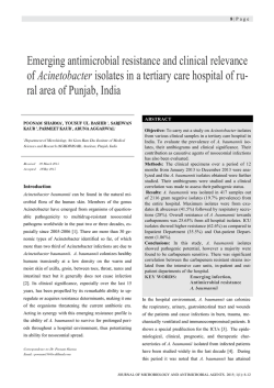
Antimicrobial Treatment of Ventilator-Associated Pneumonia David R Park MD Introduction The Importance of Ventilator-Associated Pneumonia (VAP) Strategies for Providing Optimal Antimicrobial Therapy Basic Principles of Antimicrobial Therapy Pertinent to VAP Definitions Classes of Antimicrobial Drugs, Mechanisms of Action, and Antimicrobial Spectra Pharmacokinetic and Pharmacodynamic Principles Factors That Influence Antimicrobial Drug Activity in the Lungs Penetration of Antimicrobial Drugs Into the Lungs Effect of the VAP Microenvironment on Antimicrobial Killing Antimicrobial Resistance in the Setting of VAP Prevalence of Antimicrobial Resistance in VAP Pathogens Importance of Antimicrobial Resistance in VAP A Clinical Approach to the Antimicrobial Treatment of VAP Current Opportunities and Challenges De-escalation Strategy for Antimicrobial Treatment of VAP Clinical and Bacteriological Strategies for Guiding VAP Treatment Factors to Consider in Selecting Initial Antimicrobial Therapy for VAP Data From Published Studies of VAP Etiology Local Microbiological Data Selecting Antimicrobial Therapy for VAP in Individual Patients Continuation Antimicrobial Therapy for VAP Antimicrobial Treatment of Specific “Problem Pathogens” Methicillin-Resistant Staphylococcus aureus Highly Resistant Gram-Negative Bacilli Legionnaires’ Disease Unresolved Questions About Conventional Antimicrobial Treatment of VAP Optimal Duration of Therapy Role of Combination Therapy Rotating Antimicrobial Therapy Unconventional Approaches to Antimicrobial Treatment Airway Delivery of Antimicrobial Drugs Use of Antimicrobial Drugs Lacking In Vitro Antimicrobial Efficacy Summary Ventilator-associated pneumonia is a common complication of ventilatory support for patients with acute respiratory failure and is associated with increased morbidity, mortality, and costs. Optimal antimicrobial therapy is an essential part of successful management of ventilator-associated pneumonia. Numerous safe and effective antimicrobial drugs are available, and their efficacy can be optimized by attention to basic pharmacokinetic and pharmacodynamic principles. An adequate 932 RESPIRATORY CARE • JULY 2005 VOL 50 NO 7 ANTIMICROBIAL TREATMENT OF VENTILATOR-ASSOCIATED PNEUMONIA initial empiric antimicrobial regimen is essential, because inadequate initial therapy is consistently associated with increased mortality. This regimen must be selected before final microbiology results become known, but likely pathogens and antimicrobial resistance patterns can be predicted based on published guidelines, patient-specific factors, and local epidemiologic data. Nevertheless, the initial regimen must often be broad-spectrum and typically requires combination therapy, with 2 or 3 different drugs, if there are risk factors for multidrug-resistant pathogens. The antimicrobial regimen can be narrowed or discontinued as culture and susceptibility results permit. This deescalation strategy ensures adequate initial antimicrobial therapy for most patients but lessens unnecessary antimicrobial exposure. The best diagnostic approach used to guide therapy, the optimum duration of therapy, and the roles of combination therapy, rotating therapy, and unconventional approaches to antimicrobial therapy all remain uncertain. Key words: ventilator-associated pneumonia, mechanical ventilation, treatment, nosocomial, pneumonia, antibiotic, antimicrobial, antibiotic-resistant, antimicrobial-resistant, pharmacokinetic, pharmacodynamic, review. [Respir Care 2005; 50(7):932–952. В© 2005 Daedalus Enterprises] Introduction The Importance of Ventilator-Associated Pneumonia Ventilator-associated pneumonia (VAP) is pneumonia that develops while a patient is receiving mechanical ventilation. The causes of VAP are many and varied, and antimicrobial resistance among VAP pathogens is increasingly prevalent.1 VAP is presently the most common nosocomial infection experienced by critically ill patients, especially in trauma, burn, and neurosurgical units.2 Whether VAP causes attributable mortality has been controversial because of the challenges of controlling for severity of illness, comorbidities, and other factors that may influence mortality.3,4 Nevertheless, VAP is clearly associated with increased morbidity, including prolonged duration of mechanical ventilation, prolonged length of stay, and markedly increased health care costs.3,4 Strategies for Providing Optimal Antimicrobial Therapy Optimal antimicrobial therapy of VAP is critically important, because inadequate initial antimicrobial therapy has consistently been associated with increased mortality.4,5 However, excessive antimicrobial therapy leads David R Park MD is affiliated with the Division of Pulmonary and Critical Care Medicine, Harborview Medical Center, University of Washington, Seattle, Washington. David R Park MD presented a version of this article at the 35th RESPIRATORY CARE Journal Conference, Ventilator-Associated Pneumonia, held February 25–27, 2005, in CancuВґn, Mexico. Correspondence: David R Park MD, Harborview Medical Center, Box 359762, 325 9th Avenue, Seattle WA 98104. E-mail: [email protected]. edu. RESPIRATORY CARE • JULY 2005 VOL 50 NO 7 to unnecessary treatment-related complications and costs and contributes to a further increase in the prevalence of antimicrobial resistance.4,6 This apparent paradox has led to the development of a strategy for antimicrobial treatment of VAP called “de-escalation.” According to this approach, a broad-spectrum combination antimicrobial regimen is selected initially, to ensure adequate coverage for all potential pathogens, even those with multidrug resistance. Once microbiology results are available, and after observing the clinical response, the initial empiric regimen can be narrowed or discontinued to prevent unnecessarily broad or prolonged antimicrobial use and its attendant risks and costs. This de-escalation strategy is schematized in Figure 1. The goals of this paper are to review basic principles of antimicrobial therapy that are pertinent to the management of VAP, to review the prevalence and importance of antimicrobial resistance in VAP pathogens, to describe various strategies and guidelines for choosing antimicrobial therapy for VAP, to discuss unresolved controversies in the use of conventional antimicrobial therapy, and to consider unconventional approaches to using antimicrobial therapy for VAP. Basic Principles of Antimicrobial Therapy Pertinent to VAP Definitions The terms “antimicrobial” and “antibiotic” are often used interchangeably, but have different meanings. Antimicrobial drugs are chemicals or substances that selectively inhibit the growth of microbes. Antimicrobials are distinguished from antiseptics and disinfectants by the fact that antimicrobials have sufficiently low mammalian toxicity that they can be tolerated systemically. Antibiotics are a 933 ANTIMICROBIAL TREATMENT OF VENTILATOR-ASSOCIATED PNEUMONIA Fig. 1. Schematic representation of the de-escalation strategy for antimicrobial management of ventilator-associated pneumonia. From left to right: Ventilator-associated pneumonia is suspected on the basis of clinical and radiographic features, and a variety of potentially multidrug-resistant pathogens may be responsible. After obtaining microbiology samples, broad-spectrum empiric antimicrobial therapy is initiated. Based on the microbiology and antimicrobial susceptibility results, the initial empiric antimicrobial regimen can be narrowed or even discontinued. Finally, the duration of therapy can be shortened if the clinical response and pathogen are favorable. MRSA П methicillin-resistant Staphylococcus aureus. ESBL П extended spectrum beta-lactamase producing bacilli. NF GNR П nonfermenting Gram-negative rods. naturally occurring subcategory of antimicrobial drugs that are produced by living microbes rather than by synthetic chemistry techniques. This distinction has little practical importance and is often blurred when natural antibiotic compounds are chemically modified to produce altered pharmacologic properties. Nevertheless, in this paper I will use the term antimicrobial to indicate both natural and synthetic compounds with antimicrobial activity. The main focus of this paper will be the antibacterial treatment of bacterial causes of VAP. Classes of Antimicrobial Drugs, Mechanisms of Action, and Antimicrobial Spectra All antimicrobials work by interfering with basic metabolic functions of the microbial cell, such as cell wall formation, deoxyribonucleic acid (DNA) replication, ribonucleic acid (RNA) synthesis, protein synthesis, synthesis of essential metabolites, and maintenance of cell membrane integrity. Individual antimicrobials may be bactericidal or bacteriostatic, depending on whether, at clinically achievable concentrations, they kill bacteria outright or merely inhibit bacterial growth. Antimicrobial drugs can be classified in various ways; the most practical seems to be on the basis of their structure and mechanisms of antimicrobial action. An outline of this categorization for antibacterial drugs is shown in Table 1. Many classes of antimicrobial drugs function by inhibiting bacterial cell wall formation. These include the betalactam-ring-containing penicillins, cephalosporins, 934 monobactams, and carbapenems. All of these drugs bind to bacterial penicillin binding proteins and prevent peptidoglycan cross-linking in the cell wall. The glycopeptide antibiotic vancomycin is another cell-wall-synthesis inhibitor with an unrelated structure. It works by binding to peptidoglycan precursor moieties. All of these cell wallactive drugs are bactericidal. Another large group of antimicrobial drugs interferes with bacterial protein synthesis. All work by binding to various subunits of the ribosome or ribosome-RNA complex and inhibiting protein translation. The aminoglycosides bind irreversibly and have bactericidal activity. The macrolides, tetracyclines, lincosamides, oxazolidinones, and streptogramins all bind reversibly and are generally bacteriostatic in action. However, at high concentrations they may be bactericidal for certain pathogens. A single class of antimicrobial drugs functions by interfering directly with DNA replication. The fluoroquinolones inhibit DNA gyrase and topoisomerases that are essential for DNA replication during cell division. Fluoroquinolones are bactericidal. Rifamycins such as rifampin, best known as anti-tuberculosis drugs, block RNA synthesis by inhibiting the DNAdependent RNA polymerase. Antimetabolite antimicrobials block critical bacterial metabolic pathways. For example, the sulfonamide drug and para-amino benzoic acid analog sulfamethoxazole functions by inhibiting dihydropteroate synthase and interfering with nucleic acid synthesis. Sulfonamides are bacteriostatic, but can be bactericidal when combined with a sequential inhibitor of folate metabolism such as trimethoprim. Finally, antimicrobial drugs of the polymixin class, such as colistin, insert themselves into the bacterial plasma membrane in detergent-like fashion and impair the permeability barrier function of the cell, leading to rapidly bactericidal effects. The antimicrobial spectrum of an antimicrobial drug refers to the types of pathogens that are susceptible to killing by the drug. This, in turn, depends on the drug’s mechanism of action and whether or not a specific pathogen is susceptible to attack by that mechanism. Table 2 lists commonly used antimicrobial drugs and the pathogens that usually can be treated with each drug. The microbial causes of VAP were reviewed in the previous issue of RESPIRATORY CARE.1 Pharmacokinetic and Pharmacodynamic Principles The efficacy of any antimicrobial drug depends on the concentrations of the drug that can safely be achieved and maintained in the blood and at the site of infection (pharmacokinetics), and the antimicrobial activity of the drug at RESPIRATORY CARE • JULY 2005 VOL 50 NO 7 ANTIMICROBIAL TREATMENT Table 1. OF VENTILATOR-ASSOCIATED PNEUMONIA Classification of Antimicrobial Drugs Used in the Treatment of Ventilator-Associated Pneumonia, According to Mechanisms of Action Antibiotic Class Cell Wall-Active Drugs Beta-lactams Sub-Class/Examples Mechanisms of Action Penicillins Cephalosporins Monobactams Carbapenems Vancomycin Inhibit cell wall synthesis by binding penicillin binding proteins Gentamycin Erythromycin Doxycycline Clindamycin Linezolid Quinupristin/dalfopristin Inhibit protein Inhibit protein Inhibit protein complex Inhibit protein Inhibit protein Inhibit protein DNA Synthesis Inhibitors Fluoroquinolones Ciprofloxacin Inhibit DNA synthesis by blocking DNA gyrase and topoisomerase enzymes RNA Synthesis Inhibitors Rifamycins Rifampin Inhibit DNA-dependent RNA polymerase Antimetabolites Sulfonamides Sulfamethoxazole Inhibit folic acid synthesis Miscellaneous Mechanisms Polymixins Nitroimidazole Colistin (Polymixin E) Metronidazole Disruption of cytoplasmic membrane function Direct DNA damage after reductive activation of pro-drug Glycopeptides Protein Synthesis Inhibitors Aminoglycosides Macrolides Tetracyclines Lincosamide Oxazolidinones Streptogramins Inhibit cell wall synthesis by binding peptidoglycan precursor molecules synthesis by binding irreversibly to 30S ribosomal subunit synthesis by binding reversibly to 50S ribosomal subunit synthesis by interfering with tRNA attachment to mRNA-ribosome synthesis by binding reversibly to 50S ribosomal subunit synthesis by interfering with 70S ribosomal-RNA initiation complex synthesis by binding to 50S ribosomal subunits mRNA П messenger ribonucleic acid tRNA П transfer ribonucleic acid DNA П deoxyribonucleic acid that concentration-time profile against a given pathogen (pharmacodynamics).7,8 The key concepts of pharmacokinetics are illustrated in Figure 2. The drug concentration in blood or tissues rises rapidly after a single dose, reaches a maximum concentration (Cmax), then falls steadily toward zero. Repeated doses at regular intervals lead to steadystate maximum (peak) and minimum (trough) drug levels that depend on the dose, the dosing interval, the volume of distribution, and the rate of clearance. The concentrationtime profile for any particular antimicrobial regimen can be compared against the minimum inhibitory concentration (MIC) of the drug necessary to inhibit bacterial growth by 90% (the MIC90). For some types of antimicrobial drugs, predictable bacterial killing correlates best with the ratio between the Cmax and the MIC90, or between the area under the curve (AUC) of the drug-concentration-versus-time profile and the MIC (AUC/MIC). The higher the concentration of the drug above the MIC, the more effective the killing. This RESPIRATORY CARE • JULY 2005 VOL 50 NO 7 form of pharmacodynamic response is termed concentration-dependent killing. Concentration-dependent killing is most characteristic of the effects of aminoglycoside and fluoroquinolone antimicrobials. In experimental models and in clinical studies, a Cmax/MIC ratio of Пѕ 10 or an AUC/MIC ratio of Пѕ 125 hours predicts a favorable clinical and bacteriological response.9 –12 For other classes of antimicrobial drugs, maximal killing depends not on the peak concentration of the drug, but on the proportion of time that the drug concentration exceeds the MIC. This form of response is termed concentration-independent killing, or time-dependent killing. Concentration-independent killing is characteristic of most of the cell wall-active antibiotics. In infection models and in clinical studies, inhibition of growth is likely if the drug concentration exceeds the MIC for at least 40% of the dosing interval, and a maximal bacteriological response is predicted if the drug concentration exceeds the MIC for at least 60 –70% of the dosing interval.7,9,13,14 935 ANTIMICROBIAL TREATMENT Table 2. OF VENTILATOR-ASSOCIATED PNEUMONIA Antimicrobial Spectrum of Antimicrobial Drugs Used in the Treatment of Ventilator-Associated Pneumonia Antibiotic Class Cell Wall-Active Drugs Beta-lactams Penicillins Natural penicillins Aminopenicillins Penicillinase-resistant Anti-Pseudomonal Cephalosporins 1st-generation 2nd-generation 3rd-generation Specific Examples Target Pathogens Penicillin G Ampicillin Nafcillin, Methicillin Piperacillin, Ticarcillin SP SP, EGNB, HI, and ES MSSA HRGNB except ESBL Cefazolin Cefuroxime Ceftriaxone, Cefotaxime Ceftazidime Cefepime Aztreonam Imipenem-cilastatin, Meropenem Ertapenem Vancomycin MSSA SP, HI, and EGNB SP, HI, and EGNB All GNB except ESBL SP, MSSA, and GNB except ESBL HI, EGNB, and HRGNB SP, MSSA, EGNB, ESBL, HRGNB, ES SP, MSSA, EGNB, ES MRSA, ES Gentamycin, Tobramycin, Amikacin Erythromycin Azithromycin, Clarithromycin Doxycycline Minocycline Clindamycin Linezolid Quinupristin/dalfopristin EGNR, HRGNR, MSSA SP, MSSA, LS SP, MSSA, HI, EGNR, LS LS AB, SM MSSA, Anaerobes MRSA, VRE MRSA, VRE Ciprofloxacin, Levofloxacin Moxifloxacin, Gatifloxacin EGNR, HRGNR, MSSA, LS SP, MSSA, HI, EGNR, LS RNA Synthesis Inhibitors Rifamycins Rifampin LS, MRSA Antimetabolites Sulfonamides Sulfamethoxazole (with Trimethoprim) SM, AB, MRSA, (PC) Miscellaneous Polymixins Nitroimidazoles Colistin (Polymixin E) Metronidazole HRGNB Anaerobes 4th-generation Monobactams Carbapenems Glycopeptides Protein Synthesis Inhibitors Aminoglycosides Macrolides Tetracyclines Lincosamides Oxazolidinones Streptogramins DNA Synthesis Inhibitors Fluoroquinolones SP П Streptococcus pneumoniae and other streptococci EGNB П enteric Gram-negative bacilli HI П Haemophilus influenzae ES П enterococcal species MSSA П methicillin-susceptible Staphylococcus aureus HRGNB П highly-resistant Gram-negative bacilli GNB П Gram-negative bacilli ESBL П extended-spectrum beta-lactamase producing GNB AB П Acinetobacter species MRSA П methicillin-resistant Staphylococcus aureus EGNR П enteric Gram-negative rods HRGNR П highly resistant Gram-negative rods LS П Legionella species SM П Stenotrophomonas maltophilia VRE П vancomycin-resistant enterococci PC П Pneumocystis carinii Another interesting pharmacodynamic property of certain antimicrobial drugs is that their inhibitory effects persist for some time after the drug concentration has fallen below the MIC (Fig. 3). This phenomenon is termed the “post-antibiotic effect.” Most antimicrobials exhibit some post-antibiotic effect on Gram-positive respiratory pathogens. However, betalactam drugs have no post-antibiotic effect on Gram-negative bacilli. A prolonged post-antibiotic effect is most character- 936 RESPIRATORY CARE • JULY 2005 VOL 50 NO 7 ANTIMICROBIAL TREATMENT OF VENTILATOR-ASSOCIATED PNEUMONIA Fig. 2. Basic pharmacokinetic and pharmacodynamic principles. The figure demonstrates changes in antimicrobial drug concentrations over time during regular intermittent dosing. The concentration rises rapidly to a peak (Cmax) before falling to the steadystate trough level. The proportion of time that the concentration is above the minimum inhibitory concentration (MIC) and the area under the curve (AUC) of the concentration-time plot are illustrated. See text for explanation. istic of aminoglycosides, fluoroquinolones, and carbapenems, as well as macrolides, tetracyclines, and rifampin.7 Post-antibiotic effects may be even more evident in vivo than in vitro, because bacterial morphology and expression of virulence factors can be altered by low concentrations of drug that are insufficient to inhibit bacterial growth in culture media.15–17 These effects are termed “sub-MIC activity.” Many of these changes make the bacteria more susceptible to ingestion and killing by leukocytes— effects termed “post-antibiotic leukocyte enhancement.”7,16 Fig. 3. Post-antibiotic effect. The figure indicates the effect of a single dose of antimicrobial drug on bacterial growth in 2 different circumstances. In both cases, growth is inhibited when the drug concentration exceeds the MIC. After the concentration falls below MIC, bacterial growth either resumes immediately (indicating no post-antibiotic effect, as during treatment with beta-lactam antimicrobials) or remains inhibited for a period of time (indicating a post-antibiotic effect, as occurs during treatment with aminoglycosides or fluoroquinolones). See text for further explanation. RESPIRATORY CARE • JULY 2005 VOL 50 NO 7 These pharmacokinetic and pharmacodynamic properties of antimicrobial drugs have more than academic importance. For instance, a drug that demonstrates concentration-dependent killing and a prolonged post-antibiotic effect should be administered in large, infrequent doses to achieve maximum efficacy. Once-daily dose regimens of aminoglycosides and fluoroquinolones depend on these principles. In contrast, antimicrobials that demonstrate concentration-independent killing and little or no post-antibiotic effect are most effective if maintained just above the MIC of the relevant pathogen. This necessitates the frequent dosing intervals of penicillins and most cephalosporins and underlies the motivation for studying continuous infusions of these drugs.18,19 Hypothetical dosing regimens based on these principles are depicted in Figure 4. In addition, these principles underlie the recommended drug and dosage recommendations in the joint American Thoracic Society/Infectious Diseases Society of America guidelines for the management of VAP (Table 3).4 Factors That Influence Antimicrobial Drug Activity in the Lungs Penetration of Antimicrobial Drugs Into the Lungs The pharmacologic principles outlined in the previous section apply best to a simple single-compartment model Fig. 4. The figure shows antimicrobial drug concentrations over time during 2 hypothetical intermittent dosing regimens. The solid line indicates concentrations attained using large, infrequent doses. The peak concentration is higher, but the concentration falls below the minimum inhibitory concentration (MIC) for a prolonged time. This approach is optimal when using drugs with concentrationdependent killing and prolonged post-antibiotic effects such as aminoglycosides and fluoroquinolones. The dotted line indicates concentrations attained using smaller, frequent doses. The peak concentrations are lower, but the concentration rarely falls below the MIC. This approach is best when using drugs with time-dependent killing such as beta-lactam antibiotics. The logical extension of this approach is to use continuous infusion that maintains the drug concentration above the MIC (not shown). See text for further explanation. 937 ANTIMICROBIAL TREATMENT Table 3. OF VENTILATOR-ASSOCIATED PNEUMONIA Recommended Dosages of Parenteral Antimicrobial Drugs Used for the Treatment of Ventilator-Associated Pneumonia Antimicrobial Drug Cephalosporins Ceftriaxone Cefepime Ceftazidime Carbapenems Ertapenem Imipenem Meropenem Beta-Lactam/Beta-Lactamase Inhibitors Ampicillin-Sulbactam Piperacillin-Tazobactam Aminoglycosides Gentamycin Tobramycin Amikacin Antipseudomonal Fluoroquinolones Moxifloxacin Ciprofloxacin Levofloxacin Drugs Effective Against MRSA Vancomycin Linezolid Miscellaneous Azithromycin Minocycline Colistin Recommended Dosage* 1–2 g every 24 h 1–2 g every 8–12 h 2 g every 8 h 1 g every 24 h 500 mg every 6 h, or 1 g every 8 h 1 g every 8 h 3 g every 6 h 4.5 g every 6 h 7 mg/kg every 24 h†7 mg/kg every 24 h†20 mg/kg every 24 h†400 mg every 24 h 400 mg every 8 h 750 mg every 24 h 15 mg/kg every 12 hВ§ 600 mg every 12 h 500 mg every 24 h 100 mg every 12 h 2.5–5 mg/kg/d, in divided dosesВ¶ MRSA П methicillin-resistant Staphylococcus aureus *These dosages are based on adult patients with normal renal and hepatic function. †Trough levels for gentamycin and tobramycin should be ПЅ 1 вђ®g/mL and for amikacin they should be ПЅ 4–5 вђ®g/mL. В§Trough levels for vancomycin should be 15–20 вђ®g/mL. В¶Colistin doses must be adjusted based on creatinine clearance, usually without measurement of drug levels. (Adapted from Reference 4.) such as the bloodstream. When infection involves tissues such as the lung, several additional factors come into play. First of all, penetration of antimicrobial drugs from the blood into the lung is a critical factor in determining the response to treatment. The partition of drugs between the blood and lungs is highly variable.20 –24 Some drugs appear to penetrate lung tissue primarily via diffusion (eg, betalactams, vancomycin, aminoglycosides). Others appear to be concentrated in the lung lining fluid, alveolar macrophages, and other leukocytes (eg, macrolides and fluoroquinolones).25–27 Obviously, the lung concentration of antimicrobial drugs is difficult to measure in patients with pneumonia because of the inaccessibility of the lungs for sampling. Bronchoalveolar lavage fluid concentrations have been used as a surrogate for lung tissue drug concentrations. This approach can be improved by correcting 938 for the variable dilution of lung lining fluid during the lavage procedure. Lung tissue homogenate levels are intuitively more appealing but can be misleading as to the free drug concentration in the air spaces or interstitial spaces where invading microbes are encountered. This is because lung homogenates include primarily cellular contents, so lung homogenate antimicrobial concentrations depend heavily on whether or not the drug has accumulated intracellularly.7 Recently, a novel technique using a microdialysis catheter has been used to measure interstitial antimicrobial concentrations in the peripheral lung of patients following thoracic surgery.28 –30 This and other techniques may be useful in clinical pharmacokinetics studies but seem unlikely to be adopted for routine clinical practice. One way to circumvent the problem of poor lung penetration by antimicrobial drugs is to deliver the drugs directly into the airways. This approach will be discussed in a subsequent section of this paper. Effect of the VAP Microenvironment on Antimicrobial Killing Lung tissue concentrations of antimicrobial drugs can vary considerably, compared with serum levels, as discussed above. Another set of factors influencing the antimicrobial effects of drugs in the lungs are the local conditions in the lung and VAP microenvironment. Some otherwise efficacious antimicrobial drugs such as daptomycin are unsuitable for the treatment of pneumonia because they are inactivated by surfactants in the air spaces. Others, particularly aminoglycosides, function poorly in the acidic and hypoxic environment present in an area of lung consolidation due to pneumonia.31–34 In contrast, beta-lactam antimicrobials appear to have enhanced killing effects in these conditions.33 Another key factor affecting antimicrobial killing in VAP is the role of microbes present in the endotracheal tube biofilm. The biofilm is a complex polysaccharide matrix that forms an adherent, slimy coating on the surface and within the lumen of the endotracheal tube, and on other medical devices. The biofilm may play an important role in the pathogenesis of VAP by supporting colonization of the endotracheal tube and facilitating repeated inoculation of the lower airways.35 Pathogens within the biofilm are protected from airway mucosal clearance mechanisms and are protected from systemically administered antimicrobial drugs by the wall of the endotracheal tube and by the fact that the biofilm matrix acts as a barrier to penetration by the drugs.35,36 In addition, bacteria within the biofilm adopt a sessile growth pattern and develop numerous functional and morphological changes, compared with their strain-mates grown in planktonic free-floating form in broth cultures. The most clinically important of these changes may be that biofilm growth results in markedly reduced RESPIRATORY CARE • JULY 2005 VOL 50 NO 7 ANTIMICROBIAL TREATMENT Table 4. OF VENTILATOR-ASSOCIATED PNEUMONIA Prevalence of Antimicrobial Drug Resistance Among Nosocomial ICU Pathogens in the United States in 1998 –2004 Antimicrobial-Resistant Pathogen Number of Isolates Tested Percent Resistant Methicillin-resistant Staphylococcus aureus Methicillin-resistant coagulase-negative staphylococci Vancomycin-resistant enterococci Ciprofloxacin-resistant Pseudomonas aeruginosa Levofloxacin-resistant Pseudomonas aeruginosa Ceftazidime-resistant Pseudomonas aeruginosa Piperacillin-resistant Pseudomonas aeruginosa 3rd-generation cephalosporin-resistant Enterobacter species Carbapenem-resistant Enterobacter species 3rd-generation cephalosporin-resistant Klebsiella pneumoniae 3rd-generation cephalosporin-resistant Escherichia coli Quinolone-resistant Escherichia coli Penicillin-resistant pneumococci Cefotaxime/Ceftriaxone-resistant pneumococci 22,899 13,553 14,140 13,473 5,895 12,805 11,640 5,328 4,663 7,529 12,011 11,776 1,331 854 52.9 76.6 13.9 34.8 35.3 13.9 17.5 27.7 0.7 6.2 1.3 7.3 18.9 7.5 Not shown are carbapenem-resistant Acinetobacter species. (Data from Reference 2.) susceptibility to aminoglycoside and beta-lactam antibiotics.36,37 Antimicrobial Resistance in the Setting of VAP Prevalence of Antimicrobial Resistance in VAP Pathogens The spectrum of microbial causes of VAP and the settings in which antimicrobial resistance may be encountered have recently been reviewed.1,3,4 The prevalence of antimicrobial resistance among VAP pathogens is steadily increasing.38,39 Many VAP pathogens exhibit intrinsic resistance to commonly used antimicrobial drugs, but acquired antimicrobial resistance to broad-spectrum antimicrobial drugs is becoming increasingly common. For instance, the most recent summary of data from the National Nosocomial Infection Surveillance system compared antimicrobial resistance rates of intensive care unit (ICU) pathogens recovered during the era of 1998 –2002 with those tested in 2003.2 Over this short interval, Staphylococcus aureus resistance to methicillin increased 11% and Klebsiella resistance to 3rd-generation cephalosporins increased an incredible 47%. Over the same interval, Pseudomonas resistance to imipenem, fluoroquinolones, and 3rdgeneration cephalosporins increased 15%, 9%, and 20%, respectively.2 The overall prevalence of antimicrobial resistance among nosocomial ICU pathogens during the period from 1998 –2004 is shown in Table 4. Basic mechanisms of antimicrobial resistance to commonly used drugs have been reviewed40 – 42 and are outlined in Table 5. RESPIRATORY CARE • JULY 2005 VOL 50 NO 7 Importance of Antimicrobial Resistance in VAP The increasing emergence of antimicrobial resistance has reached a crisis stage, prompting some authorities to warn of a return to the state of helplessness against serious infections known in the pre-antibiotic era.43– 46 Nowhere is antimicrobial resistance more problematic than in the ICU.38,44,47 In fact, the specter of antimicrobial resistance complicates the management of nearly every patient with VAP. Antimicrobial resistance increases the likelihood of an inadequate initial antibiotic regimen and of increased morbidity and mortality resulting from inadequate initial treatment. As a result, the mere possibility of infection due to antimicrobial-resistant pathogens necessitates broadspectrum initial empiric antimicrobial therapy, usually with a combination of drugs. This increases the costs of treatment, the occurrence of adverse drug effects, and ironically, the local prevalence of antimicrobial resistance. A Clinical Approach to the Antimicrobial Treatment of VAP Current Opportunities and Challenges The clinician responsible for antimicrobial treatment of critically ill patients with VAP has available a large armamentarium of antimicrobial drugs that are generally safe, have well-characterized mechanisms of action, and have well-understood pharmacokinetic and pharmacodynamic properties. Unfortunately for the clinician, the selection of antimicrobial therapy for VAP is a complex and challenging decision because of the diversity of microbial causes 939 ANTIMICROBIAL TREATMENT Table 5. OF VENTILATOR-ASSOCIATED PNEUMONIA Mechanisms of Resistance to Commonly Used Antimicrobial Drugs Antibiotic/Class Penicillins/Cephalosporins Resistance Mechanism Representative Pathogen(s) Altered penicillin-binding protein Beta-lactamases Extended-spectrum beta-lactamases Efflux pumps Outer-membrane porin protein deficiency Aztreonam Altered penicillin-binding protein Beta-lactamases Extended-spectrum beta-lactamases Carbapenems Altered penicillin-binding protein Outer membrane porin protein deficiency Carbapenemase Vancomycin Aminoglycosides Efflux pumps Altered peptidoglycan precursor Linezolid Efflux pumps Aminoglycoside-altering enzymes Altered ribosomal subunit Streptogramins Altered ribosomal subunit Fluoroquinolones Streptogramin acetyltransferases Efflux pumps Altered DNA gyrase or Topoisomerase IV Efflux pumps Staphylococcus aureus Pseudomonas aeruginosa Enterobacter cloacae Acinetobacter baumannii Klebsiella pneumoniae Escherichia coli Pseudomonas aeruginosa Pseudomonas aeruginosa Enterobacter cloacae Staphylococcus aureus Pseudomonas aeruginosa Enterobacter cloacae Klebsiella pneumoniae Escherichia coli Staphylococcus aureus Acinetobacter baumannii Pseudomonas aeruginosa Acinetobacter baumannii Pseudomonas aeruginosa Acinetobacter baumannii Pseudomonas aeruginosa Staphylococcus aureus Enterococci Pseudomonas aeruginosa Pseudomonas aeruginosa Staphylococcus aureus Enterococci Staphylococcus aureus Enterococci Staphylococcus aureus Staphylococcus aureus All pathogens Pseudomonas aeruginosa DNA П deoxyribonucleic acid (Adapted from References 40–42.) of VAP,1,3 the increasing prevalence of antimicrobial resistance among VAP pathogens,2,38,44,47 delays in receiving definitive microbiology and susceptibility reports, and the pressure to avoid excessive antimicrobial drug use.6 Clinical studies have consistently shown that adequate initial antimicrobial therapy for VAP is associated with lower mortality rates, compared with patients receiving inadequate initial therapy,48 –56 and changing to appropriate therapy after waiting for culture results may not reduce the risk of mortality.49 –51 These findings suggest that the selection of initial empiric antimicrobial therapy is a critically important management decision for patients with VAP. Adding to the challenge and reflecting the necessary considerations for optimal therapy for VAP, experts in the field draw a distinction between “appropriate” antimicrobial therapy and an “adequate” antimicrobial reg- 940 imen.4 Appropriate therapy means the pathogen is susceptible to the chosen antimicrobial drug. An adequate antimicrobial regimen means that appropriate antimicrobial drugs are selected, given in optimal dosages, by the correct route, in effective combinations, and for the appropriate duration. A new paradigm for antimicrobial treatment of VAP, called de-escalation, has evolved over the last decade to address these challenges and the seemingly opposed goals of adequate yet not excessive antimicrobial drug use. De-escalation Strategy for Antimicrobial Treatment of VAP De-escalation refers to a strategy of aggressive broadspectrum initial empiric antimicrobial therapy, followed by narrowing or discontinuation of antimicrobial drugs RESPIRATORY CARE • JULY 2005 VOL 50 NO 7 ANTIMICROBIAL TREATMENT OF VENTILATOR-ASSOCIATED PNEUMONIA after the results of antimicrobial susceptibility tests become available and the clinical course can be observed (see Fig. 1). Using this approach, microbiology samples are collected as soon as VAP is suspected. Antimicrobial therapy is initiated promptly and chosen to cover all likely pathogens62 in order to minimize the chance of inadequate initial therapy. After the antimicrobial susceptibility results are known, any unnecessary drugs are discontinued. Drugs with a narrow spectrum that target the known pathogens are substituted for drugs with an unnecessarily broad spectrum in order to minimize excessive antimicrobial exposure. Finally, the duration of therapy is limited. This general strategy is fully endorsed by the new American Thoracic Society/Infectious Diseases Society of America guidelines for the management of VAP.4 Clinical and Bacteriological Strategies for Guiding VAP Treatment A variety of different strategies for the management of VAP have been proposed.4,57– 61 All involve some form of clinical and microbiological assessment both to identify patients with VAP and to guide their antimicrobial treatment. Discussion of the various diagnostic approaches is beyond the scope of this review. They can be summarized as either clinical or bacteriological.4 Clinical diagnostic strategies rely on findings of fever, leukocytosis, and purulent sputum to diagnose VAP in patients with radiographic infiltrates. Gram stains and cultures of endotracheal aspirates are used to guide antimicrobial therapy but not to determine whether or not pneumonia is present. Advantages of the clinical approach include its sensitivity, simplicity, and low cost. No specialized laboratory or bronchoscopy services are needed. Disadvantages of the clinical approach include its lack of specificity, both for identifying patients with true VAP and for identifying the true VAP pathogen out of the multiple isolates that may be recovered from the trachea. As a result, many patients without VAP are treated unnecessarily, and many patients with VAP are given unnecessarily broad-spectrum antimicrobial coverage. Bacteriological diagnostic strategies also use clinical findings to identify patients with suspected VAP, but they rely on quantitative cultures to distinguish those with true infection from those with colonization. Advantages of the bacteriological approach include far better specificity, both for the diagnosis of VAP and for pinpointing the microbial etiology. Disadvantages are that specialized laboratory techniques and, usually, bronchoscopy services are needed. Also, bronchoscopy procedures are expensive and involve some risks to the patient. However, nonbronchoscopic sampling approaches are becoming more widely accepted, and even quantitative culture of the endotracheal aspirate may be an adequate quantitative bacteriological approach. RESPIRATORY CARE • JULY 2005 VOL 50 NO 7 Fagon and coworkers compared a clinical strategy (noninvasive endotracheal suction specimens and qualitative cultures) with a bacteriological strategy (invasive bronchoscopic sample collection and quantitative cultures).62 They used the initial Gram stain to tailor initial empiric therapy, they withheld immediate antimicrobial therapy if the Gram stain was negative, and they discontinued antimicrobials if quantitative cultures were below the diagnostic threshold or if qualitative cultures were negative (see Fig. 5). No other study comparing clinical and bacteriological strategies has employed all of these measures. In the end, patients in the invasive bacteriological arm received far fewer antimicrobials, less often received inappropriate antimicrobial therapy, and were less likely to die.62 I favor a similar invasive bacteriological strategy, because this approach offers the best chance to help determine which patients should be treated immediately, to select appropriate initial antimicrobial therapy, and to stop unnecessary empiric antimicrobial therapy and search for another diagnosis if quantitative culture results do not indicate pneumonia.62,63 In practice settings where bronchoscopy and/or quantitative bacterial culture services are not available, a clinical strategy including semi-quantitative endotracheal aspirate cultures is a reasonable alternative.4 Factors to Consider in Selecting Initial Antimicrobial Therapy for VAP Data From Published Studies of VAP Etiology The most simple approach to selecting initial empiric antimicrobial therapy is based on the prevalence of and risk factors for specific pathogens reported in published studies. In general, the likelihood of multidrug-resistant pathogens such as Pseudomonas aeruginosa, Acinetobacter species, Stenotrophomonas maltophilia, and methicillinresistant S. aureus (MRSA) causing VAP depends on the duration of hospitalization prior to the onset of VAP and other risk factors for multiple-drug-resistant pathogens.1,3,4 These risk factors include antimicrobial therapy in the preceding 90 days, current hospitalization duration of 5 days or longer, a high frequency of antibiotic resistance in the community or in the specific hospital unit, hospitalization for 2 days or more in the preceding 90 days, residence in a nursing home or extended care facility, home infusion therapy, chronic dialysis within 30 days, home wound care, a family member with a multidrug-resistant pathogen, and immunosuppressive disease or therapy (Table 6).4 For early-onset VAP (within 5 days of hospitalization), in the absence of risk factors for multidrug-resistant pathogens, antimicrobial-sensitive community-acquired pathogens are likely, and empiric initial antimicrobial treatment can be provided with simple monotherapy, using a nonanti-pseudomonal 3rd-generation cephalosporin, an anti 941 ANTIMICROBIAL TREATMENT OF VENTILATOR-ASSOCIATED PNEUMONIA Fig. 5. Invasive (bacteriological) and noninvasive (clinical) strategies for guiding antimicrobial treatment of ventilator-associated pneumonia. The figure shows algorithms for the management of suspected ventilator-associated pneumonia, on the basis of a noninvasive clinical strategy (A) and an invasive bronchoscopically-guided bacteriological strategy (B). (From Reference 62, with permission.) 942 RESPIRATORY CARE • JULY 2005 VOL 50 NO 7 ANTIMICROBIAL TREATMENT Table 6. OF VENTILATOR-ASSOCIATED PNEUMONIA Risk Factors for Multidrug-Resistant Ventilator-Associated Pneumonia Pathogens* Duration of current hospitalization 5 days or longer Recent antimicrobial therapy (in the preceding 90 days) Recent hospitalization (for 2 days or more in the preceding 90 days) High frequency of antibiotic resistance in the community or in the specific hospital unit Residence in a nursing home or extended-care facility Home infusion therapy Chronic dialysis (within 30 days) Home wound care Family member with a multidrug-resistant pathogen Immunosuppressive disease or therapy *Multidrug-resistant pathogens include Pseudomonas aeruginosa, Acinetobacter baumannii, Stenotrophomonas maltophilia, and methicillin-resistant Staphylococcus aureus. (Adapted from Reference 4.) Table 7. Potential Pathogens and Recommended Initial Antimicrobial Treatment for Early-Onset VentilatorAssociated Pneumonia With No Risk Factors for Multidrug-Resistant Pathogens* Potential Pathogens Streptococcus pneumoniae Haemophilus influenzae Methicillin-sensitive Staphylococcus aureus Antibiotic-sensitive enteric Gram-negative bacilli Escherichia coli Klebsiella pneumoniae Enterobacter species Proteus species Serratia marcescens Recommended Antibiotic Therapy Ceftriaxone or Levofloxacin, moxifloxacin, ciprofloxacin or Ampicillin/sulbactam or Ertapenem *Multidrug-resistant pathogens include Pseudomonas aeruginosa, Acinetobacter baumannii, Stenotrophomonas maltophilia, and methicillin-resistant Staphylococcus aureus. See Table 6 for risk factors. (Adapted from Reference 4.) pneumococcal fluoroquinolone, ampicillin/sulbactam, or ertapenem (Table 7).4 The choice of an individual antimicrobial agent in this group is determined mainly by local availability and cost factors, and by other influences, such as the allergy history. A fluoroquinolone is a sensible choice if nosocomial Legionnaires’ disease is prevalent. If VAP is late in onset (Х† 5 days after hospitalization) or if other risk factors for a multidrug-resistant pathogen are present (see Table 6), then broad-spectrum combination therapy should be given initially (Table 8).4 The need for combination antimicrobial therapy for late-onset VAP is nicely illustrated by the work of Trouillet and colleagues.64 They showed that, in 245 isolates from 135 episodes of bronchoscopically confirmed VAP, the maximum proportion of cases that could have been covered RESPIRATORY CARE • JULY 2005 VOL 50 NO 7 adequately by a single antimicrobial drug was 64% (using imipenem). The empiric regimen having the best coverage for all of the isolates was imipenem, amikacin, and vancomycin. Even then, all pathogens would have been adequately covered in only 88% of cases. Triple-combination initial antimicrobial therapy was predicted to result in both improved survival and reduced overall costs in a hypothetical decision analysis model.65 Recommended combination regimens for late-onset VAP include an antipseudomonal cell wall-active agent such as cephalosporin, carbapenem, or beta-lactam/beta-lactamase inhibitor, plus an antipseudomonal fluoroquinolone or aminoglycoside. If Legionnaires’ disease is suspected, then the regimen should include a fluoroquinolone, or a macrolide should be added. Linezolid or vancomycin should be added when MRSA is suspected. Finally, for patients who have received prior antimicrobial therapy, the use of a different antimicrobial drug class is recommended to lessen the chance of cross-resistance.4 In all cases, the antimicrobial dosage should be optimized (see Table 3). Local Microbiological Data The generic approach outlined above is effective at identifying patients at risk for multidrug-resistant pathogens. However, individual institutions may vary considerably in terms of both the prevalence of specific VAP pathogens and their antimicrobial susceptibility patterns.1,66 – 68 The practical implications of these data are that empiric antibiotic treatment decisions for patients with VAP must take into account local microbiology and antimicrobial susceptibility data, preferably using VAP-specific data.1 Selecting Antimicrobial Therapy for VAP in Individual Patients Even local microbiology data may fail to guide appropriate antimicrobial therapy in an individual patient with VAP. Ideally, clinicians would use a rapid and accurate diagnostic test that would identify both the pathogen and its antimicrobial susceptibilities as soon as VAP is suspected. Absent this ideal, we can consider the results of other microbiology data, including the results of prior airway cultures and the appearance of Gram stains of specimens obtained after VAP is suspected. Discussion of criteria for determining whether VAP is present or not is beyond the scope of this review. The focus here is on whether or not prior airway cultures or immediate Gram stain results of fresh specimens can help to predict the causative pathogen (and antimicrobial susceptibilities by inference), once the decision to treat VAP has been made. The Predictive Value of Prior Respiratory Tract Cultures. Hayon and colleagues attempted to determine the utility of prior respiratory tract cultures in a prospective 943 ANTIMICROBIAL TREATMENT Table 8. OF VENTILATOR-ASSOCIATED PNEUMONIA Potential Pathogens and Recommended Initial Antimicrobial Treatment for Ventilator-Associated Pneumonia When Late-Onset or Other Risk Factors for Multidrug-Resistant Pathogens Are Present* Potential Pathogens Pathogens listed in Table 7 plus: Multidrug-resistant pathogens Pseudomonas aeruginosa Acinetobacter species Stenotrophomonas maltophilia Enteric bacilli producing ESBL MRSA Legionella pneumophila Recommended Antibiotic Therapy Antipseudomonal cephalosporin†(cefepime or ceftazidime) or Antipseudomonal carbapenem (imipenem or meropenem) or Beta-lactam/beta-lactamase inhibitor (piperacillin-tazobactam) plus Antipseudomonal fluoroquinoloneВ§ (ciprofloxacin or levofloxacin) or Aminoglycoside (amikacin, gentamycin, tobramycin) plus linezolid or vancomycinВ¶ *Multidrug-resistant pathogens include Pseudomonas aeruginosa, Acinetobacter baumannii, Stenotrophomonas maltophilia, and methicillin-resistant Staphylococcus aureus (MRSA). See Table 6 for risk factors. †A carbapenem is recommended if infection with an extended-spectrum beta-lactamaseproducing Gram-negative bacillus is suspected. В§A fluoroquinolone is recommended if Legionnaires’ disease is a possibility. В¶Addition of linezolid or vancomycin is recommended if MRSA is prevalent locally. ESBL П extended-spectrum beta-lactamase (Adapted from Reference 4.) study of 125 episodes of bronchoscopically-confirmed VAP.69 They recorded the results of prior endotracheal aspirate and bronchoscopic cultures obtained by the treating physicians and found that confirmed VAP episodes had been preceded by a respiratory tract culture in 82% of cases. However, only 73 (33%) of 220 isolates responsible for VAP were present in these prior respiratory tract cultures. In addition, 372 (84%) of the 445 isolates from prior cultures were not implicated as causes of the subsequent episode of VAP. The authors concluded that routine microbiological studies had limited value in guiding antimicrobial therapy of VAP. However, an important limitation of this study was that the prior respiratory specimens had been obtained an average of 8 days prior to the diagnosis of VAP, and many were bronchoscopic specimens obtained during a prior episode of VAP. In a study of mechanically ventilated patients undergoing tracheostomy, isolates from pre-procedure endotracheal aspirates reflected the cause(s) of a subsequent episode of VAP in 61% of cases, overall. The predictive value im- 944 proved to 69% if only cases of VAP occurring within one week of the tracheotomy and endotracheal aspirate culture were considered.70 Perhaps more up-to-date specimens would be even more predictive. To address this hypothesis, Michel and coworkers performed a prospective study of scheduled biweekly endotracheal aspirate surveillance cultures to assess their value in predicting the causes of subsequent episodes of VAP, as confirmed by quantitative bronchoalveolar lavage culture.71 In this study the results of the most recent endotracheal aspirate culture predicted adequate antimicrobial therapy in 38 (95%) of 40 cases. For comparison, they considered hypothetical combination empiric therapy chosen based on antimicrobial susceptibility results from another center in France64 or based on the existing American Thoracic Society VAP guidelines.4 These 2 approaches, ignoring patient-specific microbiological data, would have selected adequate initial antimicrobial therapy in only 83% and 68% of cases, respectively. These results suggest that initial empiric antimicrobial therapy based on very recent endotracheal aspirate cultures should be adequate in most cases. Endotracheal aspirate cultures are not performed on a routine surveillance basis in most ICUs, and many patients with suspected VAP may not have recent endotracheal aspirate cultures available. However, this practice may be adopted more widely if the results of Michel and colleagues are confirmed at other centers. The Predictive Value of Gram Stains of Samples Obtained at the Onset of VAP. A major limitation of prior respiratory tract cultures for predicting the cause of a subsequent episode of VAP is the fact that colonization of the airways and inoculation of the lung can occur in the interval after the initial culture is obtained. In contrast, Gram stains of specimens obtained at the time of suspected VAP are current and might be useful for indicating the morphotype of the causative bacteria (eg, Gram-positive coccus vs Gram-negative bacillus). Numerous clinical studies have demonstrated that positive Gram stains or Wright-Geimsa stains (based on numbers of organisms or proportions of cells with intracellular bacteria) correlate well with quantitative culture results and are thus helpful for rapidly identifying patients with VAP. Whether the Gram stain morphotypes seen can help to predict the specific type of pathogen later identified in culture is less clear. Endotracheal aspirate Gram stains have fair sensitivity but poor specificity for predicting subsequent causes of pneumonia diagnosed by bronchoscopic quantitative culture techniques.72 Gram stains of bronchoalveolar lavage and protected-specimen-brush specimens have quite good specificity for predicting the types of pathogens subsequently isolated in culture in some series.73–75 Others RESPIRATORY CARE • JULY 2005 VOL 50 NO 7 ANTIMICROBIAL TREATMENT OF VENTILATOR-ASSOCIATED PNEUMONIA have found surprising amounts of discrepancy between the Gram stain morphotype and the identity of subsequent culture isolates.76 – 83 Whether these results are due to inherent limitations of the Gram stain or due to incorrect staining or interpretation is unclear. Few studies have directly assessed the value of the Gram stain as it impacts the appropriateness of initial antibiotic therapy for VAP. Timsit and coworkers prospectively evaluated 110 episodes of suspected VAP, finding 47 bronchoscopically confirmed cases.84 There was good correlation between the bronchoalveolar lavage fluid Gram stain and culture results; bacteria compatible with those visualized on the Gram stain were cultured in every case, and unanticipated pathogens were detected in only 4 of 47 cultures. Importantly, incorporation of the direct examination results into clinical decision making reduced the rate of incorrect initial antimicrobial therapy choices from 33% to 12%, both by discouraging antimicrobial use in those with negative Gram stains and by guiding more appropriate antimicrobial selection in those with positive Gram stains.84 Fagon and colleagues’ landmark study of invasive versus noninvasive strategies for management of suspected VAP demonstrated less antimicrobial use, lower rates of inadequate initial antimicrobial therapy, and improved survival among the subjects in the invasive diagnostic arm.62 Although not reported explicitly in the paper, these benefits are attributed, in part, to more selective identification of patients needing immediate treatment and more appropriate selection of initial antimicrobial therapy, as guided by the Gram stain results.62 In summary, results of prior respiratory-tract cultures may be reliable for predicting the cause and antimicrobial susceptibilities of VAP pathogens if the interval between the prior culture and the onset of VAP is relatively brief (a few days). The value of Gram stains of bronchoscopically obtained specimens is less certain. Correlations between Gram stain morphotype and culture results reported in the literature are unexpectedly poor, yet Gram stain results appear to guide improved management decisions. Continuation Antimicrobial Therapy for VAP Continuation antimicrobial therapy for VAP is much more straightforward than initial empiric therapy, because the antimicrobial regimen can be selected after the initial culture and susceptibility results become available. Choices of individual antimicrobial agents with equivalent in vitro susceptibilities can depend on local availability, cost, drug allergy history, and other factors.4 Continuation antimicrobial therapy does become more complicated when “problem pathogens”85 with high levels of antimicrobial resistance or unusual antimicrobial susceptibility patterns are encountered. RESPIRATORY CARE • JULY 2005 VOL 50 NO 7 Antimicrobial Treatment of Specific “Problem Pathogens” Methicillin-Resistant Staphylococcus aureus MRSA is an important cause of VAP associated with high rates of inadequate initial empiric antimicrobial therapy and poor clinical outcomes.1,86,87 Methicillin-resistant strains are usually resistant to other classes of antimicrobial drugs, and vancomycin has traditionally been the treatment of choice for VAP and other serious infections due to MRSA. The novel oxazolidinone antimicrobial linezolid is active against MRSA and achieves better tissue penetration than vancomycin, but is bacteriostatic rather than bactericidal. Nevertheless, combined analysis of 2 randomized trials comparing linezolid versus vancomycin for the treatment of nosocomial pneumonia (each in combination with aztreonam for Gram-negative coverage) suggests a therapeutic advantage of linezolid.88 In an additional analysis of the subset of patients with VAP, linezolid was associated with a significantly greater chance of bacterial eradication, clinical cure, and hospital survival, in the subgroup found to have MRSA VAP.89 The absolute mortality benefit of 22% for linezolid therapy for MRSA VAP translates into a number-needed-to-treat of 5 patients to save one life,89 and makes linezolid a cost-effective option, despite its higher cost.90 For these reasons, linezolid has become recommended therapy for MRSA VAP.4 However, in the VAP analysis, a statistically significant survival advantage was observed only in the subset of patients with MRSA VAP.89 This is biologically plausible, but randomization in the original studies was not stratified by pathogen. As it turned out, the prior duration of mechanical ventilation and proportions of patients with pleural effusion, cardiac comorbidity, and diabetes mellitus were substantially higher in the vancomycin arm of the MRSA subgroup. This may have contributed to worse outcomes in the vancomycin-treated comparator group, despite efforts to control for these factors in the analysis.89 A direct comparison of linezolid and vancomycin in patients with suspected or confirmed MRSA VAP would be valuable to confirm these preliminary results. Vancomycin and linezolid resistance among staphylococcal isolates are presently rare. Another established option for treating VAP due to MRSA (or vancomycin-resistant enterococcus) is the double streptogramin combination quinupristin/dalfopristin.91 New agents presently under investigation include tigecycline, a new glycylglycine antimicrobial derived from the tetracycline class. Tigecycline has an extremely broad spectrum of action against Gram-positive, Gram-negative, and anaerobic pathogens, with the exception of a gap in coverage for Pseudomonas.92,93 The role of tigecylcine in VAP management will be better defined after the completion of an 945 ANTIMICROBIAL TREATMENT OF VENTILATOR-ASSOCIATED PNEUMONIA ongoing phase III trial for the treatment of nosocomial pneumonia. Two new glycopeptide antimicrobials active against all Gram-positive pathogens are oritavancin and dalbavancin. These drugs are similar in activity to vancomycin but have extremely long half-lives.92,93 Highly Resistant Gram Negative Bacilli Antimicrobial therapy for highly resistant Gram-negative bacilli such as P. aeruginosa and Acinetobacter species can be particularly challenging when strains become resistant to all standard antimicrobial agents. Few of these isolates demonstrate in vitro resistance to colistin (polymixin E). However, the in vivo efficacy of colistin has been questioned. Colistin has poor in vitro killing activity and therapeutic efficacy in mouse pneumonia models utilizing carbapenem-resistant A. baumannii.94,95 Unless the level of carbapenem resistance is very high, imipenem or sulbactam appear to be the most efficacious monotherapy choices. Against highly carbapenem-resistant strains, rifampin has the best in vivo efficacy, and killing is enhanced by combination with imipenem or tobramycin.95,96 Colistin had little effect alone or in combination with rifampicin in vivo,95 but the combination demonstrated synergy in an in vitro model.97 No prospective controlled clinical trial has tested therapy for multidrug-resistant Acinetobacter VAP. Retrospective uncontrolled series suggest that ampicillin/sulbactam and imipenem/cilastatin have roughly equivalent efficacy98 and that minocycline or doxycycline can achieve reasonable clinical cure rates.99 Intravenous colistin therapy is associated with acceptable clinical outcomes from a variety of serious multidrug-resistant nosocomial Acinetobacter and Pseudomonas infections (mostly pneumonia).100 –102 Finally, colistin therapy for carbapenem-resistant Acinetobacter VAP achieved equivalent cure rates, compared to imipenem therapy for carbapenem-sensitive strains, and caused no greater toxicity.103 In the absence of clinical trial data demonstrating the superiority of any particular approach, it seems prudent to consider both the reported clinical experience and the results of experimental pneumonia studies to guide treatment. New agents such as tigecycline hold promise too, having good in vitro activity against carbapenem-resistant Acinetobacter strains.104 Airway delivery of antimicrobial drugs is another way to approach these challenging situations and will be discussed in a subsequent section of this paper. Legionnaires’ Disease Empiric antimicrobial regimens that contain fluoroquinolone or macrolide drugs may provide adequate therapy for ventilator-associated Legionnaires’ disease, but the cli- 946 nician should consider specific testing in patients with risk factors and in institutions where Legionella is known to exist. Otherwise, what might be effective initial empiric antimicrobial therapy may be stopped prematurely on the basis of negative routine bacterial cultures. The macrolide azithromycin appears to offer the best combination of antimicrobial activity and intracellular concentration. Some authorities recommend the addition of rifampin for documented cases of Legionella infection, but this may be less important when newer macrolides or fluoroquinolones are used. Finally, the duration of therapy may need to be longer than for routine VAP pathogens. A 3-week course is recommended for patients with severe forms of disease, such as VAP.105 Unresolved Questions About Conventional Antimicrobial Treatment of VAP Optimal Duration of Therapy The recommended duration of therapy for hospital-acquired pneumonia, including VAP, has traditionally been long: a minimum of 7–10 days for susceptible Haemophilus or staphylococcal infections, and 14 –21 days for more typical cases.106 The optimal duration of therapy remains unknown, but in the last several years a number of clinical studies have lent support to using shorter courses of treatment. The approaches to limiting the duration of treatment that have been evaluated have included arbitrary ultrashort-course therapy (3 days) compared with standard treatment for low-risk patients,107 arbitrary halving of the standard 2-week course of treatment,108 with extended treatment in the event of a delayed clinical response,109 and individualized discontinuation of treatment based on the clinical response.110 With each of these approaches, antimicrobial exposure was markedly reduced. With one exception, there were no increases in treatment failures or mortality. In the French study comparing 8-day and 15-day regimens there was a higher rate of recurrent pneumonia after short-course treatment in the subgroup of patients infected with nonlactose-fermenting Gram-negative rods.108 Synthesizing these reports, it seems prudent to continue antimicrobial therapy for VAP for several days after clinical improvement. Most patients shouldn’t require more than 7– 8 days of treatment. Whether shorter duration of therapy is safe in patients with highly resistant non-fermenting Gram-negative bacilli or in patients with delayed clinical responses remains unclear. And what constitutes the minimum effective duration of treatment must be determined in future studies. Another means of reducing total antimicrobial burden is by aggressive de-escalation of the antimicrobial regimen once the initial culture and susceptibility results are available. This strategy is straightforward in theory but more RESPIRATORY CARE • JULY 2005 VOL 50 NO 7 ANTIMICROBIAL TREATMENT OF VENTILATOR-ASSOCIATED PNEUMONIA difficult in practice. Rello and colleagues studied the impact of a concerted effort to perform de-escalation in a cohort of 115 patient with VAP treated with broad-spectrum initial empiric antimicrobial therapy according to a defined protocol. Overall, some degree of de-escalation was possible after reviewing quantitative microbiology results in 31% of patients. However, de-escalation was feasible in only 12.5% of late-onset VAP cases, and in only 2.7% of cases due to potentially multiple-drug-resistant pathogens.111 riod of selection pressure for resistance to any single agent. Initial studies implementing this approach showed promise by decreasing both the overall incidence of VAP and the prevalence of antimicrobial resistant strains.116 –120 However, a later study found no impact of antibiotic cycling on the acquisition of resistant pathogens after controlling for colonization at study entry.121 Mathematical modeling suggests that antimicrobial mixing may be more effective at preventing the development of resistance.122 For now, it seems prudent to await additional data before implementing an antimicrobial rotation program.123 Role of Combination Therapy When discussing combination therapy, it is important to distinguish the use of multiple antimicrobial agents in the initial empiric regimen (to ensure that a highly resistant pathogen is covered by at least one drug) from combination therapy continued intentionally after the pathogen is known to be susceptible to both agents. The former use of combination therapy is uniformly recommended, whereas the latter use remains controversial. Combination antimicrobial therapy, particularly with beta-lactam agents and aminoglycosides, has tremendous theoretical rationale, based on the potential for synergistic bactericidal activity. However, some animal models indicate interference rather than synergy.112 The potential advantages of combination therapy require clinical confirmation. Few data address the value of combination antimicrobial therapy specifically for the treatment of VAP. One study of nosocomial pneumonia comparing imipenem with imipenem plus netilmicin found no differences in clinical response rates, but significantly greater nephrotoxicity in the aminoglycoside arm.113 Two monumental metaanalyses have recently explored the value of combination antimicrobial therapy in patients with sepsis114 and Gramnegative bacteremia.115 They found no advantages to combination therapy, and increased nephrotoxicity in patients with sepsis or bacteremia. However, in the subset of bacteremic patients infected with P. aeruginosa, combination therapy (usually a beta-lactam and an aminoglycoside) reduced the risk of mortality by half. This suggests that combination therapy may be beneficial in the subgroup of patients with serious, antimicrobial-resistant infections. Whether this benefit is due to more reliable initial coverage or synergistic effects is unclear. The present recommendations are to consider combination therapy with an aminoglycoside for the initial 5 days in patients with Gramnegative bacillary VAP.4 Rotating Antimicrobial Therapy Antimicrobial rotation or cycling has been advocated as a means to lessen antimicrobial resistance rates among VAP pathogens by avoiding a continuous prolonged pe- RESPIRATORY CARE • JULY 2005 VOL 50 NO 7 Unconventional Approaches to Antimicrobial Treatment Airway Delivery of Antimicrobial Drugs The efficacy of an antimicrobial drug depends primarily on its concentration and persistence at the site of infection. The concentrations of most antimicrobial drugs in the lung are limited by the systemic levels that can be safely achieved. Airway delivery of antimicrobial drugs can generate considerably higher drug levels in the airways and lungs without concerns about systemic toxicity, particularly for drugs such as aminoglycosides. Both intratracheal colistin and aerosolized ceftazidime have been shown to reduce the incidence of VAP in high-risk patients,124,125 confirming older data from the 1970s. Others have demonstrated that aerosolized gentamycin can achieve high airway concentrations (between 350 –1250 mg/mL) and is highly effective, unlike aerosolized cephalosporins, at preventing biofilm formation in the endotracheal tube.126 Surprisingly little is known about airway delivery of antimicrobial drugs for the treatment of VAP.127 Aerosolized colistin has been used successfully to treat nosocomial pneumonia and tracheobronchitis caused by multipledrug-resistant P. aeruginosa and A. baumannii, as reported in a total of 11 cases in 2 case series.128,129 Brown and coworkers reported the only modern experimental study of airway delivery of antimicrobials for the treatment of VAP in 1990. They found that the administration of intratracheal tobramycin resulted in more effective pathogen eradication but not differences in clinical outcomes.130 Airway delivery has traditionally been achieved by direct instillation or by aerosolization. Recently, a novel approach using suspension of the drug in a perfluorocarbon emulsion has been reported as a way to improve the delivery of antimicrobials into poorly ventilated areas of pneumonia.131 Use of Antimicrobial Drugs Lacking In Vitro Antimicrobial Efficacy In vitro antimicrobial efficacy studies usually measure the effects of antimicrobial drugs on the growth of patho- 947 ANTIMICROBIAL TREATMENT OF VENTILATOR-ASSOCIATED PNEUMONIA gens on solid agar or in broth cultures. These growth conditions differ markedly from the in vivo infection site in the lungs and from other clinically relevant biological niches such as the biofilm coating endotracheal tubes and other appliances. Some pathogens may have greater in vivo efficacy than would be predicted by in vitro susceptibility testing. Macrolide antimicrobials are a good example, although there are no VAP-specific data. Extrapolating from the field of cystic fibrosis, P. aeruginosa strains isolated from cystic fibrosis patients are uniformly resistant to macrolide antibiotics, using standard assays of planktonic broth cultures. However, when growth conditions are designed to mimic conditions in the airway biofilm, macrolide drugs become quite effective.37 Macrolides also have sub-inhibitory activity against Pseudomonas, resulting in inhibition of biofilm formation, motility, and other virulence mechanisms.132 These effects may explain, in part, why treatment with macrolide antibiotics results in clinical improvement in cystic fibrosis133,134 and other forms of chronic airway infection, even when the pathogens are macrolide “resistant.” Furthermore, antimicrobial drugs may have beneficial immunomodulatory and other effects on the course of pneumonia that are unrelated to direct effects on the pathogens.135 Experimental data such as this from models of VAP may lead to new indications for old antibiotics in the treatment of VAP.136 Summary VAP is a serious complication of critical illness and is associated with increased morbidity, mortality, and costs. Optimal antimicrobial therapy is an essential part of successful VAP management. Numerous antimicrobial drugs are available and their use can be guided by basic pharmacokinetic and pharmacodynamic principles. Appropriate empiric antimicrobial therapy is essential to achieving good outcomes and must be selected before microbiology results become known. Likely pathogens and antimicrobial resistance patterns can be predicted based on published guidelines, patient-specific factors, and local epidemiologic data. Nevertheless, the initial regimen must be broad-spectrum at first, then discontinued or narrowed as culture and susceptibility results permit. This de-escalation strategy ensures adequate initial antimicrobial therapy for most patients, but lessens unnecessary antimicrobial exposure. The best diagnostic approach used to guide therapy, the optimum duration of therapy, and the roles of combination therapy, rotating therapy, and unconventional therapy all remain uncertain. REFERENCES 1. Park DR. Microbiology of ventilator-associated pneumonia. Respir Care 2005;50(6):742-765. 948 2. National nosocomial infections surveillance (NNIS) system report, data summary from January 1992 through June 2004, issued October 2004. Am J Infect Control 2004;32(8):470–485. 3. Chastre J, Fagon JY. Ventilator-associated pneumonia. Am J Respir Crit Care Med 2002;165(7):867–903. 4. Guidelines for the management of adults with hospital-acquired, ventilator-associated, and healthcare-associated pneumonia. Am J Respir Crit Care Med 2005;171(4):388–416. 5. Kollef MH. The importance of appropriate initial antibiotic therapy for hospital-acquired infections. Am J Med 2003;115(7):582–584. 6. Yu VL, Singh N. Excessive antimicrobial usage causes measurable harm to patients with suspected ventilator-associated pneumonia. Intensive Care Med 2004;30(5):735–738. 7. Craig WA. Pharmacokinetic/pharmacodynamic parameters: rationale for antibacterial dosing of mice and men. Clin Infect Dis 1998;26(1):1–10. 8. Zhanel GG. Influence of pharmacokinetic and pharmacodynamic principles on antibiotic selection. Curr Infect Dis Rep 2001;3(1): 29–34. 9. Leggett JE, Fantin B, Ebert S, Totsuka K, Vogelman B, Calame W, et al. Comparative antibiotic dose-effect relations at several dosing intervals in murine pneumonitis and thigh-infection models. J Infect Dis 1989;159(2):281–292. 10. Forrest A, Nix DE, Ballow CH, Goss TF, Birmingham MC, Schentag JJ. Pharmacodynamics of intravenous ciprofloxacin in seriously ill patients. Antimicrob Agents Chemother 1993;37(5):1073–1081. 11. Kashuba AD, Nafziger AN, Drusano GL, Bertino JS Jr. Optimizing aminoglycoside therapy for nosocomial pneumonia caused by Gramnegative bacteria. Antimicrob Agents Chemother 1999;43(3):623– 629. 12. Zelenitsky SA, Harding GK, Sun S, Ubhi K, Ariano RE. Treatment and outcome of Pseudomonas aeruginosa bacteraemia: an antibiotic pharmacodynamic analysis. J Antimicrob Chemother 2003; 52(4):668–674. 13. Craig WA. Interrelationship between pharmacokinetics and pharmacodynamics in determining dosage regimens for broad-spectrum cephalosporins. Diagn Microbiol Infect Dis 1995;22(1–2):89–96. 14. Ambrose PG, Owens RC Jr, Garvey MJ, Jones RN. Pharmacodynamic considerations in the treatment of moderate to severe pseudomonal infections with cefepime. J Antimicrob Chemother 2002; 49(3):445–453. 15. Bernabeu-Wittel M, Garcia-Curiel A, Pichardo C, Pachon-Ibanez ME, Jimenez-Mejias ME, Pachon J. Morphological changes induced by imipenem and meropenem at sub-inhibitory concentrations in Acinetobacter baumannii. Clin Microbiol Infect 2004; 10(10):931–934. 16. McDonald PJ, Wetherall BL, Pruul H. Postantibiotic leukocyte enhancement: increased susceptibility of bacteria pretreated with antibiotics to activity of leukocytes. Rev Infect Dis 1981;3(1):38–44. 17. Pruul H, Wetherall BL, McDonald PJ. Enhanced susceptibility of Escherichia coli to intracellular killing by human polymorphonuclear leukocytes after in vitro incubation with chloramphenicol. Antimicrob Agents Chemother 1981;19(6):945–951. 18. Craig WA, Ebert SC. Continuous infusion of beta-lactam antibiotics. Antimicrob Agents Chemother 1992;36(12):2577–2583. 19. Boselli E, Breilh D, Rimmele T, Poupelin JC, Saux MC, Chassard D, et al. Plasma and lung concentrations of ceftazidime administered in continuous infusion to critically ill patients with severe nosocomial pneumonia. Intensive Care Med 2004;30(5):989–991. 20. Fraschini F, Nebuloni R, Cortelazzi R, Falchi M. Antibiotics and mucous membrane: pharmacokinetic aspects. J Chemother 1991;3 Suppl 1:182–189. 21. Cunha BA, Gerding DN. Introduction: antimicrobial penetration into respiratory secretions. Semin Respir Infect 1991;6(2):67–68. RESPIRATORY CARE • JULY 2005 VOL 50 NO 7 ANTIMICROBIAL TREATMENT OF VENTILATOR-ASSOCIATED PNEUMONIA 22. Honeybourne D. Antibiotic penetration in the respiratory tract and implications for the selection of antimicrobial therapy. Curr Opin Pulm Med 1997;3(2):170–174. 23. Nix DE. Intrapulmonary concentrations of antimicrobial agents. Infect Dis Clin North Am 1998;12(3):631–646. 24. Cazzola M, Blasi F, Terzano C, Matera MG, Marsico SA. Delivering antibacterials to the lungs: considerations for optimizing outcomes. Am J Respir Med 2002;1(4):261–272. 25. Honeybourne D, Rews JM, Ashby JP, Lodwick R, Wise R. Evaluation of the penetration of ciprofloxacin and amoxycillin into the bronchial mucosa. Thorax 1988;43(9):715–719. 26. Decre D, Bergogne-Berezin E. Pharmacokinetics of quinolones with special reference to the respiratory tree. J Antimicrob Chemother 1993;31(3):331–343. 27. Olsen KM, San Pedro G, Gann LP, Gubbins PO, Halinski DM, Campbell GD Jr. Intrapulmonary pharmacokinetics of azithromycin in healthy volunteers given five oral doses. Antimicrob Agents Chemother 1996;40(11):2582–2585. 28. Herkner H, Muller MR, Kreischitz N, Mayer BX, Frossard M, Joukhadar C, et al. Closed-chest microdialysis to measure antibiotic penetration into human lung tissue. Am J Respir Crit Care Med 2002;165(2):273–276. 29. Tomaselli F, Dittrich P, Maier A, Woltsche M, Matzi V, Pinter J, et al. Penetration of piperacillin and tazobactam into pneumonic human lung tissue measured by in vivo microdialysis. Br J Clin Pharmacol 2003;55(6):620–624. 30. Tomaselli F, Maier A, Matzi V, Smolle-Juttner FM, Dittrich P. Penetration of meropenem into pneumonic human lung tissue as measured by in vivo microdialysis. Antimicrob Agents Chemother 2004;48(6):2228–2232. 31. Campbell BD, Kadner RJ. Relation of aerobiosis and ionic strength to the uptake of dihydrostreptomycin in Escherichia coli. Biochim Biophys Acta 1980;593(1):1–10. 32. Schlessinger D. Failure of aminoglycoside antibiotics to kill anaerobic, low-pH, and resistant cultures. Clin Microbiol Rev 1988;1(1): 54–59. 33. Konig C, Simmen HP, Blaser J. Effect of pathological changes of pH, PO2 and PCO2 on the activity of antimicrobial agents in vitro. Eur J Clin Microbiol Infect Dis 1993;12(7):519–526. 34. Tresse O, Jouenne T, Junter GA. The role of oxygen limitation in the resistance of agar-entrapped, sessile-like Escherichia coli to aminoglycoside and beta-lactam antibiotics. J Antimicrob Chemother 1995;36(3):521–526. 35. Sottile FD, Marrie TJ, Prough DS, Hobgood CD, Gower DJ, Webb LX, et al. Nosocomial pulmonary infection: possible etiologic significance of bacterial adhesion to endotracheal tubes. Crit Care Med 1986;14(4):265–270. 36. Adair CG, Gorman SP, Feron BM, Byers LM, Jones DS, Goldsmith CE, et al. Implications of endotracheal tube biofilm for ventilatorassociated pneumonia. Intensive Care Med 1999;25(10):1072–1076. 37. Moskowitz SM, Foster JM, Emerson J, Burns JL. Clinically feasible biofilm susceptibility assay for isolates of Pseudomonas aeruginosa from patients with cystic fibrosis. J Clin Microbiol 2004; 42(5):1915–1922. 38. Fridkin SK, Gaynes RP. Antimicrobial resistance in intensive care units. Clin Chest Med 1999;20(2):303–316. 39. Fridkin SK, Hill HA, Volkova NV, Edwards JR, Lawton RM, Gaynes RP, et al. Temporal changes in prevalence of antimicrobial resistance in 23 US hospitals. Emerg Infect Dis 2002;8(7):697–701. 40. Szabo D, Silveira F, Fujitani S, Paterson DL. Mechanisms of resistance of bacteria causing ventilator-associated pneumonia. Clin Chest Med 2005;26(1):75–79. RESPIRATORY CARE • JULY 2005 VOL 50 NO 7 41. Clark NM, Hershberger E, Zervosc MJ, Lynch JP 3rd. Antimicrobial resistance among Gram-positive organisms in the intensive care unit. Curr Opin Crit Care 2003;9(5):403–412. 42. Clark NM, Patterson J, Lynch JP 3rd. Antimicrobial resistance among Gram-negative organisms in the intensive care unit. Curr Opin Crit Care 2003;9(5):413–423. 43. Kollef M, Niederman M. Antimicrobial resistance in the ICU: the time for action is now. Crit Care Med 2001;29(4 Suppl):N63. 44. Carlet J, Ben Ali A, Chalfine A. Epidemiology and control of antibiotic resistance in the intensive care unit. Curr Opin Infect Dis 2004;17(4):309–316. 45. Nathan C. Antibiotics at the crossroads. Nature 2004;431(7011): 899–902. 46. Walsh FM, Amyes SG. Microbiology and drug resistance mechanisms of fully resistant pathogens. Curr Opin Microbiol 2004;7(5): 439–444. 47. Kollef MH, Fraser VJ. Antibiotic resistance in the intensive care unit. Ann Intern Med 2001;134(4):298–314. 48. Kollef MH, Sherman G, Ward S, Fraser VJ. Inadequate antimicrobial treatment of infections: a risk factor for hospital mortality among critically ill patients. Chest 1999;115(2):462–474. 49. Alvarez-Lerma F. Modification of empiric antibiotic treatment in patients with pneumonia acquired in the intensive care unit. ICUAcquired Pneumonia Study Group. Intensive Care Med 1996;22(5): 387–394. 50. Luna CM, Vujacich P, Niederman MS, Vay C, Gherardi C, Matera J, et al. Impact of BAL data on the therapy and outcome of ventilator-associated pneumonia. Chest 1997;111(3):676–685. 51. Rello J, Gallego M, Mariscal D, Sonora R, Valles J. The value of routine microbial investigation in ventilator-associated pneumonia. Am J Respir Crit Care Med 1997;156(1):196–200. 52. Sanchez-Nieto JM, Torres A, Garcia-Cordoba F, El-Ebiary M, Carrillo A, Ruiz J, et al. Impact of invasive and noninvasive quantitative culture sampling on outcome of ventilator-associated pneumonia: a pilot study. Am J Respir Crit Care Med 1998;157(2):371– 376. 53. Ruiz M, Torres A, Ewig S, Marcos MA, Alcon A, Lledo R, et al. Noninvasive versus invasive microbial investigation in ventilatorassociated pneumonia: evaluation of outcome. Am J Respir Crit Care Med 2000;162(1):119–125. 54. Ibrahim EH, Ward S, Sherman G, Kollef MH. A comparative analysis of patients with early-onset vs late-onset nosocomial pneumonia in the ICU setting. Chest 2000;117(5):1434–1442. 55. Iregui M, Ward S, Sherman G, Fraser VJ, Kollef MH. Clinical importance of delays in the initiation of appropriate antibiotic treatment for ventilator-associated pneumonia. Chest 2002;122(1):262– 268. 56. Dupont H, Mentec H, Sollet JP, Bleichner G. Impact of appropriateness of initial antibiotic therapy on the outcome of ventilatorassociated pneumonia. Intensive Care Med 2001;27(2):355–362. 57. Hoffken G, Niederman MS. Nosocomial pneumonia: the importance of a de-escalating strategy for antibiotic treatment of pneumonia in the ICU. Chest 2002;122(6):2183–2196. 58. Fagon JY. Hospital-acquired pneumonia: diagnostic strategies: lessons from clinical trials. Infect Dis Clin North Am 2003;17(4):717– 726. 59. Rello J, Diaz E. Pneumonia in the intensive care unit. Crit Care Med 2003;31(10):2544–2551. 60. Kollef MH. Treatment of ventilator-associated pneumonia: get it right from the start. Crit Care Med 2003;31(3):969–970. 61. Fagon JY, Chastre J. Antimicrobial treatment of hospital-acquired pneumonia. Clin Chest Med 2005;26(1):97–104. 62. Fagon JY, Chastre J, Wolff M, Gervais C, Parer-Aubas S, Stephan F, et al. Invasive and noninvasive strategies for management of 949 ANTIMICROBIAL TREATMENT 63. 64. 65. 66. 67. 68. 69. 70. 71. 72. 73. 74. 75. 76. 77. 78. 950 OF VENTILATOR-ASSOCIATED PNEUMONIA suspected ventilator-associated pneumonia: a randomized trial. Ann Intern Med 2000;132(8):621–630. Luna CM, Chirino A. Qualitative cultures in ventilator-associated pneumonia: can they be used with confidence? Crit Care 2004;8(6): 425–426. Trouillet JL, Chastre J, Vuagnat A, Joly-Guillou ML, Combaux D, Dombret MC, et al. Ventilator-associated pneumonia caused by potentially drug-resistant bacteria. Am J Respir Crit Care Med 1998; 157(2):531–539. Ost DE, Hall CS, Joseph G, Ginocchio C, Condon S, Kao E, et al. Decision analysis of antibiotic and diagnostic strategies in ventilator-associated pneumonia. Am J Respir Crit Care Med 2003;168(9): 1060–1067. Rello J, Sa-Borges M, Correa H, Leal SR, Baraibar J. Variations in etiology of ventilator-associated pneumonia across four treatment sites: implications for antimicrobial prescribing practices. Am J Respir Crit Care Med 1999;160(2):608–613. Namias N, Samiian L, Nino D, Shirazi E, O’Neill K, Kett DH, et al. Incidence and susceptibility of pathogenic bacteria vary between intensive care units within a single hospital: implications for empiric antibiotic strategies. J Trauma 2000;49(4):638–645; discussion 645–646. Babcock HM, Zack JE, Garrison T, Trovillion E, Kollef MH, Fraser VJ. Ventilator-associated pneumonia in a multi-hospital system: differences in microbiology by location. Infect Control Hosp Epidemiol 2003;24(11):853–858. Hayon J, Figliolini C, Combes A, Trouillet JL, Kassis N, Dombret MC, et al. Role of serial routine microbiologic culture results in the initial management of ventilator-associated pneumonia. Am J Respir Crit Care Med 2002;165(1):41–46. Rello J, Lorente C, Diaz E, Bodi M, Boque C, Sandiumenge A, et al. Incidence, etiology, and outcome of nosocomial pneumonia in ICU patients requiring percutaneous tracheotomy for mechanical ventilation. Chest 2003;124(6):2239–2243. Michel F, Franceschini B, Berger P, Arnal JM, Gainnier M, Sainty JM, et al. Early antibiotic treatment for BAL-confirmed ventilatorassociated pneumonia: a role for routine endotracheal aspirate cultures. Chest 2005;127(2):589–597. Blot S, Vandewoude K, Hoste E, Colardyn F. Tracheal colonization in pneumonia (letter). Chest 2000;117(4):1216. Marquette CH, Wallet F, Neviere R, Copin MC, Saulnier F, Drault JN, et al. Diagnostic value of direct examination of the protected specimen brush in ventilator-associated pneumonia. Eur Respir J 1994;7(1):105–113. Mertens AH, Nagler JM, Galdermans DI, Slabbynck HR, Weise BS, Coolen D. Diagnostic value of direct examination of protected specimen brush samples in nosocomial pneumonia. Eur J Clin Microbiol Infect Dis 1996;15(10):807–810. Prekates A, Nanas S, Argyropoulou A, Margariti G, Kyprianou T, Papagalos E, et al. The diagnostic value of Gram stain of bronchoalveolar lavage samples in patients with suspected ventilatorassociated pneumonia. Scand J Infect Dis 1998;30(1):43–47. Kollef MH, Eisenberg PR, Ohlendorf MF, Wick MR. The accuracy of elevated concentrations of endotoxin in bronchoalveolar lavage fluid for the rapid diagnosis of Gram-negative pneumonia. Am J Respir Crit Care Med 1996;154(4 Pt 1):1020–1028. Allaouchiche B, Jaumain H, Dumontet C, Motin J. Early diagnosis of ventilator-associated pneumonia. Is it possible to define a cutoff value of infected cells in BAL fluid? Chest 1996;110(6):1558– 1565. Papazian L, Autillo-Touati A, Thomas P, Bregeon F, Garbe L, Saux P, et al. Diagnosis of ventilator-associated pneumonia: an evaluation of direct examination and presence of intracellular organisms. Anesthesiology 1997;87(2):268–276. 79. Croce MA, Fabian TC, Waddle-Smith L, Melton SM, Minard G, Kudsk KA, et al. Utility of Gram’s stain and efficacy of quantitative cultures for posttraumatic pneumonia: a prospective study. Ann Surg 1998;227(5):743–751; discussion 751–755. 80. Allaouchiche B, Jaumain H, Chassard D, Bouletreau P. Gram stain of bronchoalveolar lavage fluid in the early diagnosis of ventilatorassociated pneumonia. Br J Anaesth 1999;83(6):845–849. 81. Mimoz O, Karim A, Mazoit JX, Edouard A, Leprince S, Nordmann P. Gram staining of protected pulmonary specimens in the early diagnosis of ventilator-associated pneumonia. Br J Anaesth 2000; 85(5):735–739. 82. Duflo F, Allaouchiche B, Debon R, Bordet F, Chassard D. An evaluation of the Gram stain in protected bronchoalveolar lavage fluid for the early diagnosis of ventilator-associated pneumonia. Anesth Analg 2001;92(2):442–447. 83. Davis KA, Eckert MJ, Reed RL 2nd, Esposito TJ, Santaniello JM, Poulakidas S, et al. Ventilator-associated pneumonia in injured patients: do you trust your Gram’s stain? J Trauma 2005;58(3):462– 466; discussion 466–467. 84. Timsit JF, Cheval C, Gachot B, Bruneel F, Wolff M, Carlet J, et al. Usefulness of a strategy based on bronchoscopy with direct examination of bronchoalveolar lavage fluid in the initial antibiotic therapy of suspected ventilator-associated pneumonia. Intensive Care Med 2001;27(4):640–647. 85. Chastre J, Trouillet JL. Problem pathogens (Pseudomonas aeruginosa and Acinetobacter). Semin Respir Infect 2000;15(4):287–298. 86. Rello J, Torres A, Ricart M, Valles J, Gonzalez J, Artigas A, et al. Ventilator-associated pneumonia by Staphylococcus aureus: comparison of methicillin-resistant and methicillin-sensitive episodes. Am J Respir Crit Care Med 1994;150(6 Pt 1):1545–1549. 87. Pujol M, Corbella X, Pena C, Pallares R, Dorca J, Verdaguer R, et al. Clinical and epidemiological findings in mechanically-ventilated patients with methicillin-resistant Staphylococcus aureus pneumonia. Eur J Clin Microbiol Infect Dis 1998;17(9):622–628. 88. Wunderink RG, Rello J, Cammarata SK, Croos-Dabrera RV, Kollef MH. Linezolid vs vancomycin: analysis of two double-blind studies of patients with methicillin-resistant Staphylococcus aureus nosocomial pneumonia. Chest 2003;124(5):1789–1797. 89. Kollef MH, Rello J, Cammarata SK, Croos-Dabrera RV, Wunderink RG. Clinical cure and survival in Gram-positive ventilatorassociated pneumonia: retrospective analysis of two double-blind studies comparing linezolid with vancomycin. Intensive Care Med 2004;30(3):388–394. 90. Shorr AF, Susla GM, Kollef MH. Linezolid for treatment of ventilator-associated pneumonia: a cost-effective alternative to vancomycin. Crit Care Med 2004;32(1):137–143. 91. Lamb HM, Figgitt DP, Faulds D. Quinupristin/dalfopristin: a review of its use in the management of serious gram-positive infections. Drugs 1999;58(6):1061–1097. 92. Anstead GM, Owens AD. Recent advances in the treatment of infections due to resistant Staphylococcus aureus. Curr Opin Infect Dis 2004;17(6):549–555. 93. Guay DR. Oritavancin and tigecycline: investigational antimicrobials for multidrug-resistant bacteria. Pharmacotherapy 2004;24(1): 58–68. 94. Montero A, Ariza J, Corbella X, Domenech A, Cabellos C, Ayats J, et al. Efficacy of colistin versus beta-lactams, aminoglycosides, and rifampin as monotherapy in a mouse model of pneumonia caused by multiresistant Acinetobacter baumannii. Antimicrob Agents Chemother 2002;46(6):1946–1952. 95. Montero A, Ariza J, Corbella X, Domenech A, Cabellos C, Ayats J, et al. Antibiotic combinations for serious infections caused by carbapenem-resistant Acinetobacter baumannii in a mouse pneumonia model. J Antimicrob Chemother 2004;54(6):1085–1091. RESPIRATORY CARE • JULY 2005 VOL 50 NO 7 ANTIMICROBIAL TREATMENT OF VENTILATOR-ASSOCIATED PNEUMONIA 96. Wolff M, Joly-Guillou ML, Farinotti R, Carbon C. In vivo efficacies of combinations of beta-lactams, beta-lactamase inhibitors, and rifampin against Acinetobacter baumannii in a mouse pneumonia model. Antimicrob Agents Chemother 1999;43(6):1406–1411. 97. Tascini C, Menichetti F, Bozza S, Del Favero A, Bistoni F. Evaluation of the activities of two-drug combinations of rifampicin, polymyxin b and ampicillin/sulbactam against Acinetobacter baumannii. J Antimicrob Chemother 1998;42(2):270–271. 98. Wood GC, Hanes SD, Croce MA, Fabian TC, Boucher BA. Comparison of ampicillin-sulbactam and imipenem-cilastatin for the treatment of Acinetobacter ventilator-associated pneumonia. Clin Infect Dis 2002;34(11):1425–1430. 99. Wood GC, Hanes SD, Boucher BA, Croce MA, Fabian TC. Tetracyclines for treating multidrug-resistant Acinetobacter baumannii ventilator-associated pneumonia. Intensive Care Med 2003;29(11): 2072–2076. 100. Levin AS, Barone AA, Penco J, Santos MV, Marinho IS, Arruda EA, et al. Intravenous colistin as therapy for nosocomial infections caused by multidrug-resistant Pseudomonas aeruginosa and Acinetobacter baumannii. Clin Infect Dis 1999;28(5):1008–1011. 101. Linden PK, Kusne S, Coley K, Fontes P, Kramer DJ, Paterson D. Use of parenteral colistin for the treatment of serious infection due to antimicrobial-resistant Pseudomonas aeruginosa. Clin Infect Dis 2003;37(11):154–160. 102. Michalopoulos AS, Tsiodras S, Rellos K, Mentzelopoulos S, Falagas ME. Colistin treatment in patients with icu-acquired infections caused by multiresistant gram-negative bacteria: the renaissance of an old antibiotic. Clin Microbiol Infect 2005;11(2):115–121. 103. Garnacho-Montero J, Ortiz-Leyba C, Jimenez-Jimenez FJ, BarreroAlmodovar AE, Garcia-Garmendia JL, Bernabeu-Wittel IM, et al. Treatment of multidrug-resistant Acinetobacter baumannii ventilator-associated pneumonia (VAP) with intravenous colistin: a comparison with imipenem-susceptible VAP. Clin Infect Dis 2003; 36(9):1111–1118. 104. Pachon-Ibanez ME, Jimenez-Mejias ME, Pichardo C, Llanos AC, Pachon J. Activity of tigecycline (gar-936) against Acinetobacter baumannii strains, including those resistant to imipenem. Antimicrob Agents Chemother 2004;48(11):4479–4481. 105. Roig J, Rello J. Legionnaires’ disease: a rational approach to therapy. J Antimicrob Chemother 2003;51(5):1119–1129. 106. Hospital-acquired pneumonia in adults: diagnosis, assessment of severity, initial antimicrobial therapy, and preventive strategies. A consensus statement, American Thoracic Society, November 1995. Am J Respir Crit Care Med 1996;153(5):1711–1725. 107. Singh N, Rogers P, Atwood CW, Wagener MM, Yu VL. Shortcourse empiric antibiotic therapy for patients with pulmonary infiltrates in the intensive care unit: a proposed solution for indiscriminate antibiotic prescription. Am J Respir Crit Care Med 2000; 162(2 Pt 1):505–511. 108. Chastre J, Wolff M, Fagon JY, Chevret S, Thomas F, Wermert D, et al. Comparison of 8 vs 15 days of antibiotic therapy for ventilator-associated pneumonia in adults: a randomized trial. JAMA 2003;290(19):2588–2598. 109. Ibrahim EH, Ward S, Sherman G, Schaiff R, Fraser VJ, Kollef MH. Experience with a clinical guideline for the treatment of ventilatorassociated pneumonia. Crit Care Med 2001;29(6):1109–1115. 110. Micek ST, Ward S, Fraser VJ, Kollef MH. A randomized controlled trial of an antibiotic discontinuation policy for clinically suspected ventilator-associated pneumonia. Chest 2004;125(5):1791–1799. 111. Rello J, Vidaur L, Sandiumenge A, Rodriguez A, Gualis B, Boque C, et al. De-escalation therapy in ventilator-associated pneumonia. Crit Care Med 2004;32(11):2183–2190. 112. Bernabeu-Wittel M, Pichardo C, Garcia-Curiel A, Pachon-Ibanez ME, Ibanez-Martinez J, Jimenez-Mejias ME, et al. Pharmacoki- RESPIRATORY CARE • JULY 2005 VOL 50 NO 7 113. 114. 115. 116. 117. 118. 119. 120. 121. 122. 123. 124. 125. 126. 127. 128. netic/pharmacodynamic assessment of the in-vivo efficacy of imipenem alone or in combination with amikacin for the treatment of experimental multiresistant Acinetobacter baumannii pneumonia. Clin Microbiol Infect 2005;11(4):319–325. Cometta A, Baumgartner JD, Lew D, Zimmerli W, Pittet D, Chopart P, et al. Prospective randomized comparison of imipenem monotherapy with imipenem plus netilmicin for treatment of severe infections in nonneutropenic patients. Antimicrob Agents Chemother 1994;38(6):1309–1313. Paul M, Benuri-Silbiger I, Soares-Weiser K, Leibovici L. Beta lactam monotherapy versus beta lactam-aminoglycoside combination therapy for sepsis in immunocompetent patients: systematic review and meta-analysis of randomised trials. BMJ 2004; 328(7441):668. Safdar N, Handelsman J, Maki DG. Does combination antimicrobial therapy reduce mortality in Gram-negative bacteraemia? A meta-analysis. Lancet Infect Dis 2004;4(8):519–527. Kollef MH, Vlasnik J, Sharpless L, Pasque C, Murphy D, Fraser V. Scheduled change of antibiotic classes: a strategy to decrease the incidence of ventilator-associated pneumonia. Am J Respir Crit Care Med 1997;156(4 Pt 1):1040–1048. Gruson D, Hilbert G, Vargas F, Valentino R, Bebear C, Allery A, et al. Rotation and restricted use of antibiotics in a medical intensive care unit: impact on the incidence of ventilator-associated pneumonia caused by antibiotic-resistant Gram-negative bacteria. Am J Respir Crit Care Med 2000;162(3 Pt 1):837–843. Raymond DP, Pelletier SJ, Crabtree TD, Gleason TG, Hamm LL, Pruett TL, et al. Impact of a rotating empiric antibiotic schedule on infectious mortality in an intensive care unit. Crit Care Med 2001; 29(6):1101–1108. Allegranzi B, Luzzati R, Luzzani A, Girardini F, Antozzi L, Raiteri R, et al. Impact of antibiotic changes in empirical therapy on antimicrobial resistance in intensive care unit-acquired infections. J Hosp Infect 2002;52(2):136–140. Gruson D, Hilbert G, Vargas F, Valentino R, Bui N, Pereyre S, et al. Strategy of antibiotic rotation: long-term effect on incidence and susceptibilities of Gram-negative bacilli responsible for ventilatorassociated pneumonia. Crit Care Med 2003;31(7):1908–1914. Warren DK, Hill HA, Merz LR, Kollef MH, Hayden MK, Fraser VJ, et al. Cycling empirical antimicrobial agents to prevent emergence of antimicrobial-resistant Gram-negative bacteria among intensive care unit patients. Crit Care Med 2004;32(12):2450–2456. Levin BR, Bonten MJ. Cycling antibiotics may not be good for your health. Proc Natl Acad Sci USA 2004;101(36):13101–13102. Brown EM, Nathwani D. Antibiotic cycling or rotation: a systematic review of the evidence of efficacy. J Antimicrob Chemother 2005;55(1):6–9. Rouby JJ, Poete P, Martin de Lassale E, Nicolas MH, Bodin L, Jarlier V, et al. Prevention of Gram negative nosocomial bronchopneumonia by intratracheal colistin in critically ill patients: histologic and bacteriologic study. Intensive Care Med 1994;20(3):187–192. Wood GC, Boucher BA, Croce MA, Hanes SD, Herring VL, Fabian TC. Aerosolized ceftazidime for prevention of ventilator-associated pneumonia and drug effects on the proinflammatory response in critically ill trauma patients. Pharmacotherapy 2002;22(8):972–982. Adair CG, Gorman SP, Byers LM, Jones DS, Feron B, Crowe M, et al. Eradication of endotracheal tube biofilm by nebulised gentamicin. Intensive Care Med 2002;28(4):426–431. Klepser ME. Role of nebulized antibiotics for the treatment of respiratory infections. Curr Opin Infect Dis 2004;17(2):109–112. Hamer DH. Treatment of nosocomial pneumonia and tracheobronchitis caused by multidrug-resistant Pseudomonas aeruginosa with aerosolized colistin. Am J Respir Crit Care Med 2000;162(1):328–330. 951 ANTIMICROBIAL TREATMENT OF VENTILATOR-ASSOCIATED PNEUMONIA 129. Michalopoulos A, Kasiakou SK, Mastora Z, Rellos K, Kapaskelis AM, Falagas ME. Aerosolized colistin for the treatment of nosocomial pneumonia due to multidrug-resistant gram-negative bacteria in patients without cystic fibrosis. Crit Care 2005;9(1): 53–59. 130. Brown RB, Kruse JA, Counts GW, Russell JA, Christou NV, Sands ML. Double-blind study of endotracheal tobramycin in the treatment of gram-negative bacterial pneumonia. The endotracheal tobramycin study group. Antimicrob Agents Chemother 1990;34(2): 269–272. 131. Franz AR, Rohlke W, Franke RP, Ebsen M, Pohlandt F, Hummler HD. Pulmonary administration of perfluorodecaline-gentamicin and perfluorodecaline-vancomycin emulsions. Am J Respir Crit Care Med 2001;164(9):1595–1600. 132. Wozniak DJ, Keyser R. Effects of subinhibitory concentrations of Discussion Kollef: That was a very nice review, Dave. You said something about combination antibiotic therapy really not being well established as showing any added efficacy, and in vitro, potentially showing some problems in terms of resistance. And Dennis, you may want to comment on this, since your group published a meta-analysis1 on this, and there has been another from the Cochrane group.2 There have been at least 4 or 5 in the last 4 years2 telling doctors that they probably should not use combination antibiotic therapy to treat Gramnegative infections. But those are, from my understanding, once you’ve established the infection, you know the organism, and the studies typically look at more prolonged use of combination therapy; they’re not addressing initial empiric treatment where there might be some benefit from using combination therapy. REFERENCES 1. Safdar N, Handelsman J, Maki DG. Does combination antimicrobial therapy reduce mortality in Gram-negative bacteraemia? A meta-analysis. Lancet Infect Dis 2004;4(8): 519–27. 2. Paul M, Benuri-Silbiger I, Soares-Weiser K, Leibovici L. Beta-lactam monotherapy versus beta-lactam-aminoglycoside combination therapy for sepsis in immunocompetent patients: systematic review and metaanalysis of randomised trials. BMJ 2004; 328(7441):668. Erratum in BMJ 2004; 328(7444):884. 952 133. 134. 135. 136. macrolide antibiotics on Pseudomonas aeruginosa. Chest 2004; 125(2 Suppl):62S–69S. Smith AL, Fiel SB, Mayer-Hamblett N, Ramsey B, Burns JL. Susceptibility testing of Pseudomonas aeruginosa isolates and clinical response to parenteral antibiotic administration: lack of association in cystic fibrosis. Chest 2003;123(5):1495–1502. Saiman L, Marshall BC, Mayer-Hamblett N, Burns JL, Quittner AL, Cibene DA, et al. Azithromycin in patients with cystic fibrosis chronically infected with Pseudomonas aeruginosa: a randomized controlled trial. JAMA 2003;290(13):1749–1756. Parnham MJ. Immunomodulatory effects of antimicrobials in the therapy of respiratory tract infections. Curr Opin Infect Dis 2005; 18(2):125–131. Craig WA. Proof of concept: performance testing in models. Clin Microbiol Infect 2004;10 Suppl 2:12–17. Park: I should clarify that point. I hoped to draw the distinction between initial empiric therapy and continuation therapy. I think most of us would agree that combination therapy is important and necessary at first, in order to achieve reliable coverage of potential pathogens with varying resistance patterns. The question is whether to use combination therapy once you know what the susceptibilities are. Kollef: But for many clinicians it is still very confusing, I think. Maki: You are absolutely right. Our recent meta-analysis1 looked at monotherapy versus combination therapy for Gram-negative bacteremia. We used mortality as the outcome measure, and we found that as long as the patient got one drug that was effective, if it wasn’t P. aeruginosa, there was not even the slightest signal of difference in outcome. I mean, the pooled odds ratio was 1.0. One effective drug was as good as two. On the other hand, with P. aeruginosa, interestingly, 2 drugs that were effective did much better than one. Here, the odds ratio was 0.50, with a fairly narrow confidence interval. There have been titillating data, both cohort studies and other data, suggesting that with life-threatening P. aeruginosa infections there may well be benefit with 2 effective drugs rather than one. But pseudomonas is one organism with which you can often demon- strate in vitro synergy with 2 drugs, particularly with aminoglycosides. We’re moving back toward aminoglycosides, with the diminished utility of fluoroquinolones. I think combination therapy may become even more important for pseudomonas if we’re using aminoglycosides. We recently wrote a clarification on this issue.2 I think the place for 2 effective drugs is with Pseudomonas, and I’m convinced that we will make a difference with combinations. But, in general, in the ICU, across the board, willingness to de-escalate is extremely important, and if there is anything that has driven antibiotic resistance in the ICU, it’s the tendency to start very broad regimens, and then, with the patient getting better, continuing an unnecessary broad-spectrum regimen, even though the culture results and susceptibility indicate that only 1 simple antibiotic is needed. Don’t rock the boat; we’ll continue the 3 or 4 drugs we’ve got, and 10 or 11 days later people wonder why the patient has disseminated candidiasis. REFERENCES 1. Safdar N, Handelsman J, Maki DG. Does combination antimicrobial therapy reduce mortality in Gram-negative bacteraemia? A meta-analysis. Lancet Infect Dis 2004;4(8): 519–527. 2. Maki DG, Safdar N. Combination antibiotic therapy for Pseudomonas aeruginosa bacteraemia (authors’ reply). Lancet Infect Dis 2005;5(4):193–194. RESPIRATORY CARE • JULY 2005 VOL 50 NO 7 ANTIMICROBIAL TREATMENT Solomkin: Because of the problem with aminoglycosides, I think you are right. They are relatively poor agents in the lung, and in the studies that have been done that have focused on the lung they really haven’t shown benefit to combination therapy.1 REFERENCE 1. Collins T, Gerding DN. Aminoglycosides versus beta-lactams in Gram-negative pneumonia. Semin Respir Infect 1991;6(3):136– 146. Maki: I would agree, beta-lactams do much better for Gram-negative pneumonia than do aminoglycosides alone.1 I consider aminoglycosides alone for Gram-negative pneumonia to be singularly inadequate therapy. REFERENCE 1. Collins T, Gerding DN. Aminoglycosides versus beta-lactams in Gram-negative pneumonia. Semin Respir Infect 1991;6(3):136– 146. Niederman: I would agree with that, and in the guidelines we tried to straddle the line on the combination therapy issue,1 because we said that if you started with an aminoglycoside and you found Pseudomonas, you might consider stopping it after 5 days. The other thing that’s in there, although not as a strong recommendation, for lack of data, is that you could construe aerosolized aminoglycoside therapy as combination therapy for nonbacteremic pseudomonal respiratory infection. I think this issue has not been explored enough, and a systemic antibiotic with a topical aminoglycoside for a nonbacteremic pseudomonal infection might be appropriate combination therapy. We were trying hard in the guidelines to be very specific about 2 terms that are used very imprecisely in the literature.1 I think you used the terms “appropriate” and “adequate” in the opposite way than we used them. We used the word “appropriate” to mean “just matching up bug with drug” and OF VENTILATOR-ASSOCIATED PNEUMONIA “adequate” to mean “all the other issues that go into proper therapy,” such as dosing, group penetration, and so forth. There is a lot more to getting a patient better than just choosing an antibiotic to which the organism is sensitive, and a big focus of the guideline was proper dosing. And so, not based on a lot, but to try to get some more uniformity in usage of language, we thought, somewhat arbitrarily, that “appropriate” should mean that you use the drug to which the organism is sensitive, whereas “adequate” means you did all the other things. REFERENCE 1. American Thoracic Society; Infectious Diseases Society of America. Guidelines for the management of adults with hospitalacquired, ventilator-associated, and health care-associated pneumonia. Am J Respir Crit Care Med 2005;171(4):388–416. Park: Good. Thank you. I agree that is an important distinction. MacIntyre: I’d like to go back to the issue of aerosolized antibiotics. Not so much for treating pneumonia, but a concept that Gerry Smaldone talks about, and that is aerosolizing it for tracheobronchitis. The concept is that tracheobronchitis is a precursor to fullfledged pneumonia. This might be a situation where an aerosol that coats the tracheobronchial tree might be an appropriate strategy. I realize there’s not much data on it, but I’d be interested in your thoughts. Park: That’s one setting (tracheobronchitis) where we will use antibiotic treatment despite negative quantitative cultures. If the gunk in the airway is so bad that we feel we have to treat it, we will do so. Usually, we use a combination of parenteral and inhaled antibiotics, without much supporting evidence, at least none that I am aware of, for tracheobronchitis, per se. I think this is an unusual problem in the ICU without accompanying RESPIRATORY CARE • JULY 2005 VOL 50 NO 7 pneumonia, but I’m not sure. There is no data. Maki: We’ve been using colistin as an adjunct with very multiresistant P. aeruginosa. We have no Acinetobacter, to speak of, in our hospital, virtually none, and I have always been intrigued how hospitals that are relatively similar, especially university hospitals, differ in the types of nosocomial organisms they see. On the other hand, we have a very multiresistant P. aeruginosa that is resistant to all commercial antibiotics, except that it is intermediately susceptible to cefepime. We’ve had about 20 isolates in the last 2 years, probably half infections and the rest colonizations. There’s a growing literature,1–3 and we just reviewed it because we had a knock-down, drag-out fight in our hospital about the use of nebulized colistin, particularly to lungtransplant patients, where they want to use it prophylactically in everyone. And there are reports, which I reviewed yesterday,4 showing colistinresistant P. aeruginosa being selected out by heavy use of colistin for prophylaxis that spread horizontally, with secondary cross-infection, in a British hospital. I think colistin is of adjunctive value, but I always wonder if we are fooling ourselves, because colistin is not a great antibiotic.1 It chelates calcium, it binds to proteins, and I really wonder how much we’re treating ourselves and whether it’s really making a difference or not. I have really wondered, because cohort studies with no controls report that 50% of patients did better and survived. Well, you just don’t know whether they might have done the same without aerosolized colistin. REFERENCES 1. Evans ME, Feola DJ, Rapp RP. Polymyxin B sulfate and colistin: old antibiotics for emerging multiresistant Gram-negative bacteria. Ann Pharmacother 1999;33(9): 960–967. 953 ANTIMICROBIAL TREATMENT 2. Michalopoulos AS, Tsiodras S, Rellos K, Mentzelopoulos S, Falagas ME. Colistin treatment in patients with ICU-acquired infections caused by multiresistant Gramnegative bacteria: the renaissance of an old antibiotic. Clin Microbiol Infect 2005; 11(2):115–121. 3. Falagas ME, Kasiakou SK. Colistin: the revival of polymyxins for the management of multidrug-resistant gram-negative bacterial infections. Clin Infect Dis 2005;40(9): 1333–1341. 4. Denton M, Kerr K, Mooney L, Keer V, Rajgopal A, Brownlee K, et al. Transmission of colistin-resistant Pseudomonas aeruginosa between patients attending a pediatric cystic fibrosis center. Pediatr Pulmonol 2002;34(4):257–261. Rello: I think that the last slide applies very well for colistin and A. baumannii. I disagree that colistin is a good agent for A. baumannii pneumonia. Currently, the best option is to prescribe a carbapenem. Perhaps in the future an alternative would be tigecycline. Sulbactam should be given at high doses, such as 8 g/d, with potential adverse effects, and it is bacteriostatic. It is indicated to eradicate bloodstream isolates associated with catheter-associated infections. The paper by Garnacho-Montero et al in Clinical Infectious Disease1 deserves some consideration because they reported relatively good outcomes treating patients with Acinetobacter species with low-dose colistin, which has poor lung penetration and other problems, which have been emphasized by Dr Maki. But most of these patients had polymicrobial infections, and they were not treated only with colistin. The same author reported, in Intensive Care Medicine,2 that mortality by A. baumannii was negligible in a well-designed, matched-cohort study. Therefore, what is the benefit? The problem was that some of these isolates were contaminants, and other isolates had very low virulence. I’m not sure if 10 3 colony-forming units/mL for PSB [protected specimen brush] or 104 colony-forming units/mL for BAL [bronchoalveolar lavage] is a good threshold for A. baumannii. In my opinion, acinetobacter is not “the 954 OF VENTILATOR-ASSOCIATED PNEUMONIA MRSA of Gram-negative organisms.” I prefer to consider it as the “coagulase-negative staphylococci of Gramnegatives.” In addition, a very interesting paper in the Journal of Antimicrobial Chemotherapy by Montero et al,3 which used a mouse model of pneumonia, indicated that imipenem can still be the best alternative, even for some strains with moderate carbapenem resistance, preferably combined with aminoglycosides. This study evidenced that monotherapy with colistin may not be the best option, even for strains susceptible to this agent in vitro. A combination of rifampicin and imipenem may be also advisable if resistance to rifampicin is only moderate. In summary, caution should be taken to recommend colistin alone for Acinetobacter species, whereas imipenem should remain the drug of choice. In my opinion, A. baumannii is not a very virulent pathogen, and many of these isolates are associated with contamination/colonization rather than with true infections. REFERENCES 1. Garnacho-Montero J, Ortiz-Leyba C, Jimenez-Jimenez FJ, Barrero-Almodovar AE, Garcia-Garmendia JL, Bernabeu-WittelI M, et al. Treatment of multidrug-resistant Acinetobacter baumannii ventilator-associated pneumonia (VAP) with intravenous colistin: a comparison with imipenem-susceptible VAP. Clin Infect Dis 2003;36(9):1111– 1118. 2. Garnacho J, Sole-Violan J, Sa-Borges M, Diaz E, Rello J. Clinical impact of pneumonia caused by Acinetobacter baumannii in intubated patients: a matched cohort study. Crit Care Med 2003;31(10):2478– 2482. 3. Montero A, Ariza J, Corbella X, Domenech A, Cabellos C, Ayats J, et al. Antibiotic combinations for serious infections caused by carbapenem-resistant Acinetobacter baumannii in a mouse pneumonia model. J Antimicrob Chemother 2004; 54(6):1085–1091. Park: We’ve had the same debate about the virulence of Acinetobacter. But I think all of us now have had several experiences of patients with very severe pneumonia with sepsis and bacteremia, and I’m sure that it can act like a highly virulent Gram-negative pathogen. I’m not sure if it always does, but I know that it can. I’d like to ask you to clarify. Would you use imipenem for a patient who had in vitro resistance to carbapenems? Do you have experience with that approach being effective? Rello: The effectiveness depends on the concentration on the target. If you give high doses (for example 4 g/d), they reach high concentrations in the lung. I did not have true experience with A. baumannii until 4 or 5 years ago. However, I was surprised that in some hospitals with high incidence of Acinetobacter species in Spain, most physicians tell me that it was not important to treat or not to treat this microorganism in episodes of suspected pneumonia. If you have a catheter-related infection, infection is cured by withdrawing the catheter. I remember a patient who developed positive respiratory cultures for A. baumannii, but the patient was initially treated with a cephalosporin. This patient developed ventilatorassociated pneumonia, and defervescence was slow. When we received a report from the laboratory indicating that an Acinetobacter species was isolated, we changed therapy to imipenem. Subsequently, the patient developed septic shock and died in 6 – 8 hours. Necropsy suggested that the pathogen was not Acinetobacter. Blood cultures were positive for Serratia marcescens, resistant to carbapenems. My interpretation was that the presence of A. baumannii in the respiratory culture was a colonization. The true causative pathogen was, obviously, S. marcescens. In addition, some patients who had positive blood cultures and pulmonary infiltrates not only may have pneumonia caused by acinetobacter; in my opinion, they have catheter-related infection by A. baumannii plus pulmo- RESPIRATORY CARE • JULY 2005 VOL 50 NO 7 ANTIMICROBIAL TREATMENT nary infiltrates, perhaps pneumonia by another pathogen. Niederman: I want to get back to the question of tracheobronchitis. I think it is an important question, Neil, not so much in the ICU as in the chronically ventilated patient. It needs a lot more study. We tried to address this in the guidelines, to say that the limited data that are out there show that if you have tracheobronchitis, you do worse than if you don’t have it, but nobody has shown that if you treat it, you do better than if you don’t treat it. It is unlikely that we’re ever going to get the answer that you want, because, having made a very serious effort once to design a clinical trial on nosocomial tracheobronchitis, we were told by the FDA [Food and Drug Administration] that they don’t understand what we’re talking about, and this isn’t an indication they could ever grant to an antibiotic because it’s not a well-defined disease state. OF VENTILATOR-ASSOCIATED PNEUMONIA Given that experience, unless we can educate them and get them to believe that this is a disease state that needs treatment, I don’t think we’re going to ever see the clinical trial data that you want to see. Maki: Mike, let me extend that one further. You realize that the FDA has no clinical indication for bacteremia. They have never approved any antimicrobial for bacteremia alone, or specifically line sepsis. We’ve had several meetings and argued about this, but still there is no evidence they are going to grant an indication for bacteremia alone or line sepsis. Kollef: The other comment I want to make about the tracheobronchitis issue is that there have been a number of antibiotics—lysostaphin and aminoglycosides have been looked at in vitro now—that have activity against biofilms that form in the airway as well as on surfaces. So we don’t know that this is truly an infectious problem RESPIRATORY CARE • JULY 2005 VOL 50 NO 7 and isn’t simply related to the endotracheal tube being present and some of the events occurring around the endotracheal tube. I think it may be more complicated. Plus there are viral pathogens as well that potentially can do this. We all know the story with herpes simplex virus, which we see a fair amount of in our population that is immunocompromised. So antibiotics alone may not be the answer here. Solomkin: I would just add that tracheobronchitis is of some importance in tracheostomy patients, because the thresholds for quantitative cultures change. If you use standard break points for treatment in those patients, you grossly overtreat them. I was wondering if Dr Chastre would comment on that. Chastre: This is really a difficult topic. At present we have no solid data confirming the necessity of treating mechanically ventilated patients with bronchitis in the ICU. 955
© Copyright 2026 Paperzz