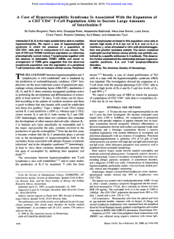
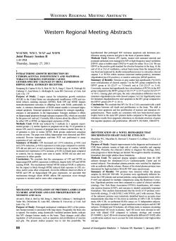
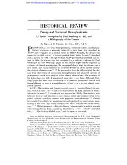
From www.bloodjournal.org by guest on December 22, 2014. For personal use only. ILA, the Human 4-1BB Homologue, Is Inducible in Lymphoid and Other Cell Lineages By Herbert Schwarz, Jean Valbracht, Julia Tuckwell, Johannes von Kempis, and Martin Lotz We recently identified a genethat isinduced by lymphocyte activation (ILA).The sequenceof the full-length 1.4-kb cDNA characterized ILA as a new member of the nerve growth factor/tumor necrosisfactor (NGFITNF) receptor family and the human homologue of the murine T-cell-specific receptor 4-IBB. Thepresent study demonstrates ILA mRNA isoforms at 4.4,4.0, and 1.8 kb in poly-A+ RNA from activated, but not from resting human peripheral blood T lymphocytes. A reverse transcriptase-polymerase chain reaction (RT-PCRI assay was used t o study tissue distribution and regulation of ILA expression. The gene was induced in T lymphocytes by phytohemagglutinin (PHA), phorbol myristate acetate (PMA), and antibody t o CD3, in B lymphocytes by PMA and antibodies t o cell surface Ig, and in blood monocytes by interleukin-1/3 (IL-lp), lipopolysaccharide (LPS), and PMA. In T lymphocytes, ILA mRNA was detectable 1.5 hours after stimulation, reachedmaximal levels at 8 hours, and declined t o background levels by 48 hours. Induction of ILA mRNA required protein synthesisand was primarily due to in- creasedtranscription. Actinomycin D reduced ILA mRNA levels in activated lymphocytes50% within 30 minutes,demonstrating a relatively short half-life of this mRNA. Analysis of nonlymphoid cells showed that ILA mRNA was not detectable in resting cells. However, in contrast t o the lymphoidspecificexpression of the murine ClBB gene, ILA was detected in nonlymphoid cells, including epithelial and hepatoma cells after stimulation with IL-lP. ILA was not detectable in several brain derived cell lines. TheILA cDNA encodes a 30-kD protein as demonstrated by in vitro translation, and this protein is immunoprecipitated by antisera that were raised against ILA peptides or a glutathione-S-transferase fusion protein. Flow cytometry showed expression of ILA protein on a subsetof activated T or B lymphocytes. In conclusion, activation-dependentexpression ofILA is found not only in T lymphocytes, but also in B lymphocytes, monocytes, and diversenonlymphoid cell types. 0 1995 by The American Societyof Hematology. T as previously described.26Chondrocytes*’ and synoviocytes** were HE BIOLOGIC ACTIVITY of cytokines as mediators isolated from tissues obtained from patients undergoing joint reof host defense responses is controlled through reguplacement or at autopsy from donors without known history of joint lated expression of cytokines, as well as their receptors. disease. Human thymocytes were isolated from tissue that was reTissue distribution of cytokine receptors and the production moved during cardiac surgery as previously described.” Human Tof soluble receptors are additional mechanisms that detercell leukemia virus (HTLV)-1 transformed T-cell lines were obtained mine cytokine function. by coculture of peripheral blood T cells with an irradiated HTLVCloning of cytokine receptor genes has resulted in the 1 -infected cell line (kindly provided by Dr W. Wachsman, University identification of several receptor families that contain strucof California, San Diego, CA). B cells from the peripheral blood of turally related receptors.’ The nerve growth factor receptor normal donors were transformed with Epstein-Barr virus (EBV) by (NGFR)/tumor necrosis factor receptor (TNFR) family is infection with EBV-containing supernatants from the B954 cell line. The following cell lines were obtained from ATCC (Rockville, characterized by the presence of 3 to 6 cysteine-rich motifs of approximately 40 amino acids in the extracellular d ~ m a i n . ~ . ~MD): U373, glioblastoma; SK-N-MC, neuroblastoma; Wen, retinoblastoma; HS683, glioma; HEp-2 epithelial; HepG2 hepatoma; Members of this family include the low-affinity NGFR; A549, lung carcinoma; U937, promonocyte; Raji, EBV-transformed TNFR-I,’.�j TNFR-II,7 CD40,’ CD30,9 CD27,’” Fas/APOB cells; HUl78, Jurkat, and Molt4, T-cell lines. 1,””’ 0X-40,13.14and 4-1BB.I’ These receptors recognize RNA preparation and Northern blot analysis. Total RNAwas soluble or cell-surface bound ligands, which also form a isolated by the acid phenol method.30 Poly A+ RNA was prepared family of structurally related proteins and mediate diverse with PolyATract mRNA isolation system (Promega, Madison, WI). cellular respon~es.~ TNF and NGF regulate cell proliferation RNA was fractionated on 1% agarose-formaldehyde gels and transand secretory functions in different cell Stimulation ferred to Hybond N membranes (Amersham, Arlington Heights, of cells through the TNFRI6 and Fas/APO- 117,” can induce IL). Gel-purified DNA fragments were labeled with 32Pby random apoptosis. CD40 is expressed on B lymphocytes and medipriming and used as probes. The 1.4-kb full-length E A cDNA was ates T-cell-dependent B-cell maturation and Ig class used as probe. s ~ i t c h . ~Stimulation ~,’~ of CD27,21,ZZ which is expressed on most peripheral blood T cells and mature thymocytes, and From the Sam and Rose Stein lnstitute for Research on Aging of the rat T-cell subset antigen OX-40 by specific antibodies and Department of Medicine, University of California. San Diego, increases lymphocyte proliferation. 4-1BB has recently been La Jolla, CA. shown to function as an accessory signaling molecule during Submitted June 23, 1994; accepted October 17, 1994. T-cell activationz3 and associate with the tyrosine kinase Supported by National Institutes of Health Grants No. CA81406 p56�ck.24 and AR39799. J.v.K. was supported byafellowship of the We recently cloned the cDNA encoding ILA, a new mem“Deutscher Akademischer Austauschdienst ( D U D ) ’ ’ . ber of the human NGFRNFR family, which is homologous Address reprint requests to Martin Lotz, MD, University of Calito 4-1BB.” The present study shows that this receptor gene fornia at San Diego, 9500 Gilman Dr, La Jolla, CA 92093-0663. can be induced in lymphoid and different nonlymphoid cell The publication costs of this article were defrayed in part by page charge payment. This article must therefore be hereby marked types. MATERIALS AND METHODS Cells and cell culture. Human peripheral blood mononuclear cells (PBMC), monocytes, T, and B lymphocytes were prepared Blood, Vol 85, No 4 (February 15). 1995: pp 1043-1052 “advertisement” in accordance with 18 U.S.C. section 1734 solely to indicate this fact. 0 1998 by The American Society of Hematology. 0006-4971/98/8804-0009$3.~~ 1043 From www.bloodjournal.org by guest on December 22, 2014. For personal use only. 1044 SCHWARZ ET AL RNA loads on thefilters were determinedby control hybridizations tor for the T7 RNA polymerase 5� to the ILA coding region, were with a full-length cyclophilin cDNA probe.” In some experiments, a used as templates. A plasmid lacking a T7 promotor was used as a G3PDHcDNAfragment was used for control hybridizations. The control. ?-cysteine was included into the reaction for detection of fragment was amplified by reverse transcriptase-polymerase chain rethe in vitro translation products by autoradiography of sodium dodeaction (RT-PCR) using G3PDH primers described below and subcy1 sulfate-polyacrylamide gel electrophoresis(SDS-PAGE)separated proteins. cloned into pGEM3z (Promega). Autoradiographs were scanned on a Microtek MSF-300G scanner Producrion of un eukuryotic ILA-Ig fusion protein. An expres(Microtek International Inc, Hsinchu, Taiwan) and bands were subse-sion vector containing the codingregion for the extracellular domain quently analyzed using NIH Image1.44 software (National Institutes of ILA and human Ig Fc constant domains was prepared and used of Health, Bethesda, MD). Relative densities of hybridization signals to transfectCOScells.FusionproteinwasisolatedonproteinA were calculated in comparison to controls (unstimulated cells) for sepharose columns from the conditioned media and shown to have both ILA and cyclophilin hybridization signals. Calculations from the expected molecular mass on silver-stained polyacrylamide gels ILA hybridizations werethen corrected on the basis of those obtained and Western blots. Sera from rabbits, which were immunized with for cyclophilin to correct for variable RNA loads. this purified protein,containedantibodies that specificallyrecognized the ILA-Ig as determined by enzyme-linked immunosorbent RT-PCR. RNA (up to 5 pg) was reverse-transcribed in a 20 p,L assay (ELISA) and immunocytochemistry. The antisera were purivolumecontaining 4 p L 5XRT-buffer(BRL), 10 mmol/L DTT, fied on protein A and the reactivity to human Fc was removed by 500 pmol/L dNTPs, I p L random hexanucleotides (2 mglmL) (Pharabsorption on human lg. macia, Alameda, CA), 200 U MoMLV-RT (BRL), and 20U RNasin Preparurion I$ f L A antibodies. Antibodies were prepared (Promega) for 60 to 120 min at 37°C. against ILA peptides and the GST-ILA fusion protein. Several pep0.1 to 3 Polymerasechainreaction(PCR)wasperformedwith pL (depending on the primers) of the RT reaction product in 25 pL tidesweresynthesized,coupledtokeyholelimpethemocyanin volume with 1 U AmpliTaq (Perkin Elmer Cetus, Emeryville, CA), (KLH) and used to immunize New Zealand white rabbits. The rabbits produced antibodies to the peptides, and the antibodies were aftinity 140 pmoUL dNTPs, 1.5 pmoVL MgClz, 10 mmol/LTris pH 8.3. purified. AntibodytopeptideILA4 (pos 49to65:Pro-Pro-Asn50 mmol/L KCI, and 10 pmollL of each primer. After a 5-minute Ser-Phe-Ser-Ser-Ala-Gly-Gly-Gln-Arg-Thr-Ser-Asp-lle-Cys) and to denaturation step at 94T, the reaction proceeded in 35 cycles of30 peptide ILAS (pos 124 to 139): Thr-Phe-Asn-Asp-Gln-Lys-Arg-Glyseconds at 94”C, 30 seconds at 55”C, and 50 seconds at 72°C. folIle-Ser-Arg-Pro-Trp-Thr-Asn-Cys) were used fortheexperiments lowed by 5 minutes at 72°C. described here. Rabbits were immunized with GST-ILA fusion proPrimers used for ILA PCR were ILA-START: GAG AAT TCC tein. After three immunizations in biweekly intervals, the sera conATG GGA AAC AGC TGT TAC, SKR6-SEN: AGG AGC AAG tained antibodies that recognizedtheGST-ILA fusion proteinon GACCTGAGACAT,SKR6-AS:AGCAGCAGGTCACAG Western blot. Theseantibodyreactivitieswere not present in the AG, ILA-BAC5’ CAT TCC CGG GTC CTT GTA GTA AC, ILApreimmune sera from the same animals. BAC3’: CGG TGA TCA TCC TGG CTC TCT CGC AGG GGC, Affinity; purification ILA-DEL1 : TGC CTG CAT ATG TCA CAG, and ILA-DEL2: CAT Affinity purijicution of peprideuntiseru. was performed by membrane affinity chromatography (MAC; AmiATGCAGGCAGACCCTGGACAAA. con, Beverly, MA) according to manufacturers’ instructions. Briefly, Primersusedforglyceraldehyde3-phosphatedehydrogenase peptides were dissolved in 0.5 mol/L NaHCO,, pH 9.0, at a concen(G3PDH) PCR were sense: TGG TAT CGT GGA AGG ACT CAT tration of 1 mgfmL. Membranes were soakedi n this peptide solution. GAC and antisense: ATG CCA GTG AGC TTC CCG TTC AGC. air dried for 2 hours at RT. treated with 1 mg/mL Na-borohydride They amplify a 190-bp fragment. in phosphate-buffered saline (PBS), pH 7.4, for 1 hour at 4°C. and Quunriturive PCR. A ILA deletionclone(ILA-DEL)wasconrinsed with PBS. Serum was diluted 1.3 in PBS and filtered through structed by PCR-based mutagenesis, which resulted in a deletion of 0.45-pm Acrodisc filters (Gelman Sciences, Ann Arbor, MI). Mem200 bp between nucleotide positions 340 to 539. branes with boundpeptidewereinserted in aMACholderand The ILA cDNA from start to the Hue111 site at position 921 was washed with PBS. The diluted serum was passed over the filters. subclonedintothe EcoRl and SmaI site of pGEM7z(Promega) washed with PBS,andeluted with 100 mmol/Lglycineand 100 between the SP6 and T7 polymerase transcription start site. A fragment containing the 5’part of ILA was amplified using a SP6 primer mmol/L NaCl in PBS. Eluted material was concentrated and reconstituted in PBS by centrifugation through Centricon 30 (Amicon). and ILA-DELI. In a parallel reaction, the 3’ ILA part was amplified Immunojluorescence staining undjow cvtomety. Cells were anwith a T7 primer and ILA-DEL2. ILA-DEL2 contained at its 5’ end alyzed using a FACScdn (Becton Dickinson, Mountain View. CA) 22 nucleotides identical to the 3� end of the S’ TLA fragment. In a and LYSIS11 software (Becton Dickinson). Laser excitation was at third PCR with the SP6 andT7 primers, the 5� and3’ ILA fragments 488 nm (argon laser) for fluorescein isothiocyanate (FITC). A total served as templates for the generation of a ILA deletion fragment, of 4 X 10’ cells were used per condition. Cells were washed three which lacksthe200nucleotideslocatedbetweenILA-DELIand times in PBS, resuspended in 100 pL staining buffer (RPM1 1640. ILA-DEL2. This fragment was subcloned into the EcoRI Hind111 and 3% FBS, 0.02% NaN,), and stained with affinity purifiedpeptide sites of pGEM4z. Serial dilutions of this deletion clone were coamplified with single ILA5 serum for 30 minutes on ice. After two washes with staining buffer. the cells were incubated with FTTC-labeled goat antirabbit stranded cDNA as described earlier. PCR products were separated IgG (1:40)(BoehringerMannheim,Indianapolis,IN) in staining on a I .3% agarose gel and stained with ethidium bromide. Pictures the cells were buffer for I hour on ice. After three more washes, were taken with a Land MP4 Polaroid camera on positivehegative analyzed by flow cytometry. Polaroid film #665 (Polaroid Corp, Cambridge, MA), scanned, and Ruugents. The recombinant human cytokines interleukin-10 (1Levaluated as described for Northern blot analysis. IO), interferon y (IFNy), and TNFcv were purchased from R & D f n virrotrunslution offLA prorein. In vitro translated ILA proSystems (Minneapolis, MN). Endotoxin content was less than 0 . 1 tein was prepared with the TNT coupled reticulocyte lysate system ng per mg of cytokine protein. Lipopolysaccharide (LPS, from Sal(Promega). ILA cDNA from position - 139 to 1021 containing the monella Minnesota), phorbol 12-myristate 13-acetate (PMA), phytocomplete ILA coding region but lacking the remaining 3� end was I site of pRc/CMV (Invitrogen, San Diego. hemagglutinin (PHA), the calcium ionophore A23187, and actinocloned into the EcoRIINot mycin D (actD) were obtained from Sigma (St Louis, MO). OKT3 CA). Two microgramsof two different plasmids, eachwith a promo- From www.bloodjournal.org by guest on December 22, 2014. For personal use only. ILA, THE HUMAN 4-186 HOMOLOGUE 1045 ILIA 1 MGNSCYNIVATLLLVLNFERTRSLQDPCSNCPAGTFCDNNRNQICSPCPP 50 4-1BB 1 MGNNCYNVW1VLLLVGCEKVGAVQNSCDNCQPGTFCRKY.NPVCKSCPP 49 ~ ~ ~ . ~ ~ ~I:. : .~ : I : . I ..I I:. : I~I I I~ .: :I . :. I . . I I I ILA 51 NSFSSAGGQRTCDICRQCKGVFRTRKECSSTSNAECIXTPGFHCLGAGCS 100 4-1BB 50 STFSSIGGQPNCNICRVCAGYFRFKKFCSSTHNAECECIEGFHCLGPQCT 99 ..Ill l l l . - l : l l l l I I I : I Ill1 I I I I : I . . I I I I I l : ~ I ~ 4-1BB 101 MCEQDCRQGQELTKKGCKiXCFGTFNDQK.RGICRPViTNCSLlXK~W 149 .ll.lll.lIlIII.III.1::IIIIII. 1:lllllllllll:lll . 100 RCEKDCRPGQELTKQGCKTCSLGTFNDQNGTGVCRPWTNCSLERSVLKT 149 ILA 150 GTKERDWCGPSPADLSPGAS.SVTPPAPAREPGHSPQIISFFLALTSTA ILA 198 l l . l ~ l l l l l i ~l l~l l .~: . :~ ~. I l l l l ~ : : '~: l l~l l l l l Fig 1. Amino acid sequence homologyof IIA and ClBB. The deduced amino acid sequencesof IIA and the murine 4-IBB were aligned,using the BestFit program of GeneticsComputerGroupInc(Madison, WII sequencoanalysis software. Identicalamino acids are indlcated by verticallines.Aminoacids with high, low and no similarity are indicated by coblanks, respectively. points, and lons, 4-1BB 150 GTIEKDWCGPPWSFSPSTTISVTPEGGP..GGHSLQVLTLFLALTS.A 196 ILIA 199 LLFLLFFLTLRFSWKRGRKKLLYIFKQPFMRPVQ'ITQEEDGCSCRFPEE 248 4-1BB 197 LLLALIFITLLFSVLKWIRKKFPHIFKQPFKKTTGAAQEEDACSCRCPQE246 ILA 249 EEGG . . . CEL 255 4-1BB 241 EEGGGGGYEL 256 1 1 : l:l:ll l l l : l : was recovered as a hybridoma supernatant and titrated for optimal Tlymphocyte proliferation. Antibody to cell surface immunoglobulin (anti-p) was purchased from Cappel (Durham, NC). RESULTS Characterization of the ILA cDNA. The 1,439-bp ILA nucleotide sequencez5 was obtained from several separate library screenings and independent sequencing reactions. The open reading frame encodes a protein of 255 amino acid(s) (aa)with a molecular mass of approximately 28 k D . Hydropathicity analysis32predicted a putative signal peptide (aa 1 to 17) and a 27 aa hydrophobic region, which is a potential transmembrane domain (aa 187 to 213), and flanked by charged residues. Following the 17 aa leader peptide, it contains an extracellular domain of 169 aa, a transmembrane region of 27 aa, and a short intracellular domain of 42 aa. Based on these features, ILA can be classified as a type I transmembrane protein. Two potential N-glycosylation sites at positions 138 and 149 are based on the presence of the NXS/T motif (with X being any amino acid except D or P). The serine at position 242 is a potential site for phosphotylation by protein kinase C. The two threonines at positions 234 and 235 are potential sites for phosphorylation by casein kinase 11. In addition, ILA contains a potential binding site for the tyrosine kinase ~56''~'. The extracellular part of L A contains three cysteine-rich repeats, characteristic of members of the NGF/TNFR superfamily. The transmembrane domain is preceded by a serinethreonine-proline rich region (aa 160 to 185), and these three aa account for 50% (14 of 27 aa) of this domain. Similar domains are found in other members of the NGF/TNFR family.'3 Comparison of ILA with sequences in the NBRF database showed that it is probably the human homologue of the murine cDNA sequence 4-1BB (Fig l), which is expressed by specific T-lymphocyte subset^.'^ The deduced amino acid Ill: .lIIlII.:.....lIII:IlII I:) IIIll1 sequences of ILA and 4-1BB display 73.6% similarity and 59.6% identity. Regulation of ILA mRNA expression in lymphocytes. ILA was not detectable in total or poly A' RNA from unstimulatednormal human peripheral blood lymphocytes from more than 20 normal donors. Mitogen stimulation of PBMC with PHA or PMA induced expression of ILA mRNA. By Northern blot analysis of poly A+ RNA, three forms of ILA mRNA could be detected at 4.4, 4.0, and 1.8 kb (Fig 2), while analysis of total RNA from stimulated PBMC showed only the 4.4-kb form. Studies on kinetics showed that ILA mRNA was rapidly induced by stimulation with PHA and PMA within 1.5 hours, increased to maximal levels by 8 hours, and declined to near background levels by 48 hours (not shown). Stimulation of lymphocytes with an antibody to CD3 also induced LLA mRNA. As compared with stimulation with PHA and PMA, the effects of anti-CD3 had a slower onset but a longer duration. L A mRNA was first detected at 4 hours, reached the maximum at approximately 19 hours, and was still strongly expressed after 48 hours (Fig 3). Induction of ILA mRNA was dependent on protein synthesis. ILA mRNA was induced in primary T lymphocytes by treatment withPHA, PMA, and OKT3. Coincubation with the protein synthesis inhibitor cyclohexamide blocked ILA mRNA synthesis in both cases (Fig 4). Treatment of activated PBMC with actD, a transcriptional inhibitor, was used to determine L A mRNA stability. Cells were stimulated with PHA (1 pg/mL) and PMA (1 ng/mL,) for 4 hours before actD (5 pg/mL) was added. Cells were collected at different time points for analysis by RT-PCR. For quantitative analysis of ILA mRNA levels, a competitive PCR was performed by coamplifying cDNA with an ILADEL, lacking a 200-bp region. Figure SA shows a titration of ILA-DEL against constant amounts of cDNA from the control and the 15- and 30-minute actD samples. Ethidium bromide stained gels showed the points of equal intensity From www.bloodjournal.org by guest on December 22, 2014. For personal use only. SCHWARZ 1046 A 1 2 3 4 k b 1 -- 4.4 4.0 - 1.8 B 1 2 3 4 ILA mRNA was not expressed in response to IL-lp (10 ng/ mL), leukemia inhibitory factor (LIF; 10 ng/mL), IFNy (500 U/mL), LPS ( 1 pg/mL) or TNFa( 1O , OO U/mL) after 7 hours (not shown). Analysis of T-cell lines showed ILAexpression in HTLVI transformed T cells and in Jurkat cells after stimulation with PHA and PMA. ILA could not be detected in the Tcell lines Hut78 andMOLT4,even after stimulation with PHA and PMA (not shown). I L A can be induced in normal B lymphocytes and is constitutivels expressed in rran.sformed B cells. In B lymphocytes purified from human peripheral blood,ILAwas detected by RT-PCR in cells that hadbeen activated with anti-p (12.5 pg/mL) or PMA ( I O ng/mL) for 6 hours. Analysis of B-cell lines showed the presence of ILA mRNA in unstimulated Raji cells, an EBV-transformed B cell line, as well as in B cells from normal donors that had been transformed byEBV in vitro. Stimulation withPMA ( I O ng/ mL) had no detectable effect on the constitutive ILA mRNA expression in EBV-transformed B cells (Fig 6 ) . I U expression in mononuclear phagocytes. In human CO Fig 2. ILA mRNA expression in lymphocytes. RNA was isolated from unstimulated cells (lanes 1 and 2) or from cultures that had been activated with PHA (l pg/mL) and PMA (1ng/mL) for11 hours (lanes 3 and 4). (A) TotalRNA (15 pg/lanes 1 and 3) or polyA' RNA (1 pg/lanes 2 and 41 was analyzed for thepresence of ILA transcripts by Northern blotting. (B) To document the amount of RNA loaded, the filters were subsequently analyzed for cyclophilin mRNA. The differences in intensity of the cyclophilinsignals in (B) are related t o the use of total RNA (lanes 1 and 3) versus poly A+ RNA (lanes 2 and 4). between target and competitor products shifting from 4 to 8 X IO4 in the sample not treated with actD to 4 X IO4 and 2 X IO4 competitor molecules in the samples treated with actD for 15 and 30 minutes, respectively. To obtain a quantitative analysis of these results, negatives of the photographs were scanned and the ratios of target to competitor products were plotted against the number of competitor molecules present in the reaction (Fig SB). The equivalence point (1:l ratio) of target to competitor products was reached at 3.2 X IO4 competitor molecules for the control, at 2.4 X IO4 competitor molecules for the 1 S-minute actD sample and at I .6 X IO4 Competitor molecules for the 30-minute actD sample. These values represent a half-life time ofILAmRNAof 30 minutes. Normal human thymocytes were studied as a source of immature T lineage cells. They exhibited a similar time course of ILA mRNA induction as peripheral blood lymphocytes when stimulated with PHA and PMA.Stimulation with PHA or PMA alone was sufficient for ILA mRNA induction. ET AL 0 4 5 7 19 24 44 hours ILA G3PDH Fig 3. Time course of ILA expression in response t o T-cell activation byanti-CD3. PBMC were stimulated with anti-CD3 (OKT3). RNA was isolated at the indicated time points and analyzed by RT-PCR using the primersILA-BAC5' and 3'. which result in amplification of a fragment with an expected size of 604 bp. To document an equal amount of template, the samples were also analyzed for G3PDH mRNA. From www.bloodjournal.org by guest on December 22, 2014. For personal use only. 1047 ILA, THE HUMAN 4-1BB HOMOLOGUE X I V lated cells but increased strongly in response to IL-Io (10 ng/mL). Among the nonlymphoid cell types tested, the lung carcinoma A549 expressed ILAmRNA constitutively (not shown). These findings demonstrate that ILA is inducible by the proinflammatory cytokine IL-Io in diverse cell types, A control 15 min ActD 30 min ActD Fig 4. Cyclohexamide inhibits ILA mRNA induction in T lymphocytes. Primary T lymphocytes were activated with PHA (2 gg/mLI, PMA (5 ng/mL), or OKT3 (1:5000 dilution of hybridomasupernatant) and cultured for 16 hours in the absence or presence of cyclohexamide (50 gg/mL). RNA was isolated and analyzed by RT-PCR using the primers ILA-BAC5' and 3'. The same templates were analyzed for G3PDH. Marker: PhiX 174 Haelll; control: unstimulated cells. blood monocytes ILA was induced by stimulation with ILIp or PMA (0.1 ng/mL) for 4 hours. The effects ofPMA were dose-dependent with induction occurring at 0.1 ng/mL. The same three mRNA isoforms of 4.4, 4.0, and 1.8 kb as in lymphocytes were also detected in monocytes (Fig 7). In addition toPMAand IL-Io, ILA was also induced in monocytes by stimulation with LPS (not shown). ILA was also found in the human premonocytic cell line U937 and in vitro-derived macrophages after induction with IL-Ip ( I O ng/mL) and PMA (10 ng/mL) (not shown). Tissue clistrihrction and repdotion of I L A expression in nonlymphoid cells. To determine whether ILA expression is restricted to the immune system, primary cells and cell lines from other tissues were examined. ILAmRNA was not detectable by Northern blotting or PCR in unstimulated human rheumatoid synoviocytes and chondrocytes and in several human tumor cell lines, including the hepatoma HepG2. However, after activation with IL-ID, ILA mRNA was detected in several nonlympoid cell types. In epithelial and hepatoma cells, ILA mRNA was not present in unstimu- B I loglo competitor (molecules) Fig 5. ILA mRNA stabilii. PHA/PMA-stimulated and actD-treated PBMC were analyzed for expression of ILA mRNA, using ILA-START and ILA-BAC3' as primers. Known amounts of an ILA-DELwere coamplified in the same reactions. (AI Products were run on a 1.30/0agarose gel and stained by ethidium bromide. IBI Plot of data obtained from scanning of photographs of (Al. Control, U; 15-minute actD, V; 30min actD, 0. From www.bloodjournal.org by guest on December 22, 2014. For personal use only. SCHWARZ ET AL 1048 primary EBV transformed 0) U -aQ c, e I 6 hour stimulation: - ILA - GSPDH PMA 10 ng/ml antiy 12.5 pg/ml Fig 6. ILA expression in primary and in EBV-transformed B lymphocytes. Primary and EBV-transformed B lymphocytes were stimulated for 6 hours and analyzed for ILA mRNA by RT-PCR using SKRGSEN and -AS as primers. To document an equal amount of template, the samples were also analyzed for GBPDH mRNA. and thus has a much wider tissue distribution than described for 4-1 To determine expression of ILA in the nervous system, several brain derived cell lines were tested for presence and inducibility of ILA. Using RT-PCR, ILA was not found in RNA from glioma (HS683) cells, resting or activated for 8 hours with IL-I(I0 U/mL) or IFNy (500 U/mL) or PMA (5 ng/mL) plus the calcium ionophore A23 187 (1 nmol/L) (not shown). Identical results were obtained for a glioblastoma and a retinoblastoma cell line (U373 and WERI, respectively) and for two neuroblastoma cell lines (SK-N-MC and IMR-32). Production of recombinant IL.4 protein and immunoprecipitation with spec@ antibodies. To characterize ILA protein, rabbit antibodies were raised against ILA peptides and GST-ILA and ILA-Ig fusion proteins. For the production of recombinant ILA protein, the full-length ILA cDNA was cloned into the expression vector pRc/CMV. RNA derived from this plasmid was translated in vitro with reticulocyte lysate in the presence of ”S-cysteine. As a specificity control, we also synthesized luciferase (luc) protein by in vitro trans- lation. Analysis of the in vitro translation products by SDSPAGE and autoradiography (no precipitation) shows the luciferase band at 60 kD and the ILA band at 30 kD as the most prominent bands (Fig 8). Aliquots of the identical in vitro translation products were precipitated with pre- and postimmune sera, generated by immunization of rabbits with the GST-ILA fusion protein or a synthetic ILA peptide. Figure 8 shows that luciferase was precipitated equally by the preimmune and immune antisera to ILA. This was due to binding of the proteins to the sepharose beads, which were used in the immunoprecipitation experiments (not shown). The antisera to GST-ILA and ILA peptide specifically precipitated the recombinant ILA protein. The low levels of ILA precipitated by the preimmune sera were also due to ILA binding to the sepharose beads. These results suggest that the ILA cDNA encodes a protein, whichis recognized by antisera, that were raised against ILA peptides or GST-ILA fusion protein. The predicted molecular mass of ILA is 28.4 k D . In vitro-translated ILA protein has an apparent mass of approximately 30 kD. Flow cytometry analysis of I L A protein expression on primary lymphocytes. Specificity and titer of the antisera for staining of native ILA proteinwas determined by immunocytochemistry. COS cells transfected with the eukaryotic expression vector pRc/CMV-ILA, but not untransfected cells, showed strong and specific staining (not shown). The same antiserum was used for flow cytometric analysis ofILA protein expression on primary lymphocytes. Lymphocytes were activated with PMA (5 ng/mL), IL-2 ( I O 0 p/mL), and OKT3 (1:5,OOO)for 48 hours. Staining was performed with affinity purified antiserum to peptide ILA5 (32 pg/mL) and FITC-labeled antirabbit IgG (Boehringer Mannheim). Approximately 25% of the activated cells showed specific staining (Fig 9). DISCUSSION Members of the NGFRNF receptor family are characterized by the presence of three to six cysteine-rich extracellular domains. ILA contains three cysteine-rich domains, and the second is composed of only 32 instead of the usual 41 to 44 amino acids. This domain structure is very similar in ILA and 4-IBB. Through the conservation of the cysteine-rich extracellular domains, the members of this receptor family share a common structure for ligand binding.Similar to other members of the NGFRNFR family, ILA contains a serinethreonine-proline-rich hinge region between the transmembrane domain and the cysteine-rich domains. For the 75-kD TNFR, it has been shown that this domain has an extended structure, which is likely to facilitate ligand binding to the cysteine-rich domains..” The interspecies identity for members of this receptor family is significantly higher than the identity of different members from the same species. For example, the human and murine type 1 and type 2 TNFR share 64% and 62% identity, respectively.3hThe identity between the human type 1 and 2 TNFRislimitedtothe ligand binding domain and is only 27%.’ Similarly, the identity of murine to human CD27 is 65%. Its identity to 4-1 BB is 39%.” The identity between 4-IBB andILA is59.6% From www.bloodjournal.org by guest on December 22, 2014. For personal use only. I U . THE HUMAN 4-1BB HOMOLOGUE 1049 A 1 2 3 4 5 6 kb .-. - 4.4 - ,R e ai .-e ai - 4.0 $ - 1.8 B 1 2 3 4 5 6 Fig 7. ILA induction in monocytes by PMA and IL-lp. Peripheral blood monocytes were stimulated with PMA and IL-lp at the indicated concentrations. (A) RNA was isolated after 4 hours and analyzed for I L A mRNA by Northern blotting. (B1 Because the load of RNA differed considerably in thisexperiment, a rehybridization of the blot with GBPDH cDNA is shown. 1, control; 2, PMA (0.1 ng/mL); 3, PMA (lnglmL1; 4, PMA (10 nglmL1; 5, PMA (50 nglmL1; 6, IL-lp 10 ng/mL. (Cl shows the results after densitometry and normalization of the ILA signals on the basis of GBPDH signals. and corresponds to the interspecies identity of other members of this receptor family. Based on this identity, ILA is the likely human counterpart of 4-1BB. Signal transduction through these receptors has only partially been characterized and diverse mechanisms appear to be used. The NGFR itself displays only low-affinity of NGF. High-affinity binding of NGF is obtained upon association with the trk oncogene product, which has tyrosine kinase activity.'* TNFR appear to use oxygen radicals, the phosphatidic acid, and the sphingomyelin pathways for intracellular signaling. The intracellular region of 4-IBB contains the consensus site for association with p56Ickoriginally identified in CD4 and CD8.39 This site appears to be functional as 4IBB and ~ 5 6 "were ~ coimmunoprecipitated with anti-41BB or anti-p56"' antibodies24suggesting the possibility that 4-1BB on ligand binding might activate the tyrosine kinase ~ 5 6 " In ~ . 4-1BB. the sequence of the potential p56ICkbinding site is CSCRCP (aa 239 to 244). The corresponding sequence in ILA, CSCRFP (aa 241 to 244), contains only one amino acid substitution. Site-directed mutagenesis showed that the two cysteine residues are the essential elements for association with p561ck.39 This sequence in ILA was confirmed with two independently isolated cDNAs and suggests that this potential signaling mechanism is conserved between 4-1BB and ILA. A threonine residue is present in the intracellular domains of CD40, Fas/APO-l, NGFR and TNFR. This residue has been implicated in CD40 signaling and may represent an additional conserved mechanism of signal transduction. In the intracellular part of ILA, as well as 4-1BB, there are two consecutive threonine residues which constitute a potential phosphorylation site for casein kinase IL4' Three different isofonns of ILA mRNA were detected in From www.bloodjournal.org by guest on December 22, 2014. For personal use only. SCHWARZ ET AL 1050 anti ILA peptideserum anti GST-ILA serum C 0 Q) C 3 E .-E 2 kD n 1 - "1 69 46 - - 30 - - 21.5 ILA L activated PBMC. The most abundant form was at 4.4 kb, and this was the only species ofILA readily detectable in total cellular RNA. Isolation of Poly A' RNA was necessary for clear detection of the 4.0 and l .8 kb isoforms. The smallest form at 1.8 kb is of sufficient size to contain the fulllength coding region, while the larger size of the two additional species is probably due to extended 3' untranslated regions. The difference between the 1.4-kb cDNA and the smallest transcript of 1.8 kb is probably due to priming of oligo dT on an internal A-rich region, because six independent cDNA clones of 1.4 kb were isolated. ILA mRNA was notdetected in nonactivated PBMC from 1 control 1 anti-ILA Fluorescence intensity Fig 9. Flow cytometry analysis of ILA protein expression on primary lymphocytes. Lymphocytes (0)resting and (B) activated for 2 days with PMA (5 nglmL),IL-2 (100 plmL1, and OKT3 (1:5000) were stained and analyzed by FACScan. (AI Antirabbit IgG (H + LI-FITC alone (B) Affinity purified peptide 5 serum (32pglmL1 + antirabbit IgG (H + L)-FITC. Fig 8. Immunoprecipitation of ILA protein. ILA and luc proteins were synthesizedby invitro translation as described in Materials and Methods. Aliquots of each reaction were analyzed by SDS-PAGE (no precipitation). Identical aliquots were precipitated with pre- and postimmune sera, generated by immunization of rabbits with the GST-ILA fusion protein, or a synthetic ILA peptide. ILA and luc proteins run at a molecular weight of 33 kD and 60 kD, respectively. more than 20 healthy donors using a sensitive PCR assay. However, ILA wasrapidly inducible byPHA, PMA,and anti-CD3. Transcripts were detected early (90 minutes) after stimulation with PHAPMA. Immediate early genes as c-jtcn and clfos are inducible within 15 minutes." ILA thus appears to be an early activation gene. A competitive PCR was performed to determine ILA mRNA stability. Fifteen and 30 minutes after addition of actD to activated PBMC, ILA mRNA decreased to 76% and 52% of control levels, respectively. Based on these values, the half-life ofILAmRNAwas calculated to be approximately 30 minutes. This characterizes ILA as a highly unstable mRNA. In activated monocytes, the half-lives of the cj u n and clfos transcripts, which are among the most unstable mRNAs, are 27 and 25 minutes, re~pectively.~' Following demonstration of ILA expression by activated PBMC, we analyzed whether ILA is similarly restricted to the expression in T lymphocytes as 4-IBB. The results showed that ILA can be inducedin all major cellular subsets of the immune system and at early, as well as late, stages of their differentiation. Its expression in all normal lymphoid cell types tested was activation-dependent. ILA was induced in blood monocytes in response to their major activators including LPS, IL-10, and PMA. In addition, it appeared that monocyte adherence is also a stimulus for the induction of this mRNA (unpublished observation). U937 cells representing immature monocytes and in vitro differentiated macrophages expressed ILA in response to PMA or IL-10. B lymphocytes wereisolatedfromperipheralblood of normal donors. In purified populations of these cells, ILA expression was induced by antibodies to cell surface Ig or PMA. EBV-transformed B-cell lines expressed ILA mRNA constitutively. Collectively, these results showthatwithin the immune system ILA is more broadly expressed than 4- From www.bloodjournal.org by guest on December 22, 2014. For personal use only. ILA, THE HUMAN 4-IBB HOMOLOGUE 1BB. This indicates that ILA may not only function as the recipient on T lymphocytes for signals derived from other cells, but potentially serves as a molecule in mediating communication among different cell types of the immune system. Of further significance to the role of L A in host defense responses is its expression in several nonlymphoid cell types. The analysis of tissue distribution performed in the present study clearly shows that ILA is inducible in human articular chondrocytes as one example of a normal mesenchymal cell type, as well as in hepatoma and epithelial tumor cell lines. It is of interest that ILA was not inducible in any of the different cell lines established from brain tumors, possibly indicating that ILA is not a product of neuronal cell types. The other members of the NGF/TNFR family differ in their tissue distribution. The rat OX-40 ismost restricted and only expressed by certain T-cell subsets. CD40 is mainly expressed by B lineage cells, while CD30 can be found on activated normal T and B cells, as well as on Hodgkin’s lymphoma cells. CD27 has been detected on T and B lymphocytes, mature thymocytes, and some chronic B lymphocytic leukemias. In contrast to these receptors that appear to be restricted to T and B lymphocytes is the broad tissue distribution of the NGFR and the two TNFR. ILA is a member of this receptor family that has a wide tissue distribution and is, in this respect, similar to the NGF and TNF receptors. Biochemical characterization of ILA protein was performed by in vitro translation of RNA, which was derived from the full-length ILA cDNA. The in vitro translation product had a molecular mass of 30 kD and wasimmunoprecipitated by antisera against GST/ILA fusion protein or ILA peptides. The predicted molecular weight of ILA is 28.4k D , which is in agreement with the size of the in vitro translated protein. The expression and inducibility of ILA protein on primary cells was demonstrated in lymphocytes where activation with known inducers of ILA mRNA also induced expression of ILA protein. ACKNOWLEDGMENT We thank J. Shin for excellent technical assistance. REFERENCES 1. Miyajima A, Kitamura T, Harada N, Yokota T, Arai K: Cytokine receptors and signal transduction. Annu Rev Immunol 10:295, 1992 2. Mallett S, Barclay AN: A new superfamily of cell surface proteins related to the nerve growth factor receptor. Immunol Today 12:220, 1991 3. Smith CA, Farrah T, Goodwin RG: The TNF receptor superfamily of cellular and viral proteins: Activation, costimulation, and death. Cell 76:959, 1994 4. Johnson D, Lanahan A, Buck CR, Sehgal A, Morgan C, Mercer E, Bothwell M, Chao M: Expression and structure of the human NGF receptor. Cell 47:545, 1986 5. Loetscher H, Schlaeger ET, Lahm HW, Pan YC, Lesslauer W, Brockhaus M: Purification and partial amino acid sequence analysis of two distinct tumor necrosis factor receptors from HL60 cells. J Biol Chem 265:20131, 1990 6 . Smith CA, Davis T, Anderson D, Solam L, Beckmann MP, Jerzy R, Dower SK, Cosman D, Goodwin RG: A receptor for tumor 1051 necrosis factor defines an unusual family of cellular and viral proteins. Science 248:1019, 1990 7. Kohno T, Brewer MT, Baker SL, Schwartz PE, KingMW, Hale KK, Squires CH, Thompson RC, Vannice JL: A second tumor necrosis factor receptor gene product can shed a naturally occurring tumor necrosis factor inhibitor. Proc Natl Acad Sci USA 87:8331, 1990 8. Stamenkovic I, Clark EA, Seed B: A B-lymphocyte activation molecule related to the nerve growth factor receptor and induced by cytokines in carcinomas. EMBO J 8:1403, 1989 9. Durkop H, Latza U, Hummel M, Eitelbach F, Seed B, Stein H: Molecular cloning and expression of a new member of the nerve growth factor receptor family that is characteristic for Hodgkin’s disease. Cell 68:421, 1992 10. Camerini D, Walz G, Loenen WA, Borst J, Seed B: The T cell activation antigen CD27 is a member of the nerve growth factor/ tumor necrosis factor receptor gene family. J Immunol 147:3165, 1991 11. ItohN, Yonehara S, Ishii A, Yonehara M, Mizushima S, Sameshima M, Hase A, Set0 Y, Nagata S: The polypeptide encoded by the cDNA for human cell surface antigen Fas can mediate apoptosis. Cell 66233, 1991 12. Oehm A, Behrmann I, Falk W, Pawlita M, Maier G, Klas C, Li-Weber M, Richards S, Dhein J, Trauth BC: Purification and molecular cloning of the APO-l cell surface antigen, a member of the tumor necrosis factohewe growth factor receptor superfamily. Sequence identity with the Fas antigen. J Biol Chem 267:10709, 1992 13. Paterson DJ, Jefferies WA, Green JR, Brandon MR. Corthesy P, Puklavec M, Williams FA: Antigens of activated rat T lymphocytes including a molecule of 50,000 Mr detected only on CD4 positive T blasts. Mol Immunol 24:1281, 1987 14. Mallett S, Fossum S, BarclayAN: Characterization of the MRC OX40 antigen of activated CD4 positive T lymphocytes-A molecule related to nerve growth factor receptor. EMBO J 9: 1063, 1990 15. Kwon BS, Weissman SM: cDNA sequences of two inducible T-cell genes. Proc Natl Acad Sci USA 86:1963, 1989 16. Beutler B, Cerami A: The biology of cachectin/TNF-a primary mediator of the host response. Annu Rev Immunol7:625, 1989 17. Yonehara S, Ishii A, Yonehara M: A cell-killing monoclonal antibody (anti-Fas) to a cell surface antigen CO-downregulatedwith the receptor of tumor necrosis factor. J Exp Med 169:1747, 1989 18. Trauth BC, Klas C, Peters AM, Matzku S, Moller P, Falk W, Debatin KM, Krammer PH: Monoclonal antibody-mediated tumor regression by induction of apoptosis. Science 245:301, 1989 19. Hollenbaugh D, Grosmaire LS, Kullas CD, Chalupny NJ, Braesch-Andersen S, Noelle RJ, Stamenkovic I, Ledbetter JA, Aruffo A: The human T cell antigen gp39, a member of the TNF gene family, is a ligand for the CD40 receptor: Expression of a soluble form of gp39 with B cell co-stimulatory activity. EMBO J 11:4313, 1992 20. Aruffo A, Farrington M, Hollenbaugh D, LiX, Milatovich A, Nonoyama S, Bajorath J, Grosmaire LS, Stemkamp R, Neubauer M: The CD40 ligand, gp39, is defective in activated T cells from patients with X-liked hyper-IgM syndrome. Cell 72:291, 1993 21. Van Lier RA, Borst J, Vroorn TM. Klein H, Van Mourik P, Zeijlemaker WP, Melief CJ: Tissue distribution and biochemical and functional properties of Tp55 (CD27), a novel T cell differentiation antigen. J Immunol 139:1589, 1987 22. Bigler RD, Bushkin Y, Chiorazzi N: S152 (CD27). A modulating disulfide-linked T cell activation antigen. J Immunol 141:21, 1988 23. Pollok K E , Kim YJ, Zhou Z , Hurtado J, KimKK. Pickard From www.bloodjournal.org by guest on December 22, 2014. For personal use only. 1052 RT, Kwon BS: Inducible T cell antigen 4-1BB. Analysis of expression and function. J Immunol 150:771, 1993 24. Kim YJ, Pollok KE, Zhou Z, Shaw A, Bohlen JB, Fraser M, Kwon BS: Novel T cell antigen 4-1BB associates with the protein tyrosine kinase p561ckl. J Immunol 151:1255, 1993 25. Schwarz H, Tuckwell J, Lotz M: A receptor induced by lymphocyte activation (ILA): A new member of the human nervegrowth factorhmor necrosis factor receptor family. Gene 134:295, 1993 26. Villiger PM, Cronin MT, Amenomori T, Wachsman W, Lotz M: IL-6 production by human T lymphocytes. Expression in HTLVl-infected but not in normal T cells. J Immunol 146:550, 1991 27. Villiger PM,Lotz M: Expression of prepro-enkephalin in human articular chondrocytes is linked to cell proliferation. EMBO J 11:135, 1992 28. Lotz M, Terkeltaub R, Villiger PM: Cartilage and joint inflammation. Regulation of IL-8 expression by human articular chondrocytes. J Immunol 148:466, 1992 29. Lotz M, Jirik F, Kabouridis P, Tsoukas C, Hirano T, Kishimot0 T, Carson DA: B cell stimulating factor Uinterleukin 6 is a costimulant for human thymocytes and T lymphocytes. J Exp Med 167:1253, 1988 30. Chomczynski P, Sacchi N: Single step method of RNA isolation by acid guanidinium thiocyanate-phenol-chloroform extraction. Anal Biochem 162:4019, 1991 31. Hasel K, Sutcliffe G: Nucleotide sequence of a cDNA for mouse cyclophilin. Nucleic Acids Res 18:105, 1990 32. Kyte J, Doolittle RF: A simple method for displaying the hydropathic character of a protein. J Mol Biol 157:105, 1982 33. Rabizadeh S, Oh J, Zhong LT, Yang J, Bitler CM, Butcher LL, Bredesen DE: Induction of apoptosis by the low-affinity NGF receptor. Science 261:345, 1993 SCHWARZ ET AL 34. Kwon BS, Kestler DP, Eshhar Z,Oh KO, Wakulchik M: Expression characteristics of two potential T cell mediator genes. Cell Immunol 121:414, 1989 35. Banner DW, D’Arcy A, Janes W, Gentz R, Schoenfeld HJ, Broger C, Loetscher H, Lesslauer W: Crystal structure of the soluble human 55 kd TNF receptor-human TNF beta complex: Implications for TNF receptor activation. Cell 73:431, 1993 36. Lewis M, Tartaglia LA, Lee A, Bennett GL, Rice GC, Wong GH, Chen EY, Goeddel DV: Cloning and expression of cDNAs for two distinct murine tumor necrosis factor receptors demonstrate one receptor is species specific. Proc Natl Acad Sci USA 88:2830, 1991 37. Gravestein LA, BlomB,Nolten LA, de Vries E,van der Horst G, Ossendorp F, Borst J, Loenen WA: Cloning and expression of murine CD27: Comparison with 4-1BB, another lymphocytespecific member of the nerve growth factor receptor family. Eur J Immunol 23:943, 1993 38. Klein R, Nanduri V, Jing SA, Lamballe F, Tapley P, Bryant S, Cordon-Cardo C, Jones KR, Reichardt LF, Barbacid M: The trkB tyrosine protein kinase is a receptor for brain-derived neurotrophic factor and neurotrophin-3. Cell 66:395, 1991 39. Shaw A, Chalupny J, Whitney JA, Hammond G, Amrein K, Kavathas P, Sefton BM, Rose JK: Short related sequences in the cytoplasmic domain of CD4 and CD8 mediate binding to the aminoterminal domain of the p561ck tyrosine proteine kinase. Mol Cell Biol 10:1853, 1990 40. Kuenzel A, Mulligan J, Sommercom J, KrebsE: Substate specificity for casein kinase I1 as deduced from studies with synthetic peptides. J Biol Chem 161:9136, 1993 41. Nakamura T, Datta R, Kharbanda S, Kufe D: Regulation of jun and fos gene expression in human monocytes by the macrophage colony-stimulating factor. Cell Growth Differ 2:267, 1991 From www.bloodjournal.org by guest on December 22, 2014. For personal use only. 1995 85: 1043-1052 ILA, the human 4-1BB homologue, is inducible in lymphoid and other cell lineages H Schwarz, J Valbracht, J Tuckwell, J von Kempis and M Lotz Updated information and services can be found at: http://www.bloodjournal.org/content/85/4/1043.full.html Articles on similar topics can be found in the following Blood collections Information about reproducing this article in parts or in its entirety may be found online at: http://www.bloodjournal.org/site/misc/rights.xhtml#repub_requests Information about ordering reprints may be found online at: http://www.bloodjournal.org/site/misc/rights.xhtml#reprints Information about subscriptions and ASH membership may be found online at: http://www.bloodjournal.org/site/subscriptions/index.xhtml Blood (print ISSN 0006-4971, online ISSN 1528-0020), is published weekly by the American Society of Hematology, 2021 L St, NW, Suite 900, Washington DC 20036. Copyright 2011 by The American Society of Hematology; all rights reserved.
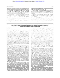
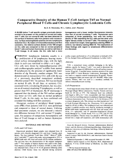
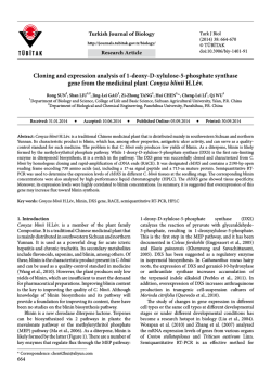
© Copyright 2026 Paperzz