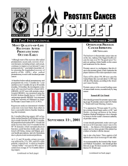
Prostate Imaging Clinical Compendium ® ® Table of Contents Introduction.....................................................................................................................................2 Studies Using Advanced MRI Imaging Techniques for the Detection and Localization of Prostate Cancer..........................................................................................................................3 Studies Using Advanced MRI Imaging Techniques for the Staging and Treatment Planning of Prostate Cancer.........................................................................................................................24 Studies Using Advanced MRI Imaging Techniques for Treatment Monitoring of Prostate Cancer...........................................................................................................................................40 Supplemental Information iCAD’s VividLook and VersaVue Enterprise Solution........................................................48 Resources.......................................................................................................................50 Glossary of Acronyms.............................................................................................51 About iCAD...................................................................................................................52 Note: Studies marked with * indicate usage of advanced image analysis/computer-aided detection technology. The content provided in this document is for informational purposes only. iCAD has compiled this information to assist providers in learning about some of the clinical uses of contrast-enhanced magnetic resonance imaging and advanced image analysis techniques for prostate imaging. This is not intended to be a comprehensive guide of all the available clinical references pertaining to MRI imaging techniques available. © 2011 iCAD, Inc. All rights reserved. iCAD, the iCAD logo, Never Stop Looking, VividLook and VersaVue are registered trademarks of iCAD, Inc. Other company, product, and service names may be trademarks or service marks of others. DMM136 Rev. C Introduction The numbers are staggering: Nearly 1 in 6 men over the age of 40 years in the U.S. will be diagnosed with prostate cancer.1 In 2010, the American Cancer Society estimates that 217,000 men in the U.S. will be diagnosed with prostate cancer and more than 32,000 will die from it.2 Prostate cancer is the second leading cause of death for men, following lung cancer.3 Clearly, there is cause for concern. Imaging of the prostate has historically been challenging for clinicians because of the complex T1 vascularity of the organ coupled with its location deep within the abdominal/pelvic cavity. Diagnostic tests such as the Prostate Specific Antigent (PSA) and digital rectal examination (DRE), which are currently used to determine the extent and behavior of prostate cancer are often unreliable or provide inaccurate results. These tests have been shown to have high false positive and false negative rates which can lead to unnecessary biopsies and/or under-diagnosis. Biopsies of the prostate are blind and random, and miss nearly 20% of cancers.4 Elevated PSA levels cause approximately 1.6 million men in the U.S. to have needle biopsies each year. Eighty percent of those biopsies are negative adding more than $2 billion in healthcare costs.5 T2 DWI iCAD VividLook In addition, prostate cancer treatments often result in many complications including impotence and incontinence in up to 80% of patients.6 Conventional treatments such as radical prostatectomy and radiation therapy frequently fail or are administered unnecessarily leaving nearly 50% of patients with one or more of the aforementioned complications. Despite what appears to be a crisis in men’s health, there is some good news. Prostate cancer is curable if it is detected at an early stage when it still confined to the prostate. Through the use of advanced imaging techniques such as magnetic resonance imaging (MRI) with dynamic contrast-enhancement (DCE), physicians can more accurately diagnose, localize, stage and treat prostate cancer. Prostate MRI provides a more thorough diagnostic assessment and allows for improved staging of the disease. Because of these benefits, the use of these advanced imaging technologies is becoming more widely accepted and used throughout the U.S. This compendium of clinical abstracts is intended to provide clinical evidence of the value of DCE MRI for imaging the prostate. The citations are provided for each abstract so that full clinical papers may be referenced if desired. American Cancer Society (http://www.cancer.org/downloads/STT/Cancer_Facts_and_Figures_2010.pdf) American Cancer Society (http://www.cancer.org/downloads/STT/Cancer_Facts_and_Figures_2010.pdf) 3 American Cancer Society (http://www.cancer.org/downloads/STT/Cancer_Facts_and_Figures_2010.pdf) 4 John Hopkins Professor Robert Getzenberg (www.usatoday.com/news/health/2007-04-25-prostate-cancer-tests_N.htm) 5 John Hopkins Professor Robert Getzenberg (www.usatoday.com/news/health/2007-04-25-prostate-cancer-tests_N.htm) 6 National Cancer Institute (www.cancer.gov/cancertopics/factsheet/pcos) 1 2 2 Prostate Imaging Detection and Localization Studies Prostate Cancer Detection and Localization Quantitative Dynamic MRI and Localisation of Non-Palpable Prostate Cancer* F. Cornud, F. Beuvon, F. Thevenin, L. Chaveinc, A. Vieillefond, A. Descazeaux, T. Flam Purpose To determine whether quantitative dynamic contrast-enhanced MRI improves the performance of T2W-MRI for the localisation of non-palpable prostate cancer (PCa) and for the estimation of tumor volume. Materials and Methods Twenty-three patients (PSA: 8.91+/-6.2ng/m) with a non-palpable cancer underwent endorectal MRI with T2W and dynamic contrast enhanced (DCE) imaging before radical prostatectomy. Each level of evaluation (apex, midportion, base) was divided in eight areas (24 areas per prostate and 552 areas for the 23 patients). Localisation and volume of tumor foci greater than 0,2cc present on the radical prostatectomy specimens were retrospectively correlated to their MR appearance on the 552 evaluated areas. The dynamic parameters included capillary permeability (K(trans)), maximum concentration of gadolinium after 60s of perfusion ([Gd]) and wash-out (K(ep)). Uni- and multivariate analysis were performed to determine which parameters were predictive of PCa. Results Mean values of K(trans), K(ep) and [Gd] were significantly higher in the 58 tumor foci greater than 0,2 cm(3) of the PZ and the TZ (all p<0.05). Logistic regression for each zone provided a value of the area under the ROC curve of 0.83 for the PZ and 0.81 for the TZ (0.7 and 0.75, respectively, for the T2W imaging), only significant for the PZ (p<0.002). Sensitivity and specificity were 79% and 77% for the PZ, 62.5 and 94% for the TZ. Above 0,2 cm(3), tumor volume on dynamic MR showed a mean difference of 51+/-100% (range: -145 to +248%). Conclusions Quantitative dynamic MRI is more accurate than T2W imaging for tumor localisation of non-palpable cancer greater than 0,2 cm(3), but the difference is only significant for the PZ. Above this volume, correlation between tumor volume measured on dynamic MRI and that on the specimen is poor. Cornud, F., et.al. Quantitative Dynamic MRI and Localisation of Non-Palpable Prostate Cancer. Prog. Urol.: 19(6), 2009, 401-413. 4 Prostate Cancer Detection and Localization Imaging of Organ-Confined Prostate Cancer: Functional Ultrasound, MRI and PET/Computed Tomography Philippe Puech, Damien Huglo, Grégory Petyt, Laurent Lemaitre, and Arnauld Villers Purpose of review To review the current status of advanced imaging techniques in identification of organ-confined prostate cancer with a focus on their impact on patient management. Recent Findings Transrectal ultrasound suffers from poor accuracy despite significant technical improvements. Generally used to distinguish cancers with extraprostatic spread, MRI is now focusing on intraprostatic prostate cancer identification. At 1.5T, the most recent high-resolution pelvic phased-array coils provide excellent imaging of the whole gland, including this challenging anterior part. Improvements in accuracy for cancer detection and volume estimation result from dynamic contrast-enhanced and diffusion-weighted imaging sequences. Histological correlations showed high sensitivity/specificity for significant volume cancers. 3T MRI scanners will improve these results. Most of the recent PET/computed tomography imaging studies use choline derivatives (11C-choline and 18 F-fluorocholine). Their results are promising but insufficient to be currently recommended in routine practice. Summary Considerable advances have been made in the identification of organ-confined prostate cancer with multiparametric MRI. Only prebiopsy MRI can provide best quality of cancer assessment and allows for targeting biopsies. It is hoped that advances in 3T MRI as well as in radiotracers for PET/computed tomography will further improve diagnosis, treatment selection, planning and outcomes. Puech, Philippe, et.al. Imaging of Organ-Confined Prostate Cancer: Functional Ultrasound, MRI and PET/Computed Tomography. Current Opinion in Urology: 19, 2009, 168-176. 5 Prostate Cancer Detection and Localization Dynamic Contrast-Enhanced Magnetic Resonance Imaging in the Evaluation of the Prostate* David Bonekamp, MD, PhD and Katarzyna J. Macura, MD, PhD Dynamic contrast-enhanced magnetic resonance imaging (DCE-MRI) is a novel MR technique that allows to interrogate pharmacokinetic processes in the tissues at a voxel level and to generate parametric maps that can be displayed for clinical interpretation. Dynamic contrast-enhanced MRI is an important imaging technique for the imaging of angiogenesis and vasculogenesis because it probes the microvascular networks at a microscopic level by being sensitive to the compartmentalization of tissue into the vascular and extravascular-extracellular space and to the diffusion of contrast molecules across the vascular endothelium and capillary boundaries. Dynamic contrast-enhanced MRI has already been shown to improve detection and localization of prostate cancer, to improve prediction of extracapsular extension, determination of tumor volume, and after treatment follow-up. In this article, we outline the fundamental principles of DCE-MRI and describe the application of DCE methods in the imaging of the prostate. Bonecamp, David et.al. Dynamic Contrast-Enhanced Magnetic Resonance Imaging in the Evaluation of the Prostate. Topics in Magnetic Resonance Imaging: Volume 19, Number 6, December 2008, 273-284. 6 Prostate Cancer Detection and Localization Computerized Analysis of Prostate Lesions in the Peripheral Zone Using Dynamic Contrast Enhanced MRI* Pieter C. Vos, Thomas Hambrock, Christina A. Hulsbergen - van de Kaa, Jurgen J. Fütterer, Jelle O. Barentsz, and Henkjan J. Huisman A novel automated computerized scheme has been developed for determining a likelihood measure of malignancy for cancer suspicious regions in the prostate based on dynamic contrast-enhanced magnetic resonance imaging (MRI) (DCE-MRI) images. Our database consisted of 34 consecutive patients with histologically proven adenocarcinoma in the peripheral zone of the prostate. Both carcinoma and non-malignant tissue were annotated in consensus on MR images by a radiologist and a researcher using whole mount step-selection histopathology as standard of reference. The annotations were used as regions of interest (ROIs). A feature set comprising pharmacokinetic parameters and a T1 estimate was extracted from the ROIs to train a support vector machine as classifier. The output of the classifier was used as a measure of likelihood of malignancy. Diagnostic performance of the scheme was evaluated using the area under the ROC curve. The diagnostic accuracy obtained for differentiating prostate cancer from non-malignant disorders in the peripheral zone was 0.83 (0.75-0.92). This suggests that it is feasible to develop a computer aided diagnosis system capable of characterizing prostate cancer in the peripheral zone based on DCE-MRI. Vos, Pieter C. et.al. Computerized analysis of prostate lesions in the peripheral zone using dynamic contrast enhanced MRI. Med. Phys.: 35, 2008, 888-899. 7 Prostate Cancer Detection and Localization Evaluation of T2-Weighted and Dynamic ContrastEnhanced MRI in Localizing Prostate Cancer Before Repeat Biopsy Alexandre Ben Cheikh, Nicolas Girouin, Marc Colombel, Jean-Marie Maréchal, Albert Gelet, Alvine Bissery, Muriel Rabilloud, Denis Lyonnet, Olivier Rouvière We assessed the accuracy of T2-weighted (T2w) and dynamic contrast-enhanced (DCE) 1.5-T magnetic resonance imaging (MRI) in localizing prostate cancer before transrectal ultrasound-guided biopsy. Ninety-three patients with abnormal PSA level and negative prostate biopsy underwent T2w and DCE prostate MRI using pelvic coil before repeat biopsy. T2w and DCE images were interpreted using visual criteria only. MR results were correlated with repeat biopsy findings in ten prostate sectors. Repeat biopsy found prostate cancer in 23 patients (24.7%) and 44 sectors (6.6%). At per patient analysis, the sensitivity, specificity, positive and negative predictive values were 47.8%, 44.3%, 20.4% and 79.5% for T2w imaging and 82.6%, 20%, 24.4% and 93.3% for DCE imaging. When all suspicious areas (on T2w or DCE imaging) were taken into account, a sensitivity of 82.6% and a negative predictive value of 100% could be achieved. At per sector analysis, DCE imaging was significantly less specific (83.5% vs. 89.7%, p<0.002) than T2w imaging; it was more sensitive (52.4% vs 32.1%), but the difference was hardly significant (p=0.09). T2w and DCE MRI using pelvic coil and visual diagnostic criteria can guide prostate repeat biopsy, with a good sensitivity and NPV. Cheikh, Alexandre, et.al. Evaluation of T2-Weighted and Dynamic Contrast-Enhanced MRI in Localizing Prostate Cancer Before Repeat Biopsy. European Radiology: 19, 2009, 770-778. 8 Prostate Cancer Detection and Localization Usefulness of Diffusion-Weighted Imaging and Dynamic Constrast-Enhanced Magnetic Resonance Imaging in the Diagnosis of Prostate Transition-Zone Cancer T. Yoshizako, A. Wada, T. Hayashi, K. Uchida, M. Sumura, N. Uchida, H. Kitagaki, M. Igawa Background Conventional T2-weighted (T2-WI) magnetic resonance imaging (MRI) has poor sensitivity for prostate transitionzone (TZ) cancer detection. Purpose To retrospectively evaluate the clinical value of diffusion-weighted MRI (DW-MRI) and dynamic contrast-enhanced MRI (DCE-MRI) in combination with T2-WI for the diagnosis of TZ cancer. Material and Methods Twenty-six TZ cancers in 23 patients with at least one tumor (tumor size >10 mm) located predominantly in the TZ were included in the study. Sixteen peripheral-zone (PZ) cancers in 12 patients with PZ cancer but without TZ cancer (control group) were selected by step-selection pathologic maps. All patients underwent MRI and radical prostatectomy. MRI was obtained by a 1.5T super-conducting system with a phased-array coil. Imaging sequences were T2-WI with fat saturation (FST2-WI), DW-MRI (single-shot echoplanar image, b=0 and 1000 s/mm2, apparent diffusion coefficient (ADC) map findings), and DCE-MRI (3D fast spoiled gradient recalled (SPGR), contrast medium (0.2 mmol/kg), total injection time 5 s, image acquisition 30, 60 and 90 s). Sensitivity, specificity, accuracy, and positive predictive value (PPV) for the diagnosis of TZ cancer were evaluated in four protocols: A) FST2-WI alone, B) FST2-WI plus DW-MRI, C) FST2-WI plus DCE-MRI, D) FST2-WI plus DW-MRI plus DCE-MRI. Results Sensitivity, specificity, accuracy, and PPV in protocol A (FST2-WI alone) were 61.5%, 68.8%, 64.3%, and 76.2%, respectively. FST2-WI plus DW-MRI (protocol B) improved the sensitivity, specificity, accuracy, and PPV. In FST2-WI plus DW-MRI plus DCE-MRI (protocol D), the number of true-negative lesions increased and false-positive lesions decreased, and the sentivity, specificity, accuracy, and PPV were 69.2%, 93.8%, 78.6%, and 94.7%, respectively. There was a significant difference between protocols A and D (p<0.05). Conclusion Adding DW-MRI to FST2-WI in the diagnosis of prostate TZ cancer increased the diagnostic accuracy. The addition of DCE-MRI may be an option to improve the specificity and PPV of diagnosing TZ cancer with FST2-WI and DW-MRI. Yoshizako, T., et.al. Usefulness of Diffusion-Weighted Imaging and Dynamic Contrast-Enhanced Magnetic Resonance Imaging in the Diagnosis of Prostate Transitional-Zone Cancer. Acta Radiologica: 10, 2008, 1207-1213. 9 Prostate Cancer Detection and Localization Prostate Cancer Screening: The Clinical Value of Diffusion-Weighted Imaging and Dynamic MR Imaging in Combination with T2-Weighted Imaging Akihiro Tanimoto, MD, Jun Nakashima, MD, Hidaka Kohno, MD, Hiroshi Shinmoto, MD, and Sachio Kuribayashi, MD Purpose To evaluate the clinical value of diffusion weighted imaging (DWI) and dynamic MRI in combination with T2weighted imaging (T2W) for the detection of prostate cancer. Materials and Methods A total of 83 patients with elevated serum prostate specific antigen (PSA) levels (4.0 ng/mL) were evaluated by T2W, DWI, and dynamic MRI at 1.5 T prior to needle biopsy. The data from the results of the T2W alone (protocol A), combination of T2W and DWI (protocol B), and the combination of T2W+DWI and dynamic MRI (protocol C) were entered into a receiver operating characteristic (ROC) curve analysis, under results of systemic biopsy as the standard of reference. Results Prostate cancer was pathologically detected in 44 of the 83 patients. The sensitivity, specificity, accuracy, and the area under the ROC curve (Az) for the detection of prostate cancer were as follows: 73%, 54%, 64%, and 0.711, respectively, in protocol A; 84%, 85%, 84%, and 0.905, respectively, in protocol B; and 95%, 74%, 86%, and 0.966, respectively, in protocol C. The sensitivity, specificity, and accuracy were significantly different between the three protocols (P 0.01). Conclusion In patients with elevated serum PSA levels, the combination of T2W, DWI, and dynamic MRI may be a valuable tool for detecting prostate cancer and avoiding an unnecessary biopsy without missing prostate cancer. Tanimoto, Akihiro, et.al. Prostate Cancer Screening: The Clinical Value of Diffusion-Weighted Imaging and Dynamic MR Imaging in Combination with T2-Weighted Imaging. Journal of Magnetic Resonance Imaging 2007; 25:146-152. 103 Prostate Cancer Detection and Localization Dynamic Contrast-Enhanced MRI of Benign Prostatic Hyperplasia and Prostatic Carcinoma: Correlation with Angiogenesis J. Ren, Y. Huan, H. Wang, Y.-J. Changa, H.-T. Zhao, Y.-L. Ge, Y. Liu, Y. Yang Aim To investigate the diagnostic and differential diagnostic values of dynamic contrast-enhanced magnetic resonance imaging (DCE MRI) in prostatic diseases, and to investigate the correlation between the parameters of SIeT curves and angiogenesis. Materials and Methods Twenty-one patients with proven prostatic carcinoma (Pca) and 29 patients with proven benign prostatic hyperplasia (BPH) were examined using DCE MRI. Diagnostic characteristics for differentiation were examined using threshold values for maximum peak time, enhancement degree, and enhancement rate. Then, the signal intensity-time curves (SIeT curves) were analyzed, and the correlations between the parameters of SIeT curves and the expression levels of vascular endothelial growth factor (VEGF) and microvascular density (MVD) were investigated. All patients underwent prostatectomy. DCE MRI and histological findings were correlated. Results Pca showed stronger enhancement with an earlier peak time, higher enhancement, and enhancement rate (p < 0.05). Regarding the type of SIeT curves, in the BPH group six were type A, 10 were type B, and 13 were type C, whereas in the Pca group, 14 were type A, six were type B, and only one was type C (Chi-square test, c2 ¼ 13.57, P < 0.005). The VEGF and MVD expression levels of Pca were higher than those of BPH. Peak time was negatively correlated with the expression levels of VEGF and MVD, whereas the enhancement degree and enhancement rate showed positive correlations (Pearson correlation, p < 0.05). Conclusion Based on T2-weighted imaging, DCE MRI curves can help to differentiate benign from malignant prostate tissue. In the present study the type C curve was rarely seen with malignant disease, but these results need confirmation. Rena, J., et.al. Dynamic Contrast-Enhanced MRI of Benign Prostatic Hyperplasia and Prostatic Carcinoma: Correlation With Angiogenesis. Clinical Radiology: 63, 2008, 153-159. 11 Prostate Cancer Detection and Localization Dynamic Contrast-Enhanced MRI of Prostate Cancer at 3 T: A Study of Pharmacokinetic Parameters* Iclal Ocak, Marcelino Bernardo, Greg Metzger, Tristan Barrett, Peter Pinto, Paul S. Albert, Peter L. Choyke Objective The objectives of our study were to determine whether dynamic contrast-enhanced MRI performed at 3 T and analyzed using a pharmacokinetic model improves the diagnostic performance of MRI for the detection of prostate cancer compared with conventional T2-weighted imaging, and to determine which pharmacokinetic parameters are useful in diagnosing prostate cancer. Subjects and Methods This prospective study included 50 consecutive patients with biopsy-proven prostate cancer who underwent imaging of the prostate on a 3-T scanner with a combination of a sensitivity-encoding (SENSE) cardiac coil and an endorectal coil. Scans were obtained at least 5 weeks after biopsy. T2-weighted turbo spin-echo images were obtained in three planes, and dynamic contrast-enhanced images were acquired during a single-dose bolus injection of gadopentetate dimeglumine (0.1 mmol/kg). Sensitivity, specificity, positive predictive value (PPV), and negative predictive value (NPV) were estimated for T2-weighted and dynamic contrast-enhanced MRI. The following pharmacokinetic modeling parameters were determined and compared for cancer, inflammation, and healthy peripheral zone: Ktrans (forward volume transfer constant), kep (reverse reflux rate constant between extracellular space and plasma, ve (the fractional volume of extracellular space per unit volume of tissue), and the area under the gadolinium concentration curve (AUGC) in the first 90 seconds after injection. Results Pathologically confirmed cancers in the peripheral zone of the prostate were characterized by their low signal intensity on T2-weighted scans and by their early enhancement, early washout, or both on dynamic contrastenhanced MR images. The overall sensitivity, specificity, PPV, and NPV of T2-weighted imaging were 94%, 37%, 50% and 89%, respectively. The sensitivity, specificity, PPV, and NPV of dynamic contrast-enhanced MRI were 73%, 88%, 75%, and 75%, respectively. Ktrans, kep and AUGC were significantly higher (p<0.001) in cancer than in normal peripheral zone. The ve parameter was not significantly associated with prostate cancer. Conclusion MRI of the prostate performed at 3 T using an endorectal coil produces high-quality T2-weighted images; however, specificity for prostate cancer is improved by also performing dynamic contrast-enhanced MRI and using pharmacokinetic parameters, particularly Ktrans and kep, for analysis. These results are comparable to published results at 1.5 T. Ocak, Iclal, et.al. Dynamic Contrast-Enhanced MRI of Prostate Cancer at 3 T: A Study of Pharmacokinetic Parameters. AJR: 189, October 2007, W192-W201. 12 Prostate Cancer Detection and Localization Dynamic Contrast Enhanced MRI in Prostate Cancer Roberto Alonzi, Anwar R. Padhani, Clare Allen Angiogenesis is an integral part of benign prostatic hyperplasia (BPH), is associated with prostatic intraepithelial neoplasia (PIN) and is key to the growth and for metastasis of prostate cancer. Dynamic contrast enhanced magnetic resonance imaging (DCE-MRI) using small molecular weight gadolinium chelates enables non-invasive imaging characterization of tissue vascularity. Depending on the technique used, data reflecting tissue perfusion, microvessel permeability surface area product, and extracellular leakage space can be obtained. Two dynamic MRI techniques (T2*-weighted or susceptibility based and T1-weighted or relaxivity enhanced methods) for prostate gland evaluations are discussed in this review with reference to biological basis of observations, data acquisition and analysis methods, technical limitations and validation. Established clinical roles of T1-weighted imaging evaluations will be discussed including lesion detection and localisation, for tumour staging and for the detection of suspected tumour recurrence. Limitations include inadequate lesion characterisation particularly differentiating prostatitis from cancer, and in distinguishing between BPH and central gland tumours. Alonzi, Roberto, et.al. Dynamic Contrast Enhanced MRI in Prostate Cancer. European Journal of Radiology: 63, 2007, 335-350. 13 Prostate Cancer Detection and Localization Dynamic Contrast Enhanced, Pelvic Phased Array Magnetic Resonance Imaging of Localized Prostate Cancer for Predicting Tumor Volume: Correlation With Radical Prostatectomy Findings Arnauld Villers, Philippe Peuch, Damien Mouton, Xavier Leroy, Charles Ballereau and Laurent Lemaitre Purpose We assessed the value of pelvic phased array dynamic contrast enhanced magnetic resonance imaging for predicting the intraprostatic location and volume of clinically localized prostate cancers. Materials and Methods Suspicious areas on prospective pre-biopsy magnetic resonance imaging in 24 patients were assigned a magnetic resonance imaging malignancy score and located with respect to anatomical features, gland side, and transition and peripheral zone boundaries. The largest surface area and volume were measured. These magnetic resonance imaging findings were compared with radical prostatectomy specimen histopathology findings. Results Histopathology maps detected 56 separate cancer foci. The largest tumor focus was located in the peripheral zone in 14 patients and in the transition zone in 10. T1-weighted dynamic contrast enhanced magnetic resonance imaging identified 30 of the 39 tumor foci greater than 0.2 cc and 27 of the 30 greater than 0.5 cc. T2-weighted sequences were suspicious in 22 of 30 foci greater than 0.2 cc that were identified by T1-weighted dynamic contrast enhanced magnetic resonance imaging sequences. Sensitivity, specificity, and positive and negative predictive values for cancer detection by magnetic resonance imaging were 77%, 91%, 86% and 85% for foci greater than 0.2 cc, and 90%, 88%, 77% and 95% for foci greater than 0.5 cc, respectively. Median focus volume was 1.37 cc (range 0.338 to 6.32) for foci greater than 0.2 cc detected by magnetic resonance imaging in the peripheral zone and 0.503 cc (range 0.337 to 1.345) for those not detected by magnetic resonance imaging (p<0.05). Corresponding median values for transition zone foci were 2.54 (range 0.75 to 16.87) and 0.435 (range 0.26 to 0.58). Conclusions Pre-biopsy pelvic phased array dynamic contrast enhanced magnetic resonance imaging is an accurate technique for detecting and quantifying intracapsular transition or peripheral zone tumor foci greater than 0.2 cc. It has promising implications for cancer detection, prognosis and treatment. Villers, Aranuld, et.al. Dynamic Contrast Enhanced, Pelvic Phased Array Magnetic Resonance Imaging of Localized Prostate Cancer for Predicting Tumor Volume: Correlation With Radical Prostatectomy Findings. The Journal of Urology: 176, 2006, 2432-2437. 14 Prostate Cancer Detection and Localization How Good is MRI at Detecting and Characterising Cancer Within the Prostate? Alexander P.S. Kirkham, Mark Emberton, Clare Allen Objectives As well as detecting prostate cancer, it is becoming increasingly important to estimate its location, size and grade. We aim to summarise current data on the efficacy of magnetic resonance imaging (MRI) in this setting. Methods Literature review of original research correlating MRI and histologic appearances. Results Estimates of the sensitivity of MRI for the detection of cancer vary widely depending on method of analysis used and the definition of significant disease. Recent estimates using T2-weighted sequences and endorectal coils vary from 60% to 96%. Several groups have convincingly shown that dynamic contrast enhancement and spectroscopy each improve detection and that the sensitivity of MRI is comparable to and may exceed that of transrectal biopsy. Specificity is not yet good enough to consider the use of MRI in screening. High-grade and large tumours are detected significantly more often with both T2 sequences and spectroscopy. Estimation of size is improved by dynamic contrast and spectroscopy, but errors of >25% are common. Conclusions The sensitivity of MRI has improved to the point that it has potential in several new areas: targeting of biopsies, monitoring of disease burden both during active surveillance and after focal therapy, and exclusion of cancer in patients with a raised prostate-specific antigen level. Kirkham, Alexander P.S., et.al. How Good is MRI at Detecting and Characterising Cancer within the Prostate? European Radiology: 50, 2006, 1163-1175. 15 Prostate Cancer Detection and Localization Localization of Prostate Cancer Using 3T MRI: Comparison of T2-Weighted and Dynamic Contrast-Enhanced Imaging Chan Kyo Kim, MD, Byung Kwan Park, MD and Bohyun Kim, MD Objective To compare dynamic contrast-enhanced imaging and T2-weighted imaging using a 3T MR unit for the localization of prostate cancer. Methods Twenty consecutive patients with biopsy-proven prostate cancer underwent both T2-weighted imaging and dynamic contrast-enhanced imaging. At T2-weighted imaging and dynamic contrast-enhanced imaging, the presence or absence of prostate cancer confined within the prostate without extracapsular or adjacent organ invasion was evaluated in the peripheral zones of base, mid-gland, and apex on each side. Final decisions on prostate cancer localization were made by consensus between two radiologists. Degrees of depiction of tumor borders were graded as poor, fair, or excellent. Results Prostate cancer was pathologically detected in 64 (53%) of 120 peripheral zone areas. The sensitivity, specificity, and accuracy for prostate cancer detection was 55%, 88% and 70% for T2-weighted imaging and 73%, 77%, and 75% for dynamic contrast-enhanced imaging, respectively. Three cancer areas were detected only by T2-weighted imaging, 15 only by dynamic contrast-enhanced imaging, and 34 by both T2-weighted imaging and dynamic contrast-enhanced imaging. A fair or excellent degree at depicting tumor border was achieved in 67% by T2weighted imaging and in 90% by dynamic contrast-enhanced imaging (P< 0.05). Conclusions Dynamic contrast-enhanced imaging at 3T MRI is superior to T2-weighted imaging for the detection and depiction of prostate cancer and thus is likely to be more useful for preoperative staging. Kim, Chan Kyo, et.al. Localization of Prostate Cancer Using 3T MRI: Comparison of T2-Weighted and Dynamic Contrast-Enhanced Imaging. Journal of Computer-Assisted Tomography: 30:1, Jan/Feb 2006, 7-11. 16 Prostate Cancer Detection and Localization Wash-In Rate on the Basis of Dynamic ContrastEnhanced MRI: Usefulness for Prostate Cancer Detection and Localization Jeong Kon Kim, MD, Seong Sook Hong, MD, Young Jun Choi, MD, Seong Ho Park, MD, Hanjong Ahn, MD, Choung-Soo Kim, MD and Kyoung-Sik Cho, MD Purpose To evaluate the usefulness of the wash-in rate based on dynamic contrast-enhanced (DCE) MRI for the detection and localization of prostate cancer. Materials and Methods In 53 patients, the wash-in rate was measured in the cancer area and in three normal areas (the peripheral zone, inner portion of the transitional zone, and outer portion of the transitional zone). On the basis of these data, parametric imaging was generated and then its accuracy for cancer detection and location was elevated compared to that of T2-weighted imaging without the use of an endorectal coil. For that purpose the entire prostate was divided into 18 segments. Results The wash-in rate value was greater in cancer tissue (9.2/second) than in three normal tissues (3.3/second, 6.7/ second and 3.2/second, respectively; P< 0.001). The sensitivity and specificity were greater on parametric imaging of the wash-in rate compared to T2-weighted imaging in the entire prostate (96% and 82% vs. 65% and 60% respectively) and the periperal zone (96% and 97% vs. 75% and 53%, P<0.05). In the transitional zone, the sensitivity was greater on parametric imaging (96%) than on T2-weighted imaging (45%, P=0.016), but the specificity was similar (51% vs. 73%; P=0.102). Conclusion The wash-in rate based on DCE-MRI is a useful parameter for prostate cancer detection and localization. Kim, Jeong Kon et.al. Wash-In Rate on the Basis of Dynamic Contrast-Enhanced MRI: Usefulness for Prostate Cancer Detection and Localization. Journal of Magnetic Resonance Imaging: 22, 2005, 639-646. 17 Prostate Cancer Detection and Localization Prostate Tissue Composition and MR Measurements: Investigating the Relationships Between ADC, T2, Ktrans, Ve and Corresponding Histologic Features* Deanna L. Langer, Theodorus H. van der Kwast, MD, Andrew J. Evans, MD, Anna Plotkin, MD, John Trachtenberg, MD, Brian C. Wilson, Masoom A. Haider, MD Purpose To investigate relationships between magnetic resonance (MR) imaging measurements and the underlying composition of normal and malignant prostate tissue. Materials and Methods Twenty-four patients (median age, 63 years; age range, 44-72 years) gave informed consent to be examined for this research ethics boards-approved study. Before undergoing prostatectomy, patients were examined with T2weighted, diffusion-weighted, T2 mapping, and dynamic constrast material-enhanced MR imaging at 1.5T. Maps of apparent diffusion coefficient (ADC), T2, volume transfer constant (Ktrans), and extravascular extracellular space (Ve) were calculated. Whole-mount hematoxylin-eosin-stained sections were generated and digitized at histologic resolution. Percentage areas of tissue components (nuclei, cytoplasm, stroma, luminal space) were measured by using image segmentation. Corresponding regions on MR images and histologic specimens were defined by singa anatomically defined segments in peripheral zone (PZ) and central gland tissue. Cancer and normal PZ regions were identified at histopathologic analysis. Each MR parameter-histologic tissue component pair was assessed by using linear mixed-effects models, and cancer versus normal PZ values were compared by using nonparametric tests. Results ADC and T2 were inversely related to percentage area of nuclei and percentage area of cytoplasm and positively related to percentage area of luminal space ( P<_ .01). These trends were reversed for Ktrans (P< .001). Ktrans had a significantly negative (P = .01) slope versus percentage area of stroma, and Ve had a positive (P = .0008) slope versus percentage area of stroma. The Ve was inversely proportional tp the percentage area of nuclei (P = .05). All MR imaging parameters (P<- .05) and the percentage areas of all tissue components (P<- .001) except stroma (P > .48) were significantly different between cancer and normal PZ tissue. 18 Prostate Cancer Detection and Localization Prostate Tissue Composition and MR Measurements: Investigating the Relationships Between ADC, T2, Ktrans, Ve and Corresponding Histologic Features* (con’t from page 18) Conclusion MR imaging-derived parameters measured in the prostate were significantly related to the proportion of specific histologic components that differ between normal and malignant PZ tissue. These relationships may help define imaging-related histologic prognostic parameters for prostate cancers. Langer, Deanna L., et.al. Prostate Tissue Composition and MR Measurements: Investigating the Relationships between ADC, T2, Ktrans, Ve, and Coresponding Histologic Features. Radiology: 255:2, May 2010, 485-494. 19 Prostate Cancer Detection and Localization Dynamic MRI and CAD vs. Choline MRS: Where is the Detection Level for a Lesion Characterisation in Prostate Cancer?* Michael Schmuecking, Carsten Boltze, Hagen Geyer, Henning Salz, Bert Schilling, Thomas G. Wendt, Karl-Heinz Kloetzer, Christiane Marx Purpose To evaluate the role of pre-interventional fused high resolution T2-weighted images with parametrically analysed dynamic contrast enhanced T1-weighted magnetic resonance (MR) images (DCE-MRI) and 1H magnetic resonance spectroscopy (MRS) for a precise biopsy for the detection of prostate cancer and for the delineation of intraprostatic subvolumes for intensity modulated radiation therapy (IMRT). Materials and Methods Inclusion criteria: pathological prostate-specific antigen values (PSA) and/or previously negative transrectal ultrasound guided biopsy. Standardised biopsy of the prostate divided into 20 regions. Image fusion of coloured parametric maps derived from DCE-MRI and MRS (single voxel spectroscopy, SVS; chemical shift imaging, CSI) with T2 images for morphological localisation using the MR-workstation, a separate CAD-workstation (CAD: computer aided diagnosis) or radiation treatment planning system. Correlation of these intraprostatic subvolumes with histology and cytokeratin-positive areas in prostatectomy species. Results DCE-MRI: Sensitivity 82%, specificity 89%, accuracy 88%, positive predictive value 61%, negative predictive value 96%. SVS: Sensitivity 55%, specificity 62%. CSI: Sensitivity 68%, specificity 67%. False positive findings due to prostatitis, adenomatous hyperplasia, false negative findings due to low signal PIN (prostatic intraepithelial neoplasis), cut-ff level for DCE-MRI: lesions smaller 3mm and less than 30% cancer cells, for SVS: lesions smaller 8 mm and less than 50% cancer cells, for CSI: lesions smaller 4 mm and less than 40% cancer cells. Our MR data are correlated with published choline PET/CT data (PET/CT: hybrid scanner of positron emission tomography and computed tomography). Conclusions DCE-MRI and MRS are helpful for a precise biopsy of the prostate. The European Society for Therapeutic Radiology and Oncology (ESTRO) guidelines for 2006 for radiation treatment planning of the prostate have to be revised, if the standardised biopsy will be replaced by a lesion-oriented biopsy. Until now it is unclear, if the parametric maps of DCE-MRI and MRS can be used for radiation treatment planning of the prostate. Schmuecking, Michael, et.al. Dynamic MRI and CAD vs. Choline MRS: Where is the detection level for a lesion characterisation in prostate cancer? International Journal of Radiation Biology: 85:9, September 2008, 814-824. 20 Prostate Cancer Detection and Localization Prostate Cancer Localization with DynamicContrast Enhanced MR Imaging and Proton MR Spectroscopic Imaging* Jurgen J. Fütterer, MD, Stijn W.T.P.J. Heijmink, MD, Tom W.J. Scheenen, Jeroen Veltman, MD, Henkjan J. Huisman, Pieter Vos, Christina A. Hulsbergen-Van de Kaa, MD, J. Alfred Witjes, MD, Paul F.M. Krabbe, Arend Heerschap, Jelle O. Barentsz, MD Purpose To prospectively determine the accuracies of T2-weighted magnetic resonance (MR) imaging, dynamic contrast material-enhanced MR imaging, and quantitative three-dimensional (3D) proton MR spectroscopic imaging of the entire prostate for prostate cancer localization, with whole-mount histopathologic section findings as the reference standard. Materials and Methods This study was approved by the institutional review board, and informed consent was obtained from all patients. Thirty-four consecutive men with a mean age of 60 years and a mean prostate-specific antigen level of 8 ng/mL were examined. The median biopsy Gleason score was 6. T2-weighted MR imaging, dynamic contrast-enhanced MR imaging, and 3D MR spectroscopic imaging were performed, and on the basis of the image data, two readers with different levels of experience recorded the location of the suspicious peripheral zone and central gland tumor nodules on each of 14 standardized regions of interest (ROIs) in the prostate. The degree of diagnostic confidence for each ROI was recorded on a five-point scale. Localization accuracy and ROI-based receiver oerating characteristic (ROC) curves were calculated. Results For both readers, areas under the ROC curve for T2-weighted MR, dynamic contrast-enhanced MR, and 3D MR spectroscopic imaging were 0.68, 0.91, and 0.80, respectively. Reader accuracy in tumor localization with dynamic contrast-enhanced imaging was significantly better than that with quantitative spectroscopic imaging (P .01). Reader accuracy in tumor localization with both dynamic contrast-enhanced imaging and spectroscopic imaging was significantly better than that with T2-weighted imaging (P .01). Conclusion Compared with use of T2-weighted MR imaging, use of dynamic contrast-enhanced MR imaging and 3D MR spectroscopic imaging facilitated significantly improved accuracy in prostate cancer localization. Futterer, Jurgen J., et.al. Prostate Cancer Localization with Dynamic Contrast-enhanced MR Imaging and Proton MR Spectroscopic Imaging. Radiology: 241:2, November 2006, 449-458. 21 Prostate Cancer Detection and Localization 3 Tesla Magnetic Resonance Imaging of the Prostate with Combined Pelvic Phased-Array and Endorectal Coils; Initial Experience(1). B. Nicolas Bloch, MD, Neil M. Rofsky, MD, Ronaldo H. Baroni, MD, Robert P. Marquis, BS, RT (MR), Ivan Pedrosa, MD, Robert E. Lenkinski, PhD Rationale and Objectives High-resolution magnetic resonance imaging of the prostate at 1.5T has gained acceptance for pretherapeutic staging of prostate cancer. The aim of this study was to evaluate the potential clinical utility of combined pelvic phased-array and endorectal coils at 3T. Materials and Methods Six volunteers were examined on 1.5T and 3T scanners with pelvic phased-array surface coil combined with a disposable endorectal prostate coil. Results We were able to acquire T2-W fast spin echo images with 1.5 mm slices, field of view 12, matrix 320 x 192, (voxel size 0.35 mm3), with excellent anatomic detail and good T2 contrast. A 1.5 mm axial slice thickness permitted highquality multiplanar reconstructions with clear visualization of small patho-anatomic structures. Dynamic contrastenhanced gradient echo images showed excellent spatial resolution (voxel size, 0.38 mm3) and temporal resolution. With this level of anatomic information in dynamic images we could clearly distinguish between intracapsular and extracapsular contrast enhancement. Conclusions Using modified T2-fast spin echo and dynamic contrast-enhanced gradient echo sequences, we obtained whole gland coverage with 35-38 µm3 resolution, without interfering artifacts, in reasonable acquisition times and staying well below the specific absorption rate guidelines. The high spatial resolution in the axial plane allowed meaningful multiplanar reconstructions. The initial results show the clinical utility of endorectal 3T for the noninvasive evaluation of the prostate with image features and quality not achievable at 1.5 T. 1 From the Department of Radiology, Beth Israel Deaconess Medical Center and Harvard Medical School, 330 Brookline Ave, boston, MA. Received march 3, 2004; accepted April 15, 2004. Address correspondence to B.N.B. e-mail: [email protected] Bloch, N., et.al. 3 Tesla Magnetic Resonance Imaging of the Prostate with Combined Pelvic Phased-Array and Endorectal Coils; Initial Experience(1). Acad Radiol.: 2004, 11(8), 863-867. 22 Prostate Cancer Detection and Localization Advancements in MR Imaging of the Prostate: From Diagnosis to Interventions. David Bonekamp, MD, PhD, Michael A. Jacobs, PhD, Riham El-Khouli, MD, Dan Stoianovici, PhD, Katarzyna J. Macura, MD, PhD Prostate cancer is the most frequently diagnosed cancer in males and the second leading cause of cancer-related death in men. Assessment of prostate cancer can be divided into detection, localization, and staging; accurate assessment is a prerequisite for optimal clinical management and therapy selection. Magnetic resonance (MR) imaging has been shown to be of particular help in localization and staging of prostate cancer. Traditional prostate MR imaging has been based on morphologic imaging with standard T1-weighted and T2-weighted sequences, which has limited accuracy. Recent advances include additional functional and physiologic MR imaging techniques (diffusion-weighted imaging, MR spectroscopy, and perfusion imaging), which allow extension of the obtainable information beyond anatomic assessment. Multiparametric MR imaging provides the highest accuracy in diagnosis and staging of prostate cancer. In addition, improvements in MR imaging hardware and software (3-T vs 1.5T imaging) continue to improve spatial and temporal resolution and the signal-to-noise ratio of MR imaging examinations. Another recent advancement in the field is MR imaging guidance for targeted prostate biopsy, which is an alternative to the current standard of transrectal ultrasonography-guided systematic biopsy. Bonekamp D., et al. Advancements in MR imaging of the Prostate: From Diagnosis to Interventions. Radiographics. 2011 May-Jun; 31(3), 677-703. 23 Prostate Cancer Staging and Treatment Planning Studies Prostate Cancer Staging and Treatment Planning Visualization of Prostate Cancer Using Dynamic Contrast Enhanced MRI: Comparison With Transrectal Power Doppler Ultrasound H. Ito, MD, K. Kamoi, MD, PhD, K. Yokoyama, MD, PhD, K. Yamada, MD, PhD, and T. Nishamura, MD, PhD This study was designed to assess the efficacy of dynamic contrast-enhanced MRI (DCE-MRI), in comparison with power Doppler ultrasound (PDUS), for visualizing prostate cancer. 111 men suspected of having prostate cancer underwent imaging before undergoing octant biopsy. Subsequently, 31 cancer-positive patients were enrolled in this study. DCE-MRI was obtained using a three-dimensional fast-field echo sequence, which assured wide coverage of the prostate gland. The transrectal PDUS were scored according to the degree of power Doppler flow signals. The time intensity curve types for the DCE-MRI and the PDUS scores were compared with the histopathologic results for each region. The time intensity curves were correlated significantly with PDUS scores (p,0.001). Using PDUS, the overall sensitivity, specificity and accuracy of cancer visualization in peripheral zones were 69%, 61% and 66%, respectively. Using DCE-MRI, the corresponding values were 87%, 74% and 82%. In the inner gland, using PDUS, the overall sensitivity, specificity and accuracy were 68%, 94% and 83%, respectively. Using DCE-MRI, the corresponding values were similar (68%, 86% and 78%). DCE-MRI was significantly more sensitive than transrectal PDUS in peripheral zones (p,0.05). In conclusion, both transrectal PDUS and DCEMRI can be used to demonstrate hypervascularity in many prostate cancers. DCE-MRI was significantly more sensitive than PDUS for visualizing of prostate cancers without loss of specificity in the peripheral zone. Ito, H., et.al. Visualization of Prostate Cancer Using Dynamic Contrast-Enhanced MRI: Comparison With Transrectal Power Doppler Ultrasound. The British Journal of Urology: 76, 2003, 617-624. 25 Prostate Cancer Staging and Treatment Planning Use of Dynamic Gadolinium-Enhanced Perfusion MRI of Prostate Cancer to Assess Response to CyberKnife Radiosurgery* Russell N. Low, MD, Donald Fuller, MD, Naira Muradyan, PhD Purpose To evaluate the utility of dynamic gadolinium-enhanced perfusion MRI for preoperative planning and to monitor response to CyberKnife® radiosurgery in patients with prostate cancer. Materials and Methods Fifty-four patients with proven prostate cancer treated with CyberKnife radiosurgery using the virtual HDR® peripheral zone dose-escalation method underwent pre-CyberKnife MRI. MRI included thin section high resolution T2-weighted imaging, dynamic gadolinium-enhanced perfusion MRI, and delayed high resolution SGE gadoliniumenhanced imaging. Prostate volume was calculated. iCAD’s workstation was used to generate color permeability maps showing perfusion of the prostate gland and enhancing tumors. For identified tumors, permeability values were calculated. Follow-up MRI has been performed at 2 months for 25 patients, 6 months for 29 patients, and 12 months for 9 patients. Repeated measurements of prostate volume and permeability values of visualized prostate tumors have been performed. The percent change in these parameters was calculated for each patient at 2, 6, and 12 months. MRI findings were correlated with serial serum PSA values. Results The perfusion MRI depicted focal enhancing tumors in the prostate gland which correlated with biopsy results in 49 (.91) patients. In five patients focal lesions were not identified on the perfusion images. For the 49 patients with focal lesions the pre-CyberKnife mean permeability of the tumor on the perfusion MRI was 2.04 min-1. Followup MRI demonstrated resolution of tumors on the perfusion images with decrease in the size and degree of enhancement. For the entire studied population the mean tumor permeability value decreased to 1.14 at 2 months, 0.37 at 6 months, and 0.13 at 12 months. Evaluating sequential changes in individual patients, we observed a 36% mean decrease in tumor permeability values at 2 months, 75% at 6 months, and 81% at 12 months. There are several examples of virtually ablated perfusion in the peripheral zone, spatially coinciding with the 125%-150% isodose line, manifest by 12 months post-treatment. The volume of the prostate gland showed a corresponding decrease following CyberKnife radiotherapy with a 29% decrease at 2 months, 29% decrease at 6 months, and 37% decrease at 12 months. At 12 months after CyberKnife radiosurgery prostate glands have shown decrease in gland size, resolution of tumor enhancement, and marked decrease in overall enhancement of the prostate gland. Corresponding mean PSA values in this cohort measured 8.46 ng/mL pre-treatment, 2.86 ng/mL 2 months posttreatment, 1.59 ng/mL 6 months post-treatment, and 1.44 ng/mL 12 months post-treatment. (con’t on page 25) 26 20 Prostate Cancer Staging and Treatment Planning Use of Dynamic Gadolinium-Enhanced Perfusion MRI of Prostate Cancer to Assess Response to CyberKnife Radiosurgery (con’t from page 25) Conclusions Dynamic gadolinium-enhanced perfusion MR imaging is a useful tool to monitor response of prostate cancer to CyberKnife radiosurgery providing both qualitative and quantitative data. Routinely, by 12 months after CyberKnife radiosurgery, prostate glands have shown a decrease in gland size, resolution of tumor enhancement, and marked decrease in overall enhancement of the prostate gland, particularly within the peripheral zone, where some cases demonstrate essentially zero perfusion. Low, Russell, et.al., Use of Dynamic Gadolinium-Enhanced Perfusion MRI of Prostate Cancer to Assess Response to CyberKnife Radiosurgery. Presented at CyberKnife User’s Meeting, February 9, 2009. 27 Prostate Cancer Staging and Treatment Planning Accurate Determination of Extracapsular Extension with High-Spatial-Resolution Dynamic ContrastEnhanced and T2-Weighted MR Imaging—Initial Results* B. Nicolas Bloch, MD, Edna Furman-Haran, PhD, Thomas H. Helbich, MD, Robert E. Lenkinski, PhD, Hadassa Degani, PhD, Christian Kratzik, MD, Martin Susani, MD, Andrea Haitel, MD, Silvia Jaromi, MD, Long Ngo, PhD, Neil M. Rofsky, MD Purpose To prospectively compare the sensitivity and specificity of high-spatial-resolution dynamic contrast material– enhanced magnetic resonance (MR) imaging with those of high-spatial-resolution T2-weighted MR imaging, performed with an endorectal coil (ERC), for assessment of extracapsular extension (ECE) and staging in patients with prostate cancer, with histopathologic findings as reference. Materials and Methods The study was approved by the institutional internal review board; a signed informed consent was obtained. MR imaging of the prostate at 1.5 T was performed with combined surface coils and ERCs in 32 patients (mean age, 65 years; range, 42–78 years) before radical prostatectomy. High-spatial-resolution T2-weighted fast spin-echo and high-spatial-resolution dynamic contrast-enhanced three-dimensional gradient-echo images were acquired with gadopentetate dimeglumine. Dynamic contrast-enhanced MR images were analyzed with a computergenerated color-coded scheme. Two experienced readers independently assessed ECE and tumor stage. MR imaging–based staging results were compared with histopathologic results. For the prediction of ECE, sensitivity, specificity, positive predictive value (PPV), and negative predictive value (NPV) were calculated. Staging accuracy was determined with the area under the receiver operating characteristic curve (AUC) by using the Wilcoxon-MannWhitney index of diagnostic accuracy. Results The mean sensitivity, specificity, PPV, and NPV for assessment of ECE with the combined data sets for both readers were 86%, 95%, 90%, and 93%, respectively. The sensitivity of MR images for determination of ECE was significantly improved for both readers (25%) with combined data sets compared with T2-weighted MR images alone. The combined data sets had a mean overall staging accuracy for both readers of 95%, as determined with AUC. Staging results for both readers were significantly improved (P<.05) with the combined data sets compared with T2weighted MR images alone. (con’t on page 27) 28 Prostate Cancer Staging and Treatment Planning Accurate Determination of Extracapsular Extension with High-Spatial-Resolution Dynamic ContrastEnhanced and T2-Weighted MR Imaging—Initial Results* (con’t from page 26) Conclusions The combination of high-spatial-resolution dynamic contrast-enhanced MR imaging and T2-weighted MR imaging yields improved assessment of ECE and better results for prostate cancer staging compared with either technique independently. Bloch, B. Nicolas, et.al. Accurate Determination of Extracapsular Extension with High-Spatial-Resolution Dynamic Contrast-enhanced and T2-weighted MR Imaging—Initial Results. Radiology: 245, October 2007, 176-185. 29 Prostate Cancer Staging and Treatment Planning MRI and Prostatic Cancer: Measurements of Kinetic Perfusion Parameters of Gadolinium with a Computerized-Aided Detection Tool (CAD) J.-L. Sauvain, P. Palascak, W. Gomez, N. Nader, J.-M. Bremon, P. Bloqueau, L. Jung, P. Maniere, R. Papavero, P. Rhomer Objectives To assess with a CAD in the peripheral (ZP) and transitional (ZT) zones the areas with modifications of the kinetic parameter Kep (ratio of exchanges between vascular compartment and extravascular extracellular spaces) in prostatic cancers with DCE MRI before radical prostatectomy. Methods Forty-two consecutive patients (mean age 67 years, mean PSA: 8.9 ng/ml) with a prostatic cancer proved after a set of 12 biopsies underwent, before radical prostatectomy, a dynamic MRI (1.5T) with a surface coil after injection of gadolinium. We look with a CAD for foci of voxels with an abnormal Kep in ZP and/or in ZT. Foci of abnormal voxels computerized were compared with histological results of radical prostatectomies: prostates were shared in 12 sectors (six peripheral and six central) and a total of 504 sectors were studied. The links between prostatic capsule and foci of voxels with elevated Kep were systematically evaluated. The location and the local extension of the various cancerous foci were estimated. A comparison with the results of T2W and T1 DCE MRI sequences without the use of CAD was made. Results Eighty-eight percent of investigated patients revealed at least a cancerous focus associated with a group of pathological voxels. Hundred and seventy-eight of the 504 investigated prostatic sectors revealed a cancerous lesion after radical prostatectomy (RP) and 116 a focus of voxels with a pathological Kep being linked to 71 isolated lesions, some of them filling several sectors (47 peripheral and 24 transitional). The automatic research with the software of foci of voxels with a parameter Kep more than 2,2 per minute to detect a cancerous lesion had a sensitivity by sector less than the reading without CAD (69% in ZP and 58% in ZT against respectively, 85 and 66% (p<0.01) but seemed more specific: 98% in ZP and 95% in ZT against respectively, 80 and 82% (p<0.01). After RP, 16 cancers were classified Pt2, 10Pt2$+ and 15 Pt3. The CAD had a better accuracy (74%) that T2W MRI (60%) to look for an extracapsular extension (EPE) or a rist of positive margins: 86% of extraprostatic extension and 60% of positive margins were near a focus of pathological voxels. (con’t on page 29) 30 Prostate Cancer Staging and Treatment Planning MRI and Prostatic Cancer: Measurements of Kinetic Perfusion Parameters of Gadolinium with a Computerized-Aided Detection Tool (CAD) (con’t from page 28) Conclusions CAD allowed a computerized qualitative and quantitative study of DCE MRI. It identified and localized with a good specificity the significant foci. A focus of voxels with elevated Kep against the capsule increased significantly the risk of an extraprostatic extension or a positive margin after radical prostatectomy. Sauvain, J-L., et.al. MRI and prostatic cancer: Measurements of kinetic perfusion parameters of gadolinium with a computerized-aided diagnostic tool (CAD). Prog. Urol.: 2009, doi: 10.1016. 31 Prostate Cancer Staging and Treatment Planning Gadolinium-Enhanced MRI in the Evaluation of Minimally Invasive Treatments of the Prostate: Correlation with Histopathologic Findings Benjamin T. Larson, Joseph M. Collins, Christian Huidobro, Alberto Corica, Santiago Vallejo, David G. Bostwick Objectives To explore the use of magnetic resonance imaging (MRI) with gadolinium enhancement as a noninvasive method to image the extent of ablation after minimally invasive treatment. Minimally invasive methods for ablating prostatic tissue have emerged as a viable option in the treatment of prostate disease. As these devices enter the mainstream of patient care, imaging methods that verify the exact location, extent, and pattern of ablation are needed. Methods Nineteen patients with prostate cancer were evaluated. All received some type of minimally invasive treatment, post-treatment gadolinium-enhanced MRI sequences, and radical retropubic prostatectomy for histopathologic evaluation. Visual comparisons of gadolinium defects and areas of coagulation necrosis as seen on histopathologic evaluation were made by us. Volumetric and two-dimensional area measurements of the ablation lesions were also compared for correlation between the MRI and histopathologic results. Results Gadolinium-enhanced MRI could be matched to histopathologic findings by visual comparison in 17 of the 19 cases. Surgically distorted histopathological specimens and a small periurethral lesion caused 2 patients to have MRI and histopathological results that could not be matched. Complete volumetric measurements were available for 16 of the 19 patients and correlated strongly (r = 0.924). The two-dimensional area data for all patients also showed significant correlation (r = 0.886). Conclusions Correlation with histopathologic findings showed gadolinium-enhanced MRI to be useful for determining the location, pattern, and extent of necrosis caused within the prostate by minimally invasive techniques. Gadoliniumenhanced MRI gives the urologist a useful tool to evaluate the effectiveness of new minimally invasive therapies. Larson, Benjamin T., et.al. Gadolinium-Enhanced MRI in the Evaluation of Minimally Invasive Treatments of the Prostate: Correlation with Histopathological Findings. Urology: 62, 2003, 900-904. 32 Prostate Cancer Staging and Treatment Planning Dynamic Contrast Enhanced MRI of Prostate Cancer: Correlation with Morphology and Tumour Stage, Histological Grade and PSA* Anwar R. Padhani, Connie J. Gapinski, David A. Macvicar, Geoffrey J. Parker, John Suckling, Patrick B. Revell, Martin O. Leach, David P. Dearnaley, Janet E. Husband Aim To quantify MRI enhancement characteristics of normal and abnormal prostatic tissues and to correlate these with tumour stage, histological grade and tumour markers. Materials and Methods Quantitative gradient recalled echo MR images were obtained following bolus injection of gadopentetate dimeglumine in 48 patients with prostate cancer. Turbo spin-echo T2-weighted images at the same anatomical position were reviewed for the presence of tumours (45 regions), normal peripheral zone (33 regions), and normal appearing central gland (30 regions). Time-signal intensity parameters (onset time, mean gradient and maximal amplitude of enhancement and wash-out score) and modeling parameters (permeability surface area product, lesion leakage space and maximum gadolinium concentration) were correlated with tumour stage, histological grade (Gleason score) and serum prostatic specific antigen (PSA) levels. Results Significant differences were noted between peripheral zone and tumour with respect to signal intensity and modeling parameters (P=0.0001), except onset time. No differences between central gland and tumour enhancement values were seen. There was weak correlation between MRI tumour stage and tumour vascular permeability (r2 = 12%; P=0.02) and maximum tumour gadolinium concentration (r2=14%; P=0.015). However, no significant correlations were seen with Gleason score or PSA levels. Conclusion Quantification of MR contrast enhancement characteristics allows tissue discrimination in prostate cancer consistent with known variations in microvessel density estimates. Padhani, Anwar, et.al. Dynamic Constrast Enhanced MRI of Prostate Cancer: Correlation with Morphology and Tumour Stage, Histological Grade and PSA. Clinical Radiology: 55, 2000, 99-109. 33 Prostate Cancer Staging and Treatment Planning Prostate MRI and Surgical Planning in RoboticAssisted Laparoscopic Prostatectomy T. McClure, D. Margolis, A. Thomas, S. Raman, R. Reiter Objective Conventional nerve sparing radical prostatectomy relies on haptic feedback in the assessment of the neurovascular bundle (NVB). NVB preservation improves postoperative rates of both continence and potency. Robotic-assisted laparoscopic prostatectomy (RALP) touts improved visualization of the NVB but at a cost of loss of haptic feedback, which may lead to increased rates of positive surgical margins. Prostate MR has previously demonstrated its efficacy in the staging of prostate cancer. Therefore, it may give the robotic surgeon additional information to compensate for this loss of haptic feedback. We investigate the utility of endorectal MR of the prostate in changing surgical the decision making with regards to nerve sparing RALP. Materials and Methods After Institutional Review Board approval, the charts of 104 men with biopsy-proved prostate cancer who underwent preoperative endorectal prostate MR prior to RALP were evaluated. 1.5T MRI included T2WI, diffusion-weighted imaging, dynamic contrast enhanced imaging, and MR spectroscopy. A single surgeon then determined the preoperative plan: initially on clinical information and then on clinical information and the final MR report. Any changes in the surgical plan with regards to NVB preservation and actual surgical technique were noted. Results Twenty nine of 104 patients had the nerve sparing technique changed because of MRI. Of patients for whom the plan was changed, 17 of 29 (49%) underwent nerve sparing surgery and 12 of 29 (40%) patients had their plan changed to nonnerve sparing surgery. The rate of positive margins was 6.7% (7/104 patients). Six of 7 (87%) patients with positive margins were seen in patients with MR images that made no change in the operative plan. One of seven patients (14%) had a positive margin in which the MRI changed operative plan to nerve sparing surgery In patients that had their surgical plan changed to nonnerve sparing techniques zero had positive margins. Conclusion RALP is limited by a lack of haptic feedback, a component urologic surgeons use to evaluate the NVBs and determine if a nerve sparing technique is possible. Preoperative prostate MRI helps the robotic surgeon compensate for this lack of tactile feedback to optimize nerve sparing technique without compromising oncological outcome. MRI of the prostate may help the urologic surgeon with operative planning by optimizing NVB sparing without compromising oncologic outcome. McClure, Timothy, et.al. Prostate MRI and Surgical Planning in Robotic-Assisted Laparoscopic Prostatectomy. Presented at the 110th Annual Meeting of the American Roentgen Ray Society: May, 2010. 34 Prostate Cancer Staging and Treatment Planning Multiparametric MRI is Helpful to Predict Tumor Focality, Stage, and Size in Patients Diagnosed With Unilateral Low-Risk Prostate Cancer.* NB Delongchamps, F Beuvon, D Eiss, T Flam, N Muradyan, M Zerbib, M Peyromaure, F Cornud. To study the staging accuracy of multiparametric magnetic resonance imaging (MRI) in patients showing unilateral low-risk cancer on prostate biopsy. A total of 58 consecutive patients with low-risk cancer (D’Amico classification) and unilateral cancer involvement on prostate biopsies were included prospectively. All patients underwent multiparametric endorectal MRI before radical prostatectomy, including T2-weighted (T2W), diffusion-weighted (DW) and dynamic contrast enhanced (DCE) sequences. Each gland was divided in eight octants. Tumor foci >0.2 cm3 identified on pathological analysis were matched with MRI findings. Pathological examination showed tumor foci >0.2 cm3 in 50/58 glands (86%), and bilateral tumor (pathological stage≥pT2c) in 20/58 (34%). For tumor detection in the peripheral zone (PZ), T2W+DWI+DCE performed significantly better than T2W+DWI and T2W alone (P<0.001). In the transition zone (TZ), only T2W+DWI performed better than T2W alone (P=0.02). With optimal MR combinations, tumor size was correctly estimated in 77% of tumor foci involving more than one octant. Bilateral tumors were detected in 80% (16/20) of cases. In patients with unilateral low-risk prostate cancer on biopsy, multiparametric MRI can help to predict bilateral involvement. Multiparametric MRI may therefore have a prognostic value and help to determine optimal treatment in such patients. Delongchamps NB., et.al. Multiparametric MRI is Helpful to Predict Tumor Focality, Stage, and Size in Patients Diagnosed with Unilateral Low-Risk Prostate Cancer. Prostate Cancer Prostatic Dis. 2011 Sep;14(3):232-237. 35 Prostate Cancer Staging and Treatment Planning Accuracy of 3-Tesla Magnetic Resonance Imaging for the Staging of Prostate Cancer in Comparison to the Partin Tables H. Augustin, G.A. Fritz, Ehammer, M. Auprich & K. Pummer Background An accurate prediction of final pathological stage is essential for proper patient selection to determine who will experience maximum benefit from radical prostatectomy. In this context, the Partin tables represent one of the most widely used statistical prediction tools. Purpose To compare the accuracy in predicting pathological stage in patients intended for radical prostatectomy between 3-Tesla (T) magnetic resonance imaging (MRI) and the Partin tables in a prospective trial. Material and Methods 27 men with staging results from 3T MRI using a phased-array coil were compared in terms of staging accuracy with whole-mount-section histopathologic analyses as the standard of reference. Probabilities for pathological stages were estimated according to the Partin tables. Spearman rank correlation and discriminant analysis were calculated to assess relationships. Conclusion 3T MRI showed a high accuracy for the staging of clinically localized prostate cancer, and it was significantly more accurate in predicting the final pathological stage than the Partin tables. Augustin H., et.al. Accuracy of 3-Tesla Magnetic Resonance Imaging for the Staging of Prostate Cancer in Comparison to the Partin Tables. Acta Radiologica: Mar. 2009, 562-569. 36 Prostate Cancer Staging and Treatment Planning Accuracy of High Resolution (1.5 Tesla) Pelvic Phased Array Magnetic Resonance Imaging (MRI) in Staging Prostate Cancer in Candidates for Radical Prostatectomy: Results From a Prospective Study Raphaël Renard-Penna MD, Morgan Rouprêt MD, PhD, Eva Comperat MD, PhD, Amine Ayed MD, Matthew Coudert MA, Pierre Mozer MD, PhD, Evanguelos Xylinas MD, Marc Oliver Bitker, MD, PhD, Phillippe Grenier MD, PhD Objective To evaluate the role of pelvic phased array MRI in staging prostate cancer (CaP). Materials and Methods We prospectively collected data over 12 months on CaP patients who underwent preoperative MR imaging with a pelvic phased array before radical prostatectomy. MR images were analyzed prospectively by 2 radiologists. MR imaging findings were then correlated with pathologic findings. Results Overall, 101 patients were included with a mean PSA level of 8 (range 1.8-30). Reader 1 (AUC 0.895, 95% CI 0.7910.999) had a higher performance than reader 2 (AUC 0.687, 95% CI, 0.555-0.819) and than DRE (AUC 0.728, 95% CI, 0.599-0.857) in discriminating T2 from T3 CaP (P = 0.01). The k-index of inter-observer agreement was 0.56. A model that combines MRI findings, DRE, PSA, and Gleason score was the most competitive for staging (AUC 0.895, 95% CI, 0.791-0.999). For the multivariate analysis, 3 criteria were significantly associated with extracapsular extension: asymmetry of the neuro-vascular bundles (P = 0.001), asymmetric enhancement of neurovascular bundles (P = 0.02), and bulging of the capsule (P = 0.0003). Conclusion Pelvic phased array MRI presented satisfying results in its ability to adequately stage CaP and notably in detecting the extracapsular extension of tumors. It is likely to provide reliable information but rather in the hands of an experienced radiologist. Renard-Penna R., et.al. Accuracy of High Resolution (1.5 Tesla) Pelvic Phased Array Magnetic Resonance Imaging (MRI) in Staging Prostate Cancer in Candidates for Radical Prostatectomy: Results From a Prospective Study. J.UrolOnc. 2011, 562-569. 37 Prostate Cancer Staging and Treatment Planning MRI for Men Undergoing Active Surveillance or With Rising PSA and Negative Biopsies. Orit Raz, Masoom Haider, John Trachtenberg, Dan Leibovici, Nathan Lawrentschuk The role of imaging in treatment decision-making for patients with prostate cancer is to characterize the cancer already diagnosed on biopsy, to determine tumor location, to assess tumor volume, and to exclude more-extensive disease. MRI is currently the most established imaging modality for this purpose, with the highest sensitivity and specificity for detection and staging of prostate tumors. The development and wider adoption of active surveillance and focal treatment approaches would also benefit from accurate localization of cancer. As such, 3T MRI and multiparametric approaches are being developed as tools for the localization and staging of prostate cancer. Men wishing to commence or remain on active surveillance might benefit by having larger cancers identified before embarking on this management strategy. MRI might have its greatest role in patients where there is a discrepancy between PSA and biopsy results suggesting a potential missed prostate tumor. Raz O., et.al. MRI for Men Undergoing Active Surveillance or With Rising PSA and Negative Biopsies. Nat Rev Urol; 2010 Oct, 7(10):543-551. 38 Prostate Cancer Staging and Treatment Planning Visibility of Different Seeds in CT and MRI in PostPlanning Procedures After Permanent Seed Implants. U. Maurer, G. Maurer, F. Bialas, B. Spornhauer, K. Lang, J. Zumbe Purpose Treatment post-planning has become an integral step in the process of prostate brachytherapy. The aim of this study is a comparison of seeds from different vendors in CT and MRI based post-planning. Different MRI field strength and parameters were used. Methods and Materials Since May 2003 and after establishment of MRI in post-planning procedures we used this method in all patients without contraindication. Planning system VARISEED 7.1 was used in all cases for pre plan, intraoperative planning and postplanning procedures. This test included the 6,711 model of Oncura, S06 and S17 model from BEBIG as well as the STM1251 from Bard. The seed detection was realized by the automatic seed finder integrated in the software for CT studies. On MRI seed detection was done manually. Results On CT all types of seeds could be detected with the use of automatic seed finder. We found no major differences in seed detection with the exception of BEBIG S17 model that showed larger artifacts than the other seeds. On MRI the 6,711 and the S06 model were not seen on the TSE images. For those it was necessary to run a FFE sequence as well. After image fusion of both sequences it was possible to do post-implant procedures with MRI alone. The S17 and the STM1251 is seen on the TSE clearly. Both showed a so called Halo in the central area of the seed that simplifies the seed recognition on the 1.5 Tesla machine. The FFE sequence leads into an over expression of these two models and differentiation between the seeds in one slice was not always possible, either on the 3.0 or 1.5T machine. MRI based post-planning results revealed a D90 of 159.8 Gy (±13.6 Gy) in 17 patients. The parameters V100 and NDR (natural dose ratio) resulted in 93.2% (±2.7%)and 1.38 (±0.3). If image fusion was performed with two transversal MRI data sets the transformation error RMS (root mean square error) was below 1 mm (0.69 mm ± 0.3 mm). Conclusion MRI in terms of seed implant post-planning methods leads to optimized quality results and high precision. In MRI images shape and size of theprostate are marked excellent, which is an advantage in comparison to CT based post-planning. In this study best results were achieved with STM1251 and S17 on a 1.5T machine. Maurer U., et.al. Visibility of different Seeds in CT and MRI in Post-Planning Procedures After Permanent Seed Implants. 2007 Prostate Cancer Symposium. Abstract No: 374. 39 Prostate Imaging Treatment Monitoring Studies Prostate Cancer Treatment Monitoring CyberKnife Radiotherapy of Osseous Tumors: Use of Dynamic Gadolinium-Enhanced Perfusion MR Imaging for Planning and Monitoring Response to Therapy* Russell N. Low, MD, Donald Fuller, MD, Neeraj Panchal, MD Purpose To describe our preliminary experience using dynamic contrast enhanced MRI for treatment planning and to monitor response of osseous tumors to CyberKnife® stereotactic radiotherapy. Method and Materials Ten patients with tumors of osseous structures including pelvis (4), spine (4), and skull base (3) were referred for MRI prior to robotic CyberKnife radiotherapy. MRI included T1, fat surpressed T2, dynamic gadolinium-enhanced perfusion imaging, and delayed fat surpressed 2D and 3D SPGR imaging. To date six patients have been evaluated with repeat MR imaging 1-6 months after CyberKnife treatment. The dynamic contrast enhanced (DCE) perfusion MR imaging was performed with a temporal resolution of 10-19 seconds using a 3D FSPGR sequence (TR 2-3 msec, TE min, 160x160, 3-5 mm thickness, 2 NEX). MR imaging was started 20 seconds after the injection of 0.1 mmol/kg gadolinium chelate and was continued for 7 min. DCE data set was processed on a CAD Sciences (iCAD) workstation with calculation of tumor permeability values, tumor extracellular volumes, and generation of colorized permeability maps. The pre and post CyberKnife anatomic imaging were separately evaluated by two observers without the DCE to determine response to therapy. Tumor size, location, and degree of delayed enhancement were noted. The DCE imaging was then evaluated for changes in the quantitative tumor permeability value and in the colorized permeability map. Results In the six patients with follow up MR examinations review of anatomic MR images showed no change in tumor size, location, or degree of delayed enhancement in all 6 patients. On the DCE images the pre-treatment mean 50% and 90% permeability values were 4.78 and 11.85 compared to 1.06 and 1.95 on the post-treatment DCE MRI. On the DCE all six patients demonstrated a decrease in tumor perfusion with an average 79% decrease in the 50% permeability value and 75% decrease in 90% permeability value of the tumor. Interval decrease in tumor perfusion was evident on the colorized permeability maps with decrease of resolution of the highly vascular areas within the tumors. Conclusion Dynamic gadolinium-enhanced perfusion MR imaging is an effective means to evaluate osseous tumors before and following CyberKnife radiotherapy. Low, Russell, et.al., Radiotherapy of Osseous Tumors: Use of Dynamic Gadolinium-Enhanced Perfusion MR Imaging for Planning and Monitoring Response to Therapy. Presented at RSNA, November 30, 2008. 41 Prostate Cancer Treatment Monitoring Endorectal and Dynamic Contrast-Enhanced MRI for Detection of Local Recurrence After Radical Prostatectomy* Emanuele Casciani, Elisabetta Polettini, Enrico Carmenini, Irene Floriani, Gabriele Masselli, Luca Bertini, Gian Franco Gualdi Objective The objective of our study was to evaluate the sensitivity and specificity of endorectal MRI combined with dynamic contrast-enhanced MRI to detect local recurrence after radical prostatectomy. Materials and Methods A total of 51 patients who had undergone radical prostatectomy for prostatic adenocarcinoma 10 months to 6 years before underwent a combined endorectal coil MRI and dynamic gadolinium-enhanced MRI before endorectal sonographically guided biopsy of the prostatic fossa. The MRI combined with MR dynamic imaging results were correlated with the presence of recurrence defined as a positive biopsy result or reduction in prostate-specific antigen level after radiation therapy. The pre and post CyberKnife anatomic imaging were separately evaluated by two observers without the DCE to determine response to therapy. Tumor size, location, and degree of delayed enhancement were noted. The DCE imaging was then evaluated for changes in the quantitative tumor permeability value and in the colorized permeability map. Results Overall data of 46 (25 recurred, 21 nonrecurred) out of 51 evaluated patients were analyzed. All recurrences showed signal enhancement after gadolinium administration and, in particular, 22 of 24 patients (91%) showed rapid and early signal enhancement. The overall sensitivity and specificity of MR dynamic imaging was higher compared with MRI alone (88%, [95% CI] 69–98% and 100%, 84–100% compared with 48%, 28–69% and 52%, 30–74%). MRI combined with dynamic imaging allowed better identification of recurrences compared with MRI alone (McNemar test: chi-square1 = 16.67; p = < 0.0001). Conclusion MRI combined with dynamic contrast-enhanced MRI showed a higher sensitivity and specificity compared with MRI alone in detecting local recurrences after radical prostatectomy. Casciani, Emanuele, et.al., Endorectal and Dynamic Constrast-Enhanced MRI for Detection of Local Recurrence After Radical Prostatectomy. AJR: 190, May 2008, 1187-1192. 42 Prostate Cancer Treatment Monitoring MRI Techniques for Prediction of Local Tumor Progression After High-Intensity Focused Ultrasonic Ablation of Prostate Cancer Chan Kyo Kim, Byung Kwan Park, Hyun Moo Lee, Sam Soo Kim, EunJu Kim Objective The purpose of this study was to evaluate the diagnostic performance of dynamic contrast-enhanced MRI (DCEMRI) and of T2-weighted MRI with diffusion weighted imaging (DWI) for predicting local tumor progression after high-intensity focused ultrasonic ablation of localized prostate cancer. Methods and Materials Twenty-seven patients who had increased levels of prostate-specific antigen after high-intensity focused ultrasonic ablation underwent MRI and endorectal biopsy. The MR images and biopsy results were correlated for six prostate sectors. Residual or recurrent prostate cancer after treatment was defined as local tumor progression if the biopsy results showed cancer foci. Two readers blinded to the clinical findings and biopsy results used a 5-point scale to independently assess DCE-MR images and T2-weighted and diffusion-weighted MR images. The results were compared by use of the McNemar test with Bonferroni correction, generalized estimating equations, and receiver operating characteristic analysis. Results After high-intensity focused ultrasonic ablation, local tumor progression was pathologically detected in 54 (33%) of 162 sectors in 18 patients. The sensitivities of DCE-MRI and T2-weighted MRI with DWI were 80% and 63% for reader 1 (p = 0.004) and 87% and 70% for reader 2 (p = 0.004). The specificities of DCE-MRI and T2-weighted MRI with DWI were 68% and 78% for reader 1 (p = 0.002) and 63% and 74% for reader 2 (p < 0.001). The accuracy rates of DCE-MRI and T2-weighted MRI with DWI were 72% and 73% for reader 1 (p > 0.05) and 71% and 73% for reader 2 (p > 0.05). The areas under the receiver operating characteristic curve for DCE-MRI and T2-weighted MRI with DWI were 0.77 and 0.77 for reader 1 and 0.85 and 0.81 for reader 2. Conclusion For prediction of local tumor progression of prostate cancer after high-intensity focused ultrasonic ablation, DCEMRI was more sensitive than T2-weighted MRI with DWI, but T2-weighted MRI with DWI was more specific than DCE-MRI. Kim, Chan Kyo., et.al. MRI Techniques for Prediction of Local Tumor Progression After High-Intensity Focused Ultrasonic Ablation of Prostate Cancer. AJR: 190, 2008, 1180-1186. 43 Prostate Cancer Treatment Monitoring Dynamic Contrast-Enhanced Magnetic Resonance Imaging for Localization of Recurrent Prostate Cancer After External Beam Radiotherapy Masoom A. Haider, MD, Peter Chung, MD, Joan Sweet, MD, Ants Toi, MD, Kartik Jhaveri, MD, Cynthia Ménard, MD, Padraig Warde, MD, John Trachtenberg, MD, Gina Lockwood, M. Math., Michael Milosevic, MD Purpose To compare the performance of T2-weighted (T2w) imaging and dynamic contrast-enhanced (DCE) magnetic resonance imaging (MRI) of the prostate gland in the localization of recurrent prostate cancer in patients with biochemical failure after external beam radiotherapy (EBRT). Methods and Materials T2-weighted imaging and DCE MRI were performed in 33 patients with suspected relapse after EBRT. Dynamic contrast-enhanced MRI was performed with a temporal resolution of 95 s. Voxels enhancing at 46 s after injection to a greater degree than the mean signal intensity of the prostate at 618 s were considered malignant. Results from MRI were correlated with biopsies from six regions in the peripheral zone (PZ) (base, mid, and apex). The percentage of biopsy core positive for malignancy from each region was correlated with the maximum diameter of the tumor on DCE MRI with a linear regression model. Results On a sextant basis, DCE MRI had significantly better sensitivity (72% [21 of 29] vs. 38% [11 of 29]), positive predictive value (46% [21 of 46] vs. 24% [11 of 45]) and negative predictive value (95% [144 of 152] vs. 88% [135 of 153] than T2w imaging. Specificities were high for both DCE MRI and T2w imaging (85% [144 of 169] vs. 80% [135 of 169]). There was a linear relationship between tumor diameters on DCE MRI and the percentage of cancer tissue in the corresponding biopsy core (r = 0.9, p < 0.001), with a slope of 1.2. Conclusions Dynamic contrast-enhanced MRI performs better than T2w imaging in the detection and localization of prostate cancer in the peripheral zone after EBRT. This may be helpful in the planning of salvage therapy. Haider, Masoom A., et.al. Dynamic Constrast-Enhanced Magnetic Resonance Imaging For Localization of Recurrent Prostate Cancer After External Beam Radiotherapy. Int.J. Radiation Oncology Biol. Phys.: 70, 2008, 425-430. 44 Prostate Cancer Treatment Monitoring Recurrent Prostate Cancer After External Beam Radiotherapy: Value of Contrast-Enhanced Dynamic MRI in Localizing Intraprostatic Tumor — Correlation with Biopsy Findings Olivier Rouvière, Olivier Valette, Stéphanie Grivolat, Catherine Colin-Pangaud, Raymonde Bouvier, Jean Yves Chapelon, Albert Gelet, Denis Lyonnet Objectives To assess the accuracy and interobserver variability of T2-weighted (T2W) and contrast-enhanced dynamic (CEDyn) magnetic resonance imaging (MRI) in predicting the results of transrectal biopsy in patients with suspected recurrent prostate cancer after external beam radiotherapy. Methods A total of 22 patients with increasing prostate-specific antigen levels after external beam radiotherapy for prostate cancer underwent T2W and CE-Dyn MRI of the prostate. The CE-Dyn sequence (acquisition time 30 seconds) was repeated three times after the injection of gadolinium. All patients underwent subsequent transrectal biopsy. Three independent readers interpreted the MRI scans. The MRI and biopsy results were correlated in 10 prostate sectors (the sextants of the peripheral zone, the two transitional zones, and the two seminal vesicles.) Results Biopsy cores were obtained in 147 prostate sectors. Of these, 63 were positive for cancer in 19 patients. On the T2W images, the three readers interpreted as postitive for cancer 15, 15, and 13 of the 19 patients showing cancer at biopsy. They interpreted as negative 3, 0, and 1 of the 3 patients showing no cancer at biopsy. On CE-Dyn images, the three readers correctly classified all the patients as positive or negative for cancer. The T2W and CE-Dyn MRI findings were concordant with biopsy results in, respectively, 81 to 95 and 107 to 117 prostate sectors (P<0.001 and P<0.01 for readers 1 and 2 and was nonsignificant for reader 3.) The interobserver agreement was better for CE-Dyn images (kappa = 0.63 to 0.70) than for the T2W images (kappa = 0.18 to 0.39). The MRI-calculated tumor volumes and the mean biopsy core invasion rates were significantly correlated on the CE-Dyn images for all readers. They correlated significantly on T2W images only for one reader. Conclusions CE-Dyn MRI depicts the intraprostatic distribution of recurrent cancer after external beam radiotherapy more accurately and with less interobserver variability than T2W MRI. Rouviere, Olivier, et.al. Recurrent Prostate Cancer After External Beam Radiotherapy: Value of Contrast-Enhanced Dynamic MRI in Localizing Intraprostatic Tumor - Correlation with Biopsy Findings. Urology: 63, 2004, 922-927. 45 Prostate Cancer Treatment Monitoring Antivascular Effects of Neoadjuvant Androgen Deprivation for Prostate Cancer: An in Vivo Human Study Using Susceptibility and Relaxivity Dynamic MRI. Roberto Alonzi, F.R.C.R., MD, Anwar R. Padhani, F.R.C.R., MD, N. Jane Taylor, PhD., David J. Collins, BA, James A. D’Arcy, M.Phys., J. James Stirling, M.Sc., Michele I. Saunders, F.R.C.R., MD, and Peter J. Hoskin, F.R.C.R., MD Purpose The antivascular effects of androgen deprivation have been investigated in animal models; however, there has been minimal investigation in human prostate cancer. This study tested the hypothesis that androgen deprivation causes significant reductions in human prostate tumor blood flow and the induction of hypoxia at a magnitude and in a time scale relevant to the neoadjuvant setting before radiotherapy. Methods and Materials Twenty patients were examined, each with five multi-parameter magnetic resonance imaging scans: two scans before the commencement of androgen suppression, one scan after 1 month of hormone treatment, and two further scans after 3 months of therapy. Quantitative parametric maps of the prostate informing on relative blood flow (rBF), relative blood volume (rBV), vascular permeability (transfer constant [Ktrans]), leakage space (ve) and blood oxygenation (intrinsic relaxivity [R2*]) were calculated. Results Tumor blood volume and blood flow decreased by 83% and 79%, respectively, in the first month (p < 0.0001), with 74% of patients showing significant changes. The proportion of individual patients who achieved significant changes in T1 kinetic parameter values after 3 months of androgen deprivation for tumor measurements was 68% for Ktrans and 53% for ve By 3 months, significant increases in R2* had occurred in prostate tumor, with a rise of 41.1% (p < 0.0001). Conclusion Androgen deprivation induces profound vascular collapse within 1 month of starting treatment. Increased R2* in regions of prostate cancer and a decrease in blood volume suggest a reduction in tumor oxygenation. Alonzi R., et al. Antivascular Effects of Neoadjuvant Androgen Deprivation for Prostate Cancer: An In Vivo Human Study Using susceptibility and Relaxivity Dynamic MRI. Int J Radiat Oncol Biol Phys: 2011, 80(3):721-727. 46 Supplemental Information Supplemental Information: VividLook and VersaVue Enterprise Computer-Enhanced Prostate MR Analysis Streamlines Workflow and Provides Robust Information VividLook®, iCAD’s MR Prostate analysis solution, provides more diagnostic information by utilizing all available time points and creating colorized images based on signal changes defined by tumor physiology. All-Time Point (ATP) analysis is combined with an advanced pharmacokinetic model that calculates numerical values of key physiological parameters allowing the user to discern the different contrast enhancement profiles in malignant versus benign tumors. VersaVue™ Enterprise image review and analysis software provides maximum functionality facilitating the analysis of ATP colorized images and quantitative data. Using VersaVue, users can view contrast enhancement curves, dynamic images and histograms of key lesion parameters. VersaVue’s automation of critical manual steps results in a radical reduction in the time required to generate ROI curves and a significant improvement in streamlining the radiologist’s workflow and diagnosis. iCAD’s solution also allows the user to create standard and customized reports with detailed and comprehensive information critical to the identification and analysis of abnormalities. Colorized images within the report assist clinicians to effectively communicate options to referring physicians and their patients. VividLook image clearly defines the enhancing bilateral prostate cancers. VividLook and VersaVue are specifically designed to improve the analysis workflow, interventional planning, and reporting of prostate MR results. Increased diagnostic confidence through an advanced post-processing algorithm powered by ATP technology. ATP analyzes ALL available time points in dynamic contrast enhanced MR images of the prostate. The algorithm conducts a continuous analysis of the entire data set versus an analysis of a few discrete time points. As a result, the performance of ATP is independent of dynamic protocol changes and of contrast injection timing. Because it uses all available time points, ATP analyzes contrast enhancement from MRI datasets in a manner that is less affected by noise or other external factors such as patient motion. ATP uses the whole signal enhancement curve to perform pharmacokinetic analysis on a voxel-by-voxel basis. The physiological parameters are calculated and used for tissue differentiation and may aid in distinguishing benign versus malignant lesions. Based on the value of those parameters, voxels are colorized with a specific color hue and intensity that helps distinguish tissue and lesions. 48 T2-weighted image shows ill-defined low intensity lesions in the peripheral zone bilaterally. Supplemental Information: VividLook and VersaVue Enterprise (con’t) Thin client architecture provides faster data access and read anywhere capability. VividLook’s thin client architecture is specifically designed to provide users with increased speed and greater flexibility. The solution offers on demand viewing of studies which minimizes time to begin study interpretation. No additional hardware is required at each reading location, allowing case review any time, anywhere. In addition, VividLook and VersaVue provide the most powerful and flexible DICOM connectivity solutions – enhancing workflow and enabling seamless integration with all leading acquisition systems, review workstations, and PACS solutions. Flexible integration options can send and receive original and parametric images to and from any DICOM viewer or PACS device in your network. VividLook enhances imaging workflow and increases your diagnostic confidence while reducing operating costs and increasing patient value. Full PACS storage and retrieval capabilities Calculates physiological parameters of lesions and generates histograms: - Extracellular Volume Fraction - Vascular Permeability VersaVue image review and analysis software facilitates the analysis of ATP colorized images and quantitative data. Advanced hanging protocol configuration options Integrated 3D/4D displays for rapid, efficient interpretation - View dynamic contrast changes on any single slice on the fly Colorized image overlays on any underlying MR sequence One-click ROI Customizable analysis and reporting tools - Reports, images, charts can all be archived to PACS - Lesion analysis summary for easier dictation Time-intensity curve generated by VersaVue Enterprise review software. 49 Supplemental Information: Prostate Cancer Resources Websites Admetech http://www.admetech.org/ American Cancer Society http://www.cancer.org/docroot/home/index.asp Prostate Cancer Foundation http://www.prostatecancerfoundation.org/ National Institutes of Health http://www.cancer.gov/cancerinfo/types/prostate National Cancer Institute http://www.cancer.gov/cancertopics/wyntk/prostate National Prostate Coalition www.zerocancer.org/index.html American Urological Association Foundation www.urologyhealth.org National Prostate Cancer Coalition www.4npcc.org Publications Bard, Robert L. Dynamic Contrast-Enhanced MRI Atlas of Prostate Cancer. New York: Springer, 2009. 50 Supplemental Information: Glossary of Prostate Imaging Acronyms ATPAll-Time Points AUGC Area under the gadolinium concentration curve BPH Benign prostatic hyperplasia DCE or CE-Dyn Dynamic contrast-enhanced DRE Digital rectal exam DWI Diffusion weighted imaging EBRT External beam radiotherapy ECEExtracapsular extension EVF Extracellular volume fraction KEP Contrast efflux rate constant (also known as permeability/EVF) KTRANS Contrast influx rate constant (also known as permeability from blood to lesion tissue leakage space) MRI Magnetic resonance imaging NPV Negative predictive value PACS Picture archiving and communications system PDUS Power Doppler ultrasound PPV Positive predictive value PSA Prostate specific antigen PZPeripheral zone ROI Region of Interest T1W T1 weighted imaging T2W T2 weighted imaging TRUSTransrectal ultrasound TZTransitional zone Ve Fractional volume of extracellular space per unit volume of tissue (also known as extracellular volume fraction) 51 About iCAD iCAD, Inc. is an industry-leading provider of advanced image analysis and workflow solutions that enable healthcare professionals to better serve patients by identifying pathologies and pinpointing cancer earlier. iCAD offers a comprehensive range of high-performance, upgradeable Computer-Aided Detection (CAD) systems and workflow solutions for mammography (film-based, digital radiography (DR) and computed radiography (CR)), Magnetic Resonance Imaging (MRI), and Computed Tomography (CT). Since receiving FDA approval for the Company’s first breast cancer detection product in 2002, over 3,100 iCAD systems have been placed in healthcare practices worldwide. iCAD’s solutions aid in the early detection of the most prevalent cancers including breast, prostate and in the future, colon and lung cancer. For more information, call (877) iCADnow or visit www.icadmed.com. 52 ® 98 Spit Brook Road, Suite 100 Nashua, NH 03062 +1 866 280 2239 toll free +1 937 431 1464 phone [email protected] email www.icadmed.com
© Copyright 2026 Paperzz