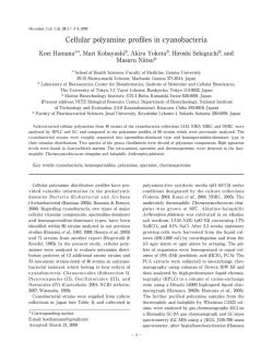
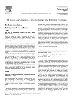
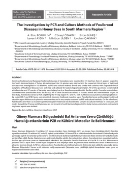
261 Extended Virulence Genotypes ofEscherichia coli Strains from Patients with Urosepsis in Relation to Phylogeny and Host Compr omise James R. Johnsonand AdamL. Stell VA Medical Center and Department of Medicine, University of Minnesota, Minneapolis, Minnesota Among 75 urosepsis isolates of Escherichia coli,29 virulence factor (VF) genes were detected by use of a novel polymerase chain reaction (PCR) assay. Compared with probe hybridization, the PCR assay’s specificity was 100% and sensitivity 97.1%. fyuA (yersiniabactin: overall prevalence, 93%), traT (serum resistance, 68%), and a pathogenicity-associated island marker (71%) occurred in most strains from both compromised and noncompromised hosts. Present in !20% of strains each were sfaS, focG (F1C fimbriae), afa/dra, bmaE (M fimbriae), gafD (G fimbriae), cnf1, cdtB (cytolethal distending toxin), cvaC (colicin V), and ibeA (invasion of brain endothelium). Different VFs were variously confined to virulence-associated phylogenetic group B2 (as defined by multilocus enzyme electrophoresis); concentrated in group B2, but with spread beyond; or concentrated outside of group B2. These findings provide novel insights into the VFs of extraintestinal pathogenic E. coli and demonstrate the new PCR assay’s utility for molecular epidemiological studies. Escherichia coli is the major cause of extraintestinal infections such as neonatal meningitis, gram-negative bacteremia, pyelonephritis, cystitis, and prostatitis [1–4]. Paradoxically, it also is the predominant facultative member of the normal human intestinal flora [5, 6]. Extraintestinal pathogenic E. coli and commensal E. coli typically differ with respect to phylogenetic background and virulence attributes. Pathogenic E. coli strains derive chiefly from phylogenetic group B2 (and to a lesser extent group D), as defined by multilocus enzyme electrophoresis [5, 7–11]. Commensal E. coli, in contrast, are characteristically from phylogenetic group A [5, 7, 8, 11]. Groups B2 and D comprise diverse evolutionary lineages that, because of their consistent associations with various extraintestinal infection syndromes, have come to be regarded as “virulent clones,” as traditionally defined based on O:K:H serotypes [9, 12–18]. A hallmark of such virulent E. coli clones is their possession of specialized virulence factors (VFs), traits that confer pathogenic potential and characteristically are infrequent among commensal strains [1, 2, 8, 19–21]. Recognized VFs of extraintestinal E. coli include diverse adhesins (e.g., P fimbriae, S and F1C fimbriae, Dr-antigen specific adhesins, and type 1 fimbriae (which, unlike other VFs, are present in nearly all E. coli), toxins (e.g., hemolysin and cytotoxic necrotizing factor), Received 13 August 1999; revised 27 September 1999; electronically published 23 December 1999. Presented in part: 36th Annual Meeting of the Infectious Diseases Society of America, Denver, CO, November 1998 (abstract 77). Financial support: VA Merit Review and NIH (DK-47504) to J.R.J. Reprints or correspondence: Dr. James R. Johnson, University of Minnesota Department of Medicine, Infectious Diseases (111F), Minneapolis VA Medical Center, 1 Veterans Dr., Minneapolis, MN 55417 (johns007@ maroon.tc.umn.edu). The Journal of Infectious Diseases 2000; 181:261–72 ᧠2000 by the Infectious Diseases Society of America. All rights reserved. 0022-1899/2000/18101-0033$02.00 siderophores (e.g., the aerobactin system), polysaccharide coatings (e.g., group II and group III capsules and lipopolysaccharide [LPS]), and invasins (e.g., IbeA, also called Ibe10) [19–23]. These VFs facilitate colonization and invasion of the host, avoidance or disruption of host defense mechanisms, injury to host tissues, and/or stimulation of a noxious host inflammatory response. Informed selection of VFs to be targeted for prevention of extraintestinal E. coli infections (with their associated morbidity, mortality, and increased health care costs [24, 25]) requires knowledge of which VFs are prevalent in specific clinical syndromes and host populations, as revealed by epidemiological studies [26]. Because host compromise can reduce the pathogenic importance of certain VFs [19, 27], it is also important to identify VFs that remain prevalent even among compromised hosts. Furthermore, because pathogenic behavior is predicted both by VF repertoire and by phylogenetic background [8, 15, 21, 28, 29], clonal associations of VFs must be evaluated. During the past decade, new VFs have been described in E. coli [23, 30–35], and certain established VFs have attracted renewed attention [24, 36–45]. In addition, pathogenicityassociated islands (PAIs); that is, blocks of (known and suspected) VF genes that provide a mechanism for coordinate horizontal transfer of VF genes between lineages, and even between species, have emerged as a unifying characteristic of diverse pathogenic bacteria, including extraintestinal E. coli [21, 46–52]. The epidemiological studies called for by these recent discoveries should be facilitated by advances in polymerase chain reaction (PCR) technology and by the increasing availability of VF gene-sequence data, innovations that have permitted the development of PCR assays as an alternative to traditional DNA-probe hybridization methods for VF gene detection [36, 53–59]. In the present study, we developed and validated a multiplex PCR assay for 29 known and suspected Copyright ©2001. All Rights Reserved. 262 Johnson and Stell VF gene regions of extraintestinal E. coli. We then used this new assay to define the prevalence, phylogenetic distribution, and associations with host compromise of these VF sequences among 75 well-characterized E. coli blood isolates from patients with urosepsis. Methods Primers. Primers’ sequences (table 1) were as published, provided by other investigators, or selected de novo from available nucleotide sequences with the assistance of the application AMPLIFY (William Engels, University of Wisconsin Genetics Department, Madison, WI). For enhanced detection of partial copies of pap, primers specific for multiple regions within the pap operon were included, as were previously published primers specific for the 3 papG alleles [55]. In addition to the central (consensus) region of the sfa/foc operon, as detected by published sfa primers [53], the adhesin genes of S fimbriae (sfaS) and F1C fimbriae (focG) were separately targeted. Dr family adhesins were detected by use of (consensus) afa/dra primers [53]. Three other mannose-resistant adhesins of extraintestinal E. coli—that is, glucosaminyl-specific G fimbriae (gaf), M blood group antigen-specific M fimbriae (bma), and nonfimbrial adhesin-1 (nfa)—were also targeted, as was fimH, which encodes the mannose-specific adhesin subunit of type 1 fimbriae. Toxins genes included were hlyA and cnf1, which have known associations with extraintestinal pathogenic E. coli [54], and cdtB (cytolethal distending toxin), which, to date, has been studied primarily in enteric E. coli and in genera other than Escherichia (the cdtB primers were designed and validated by Eric Oswald) [30, 31, 35, 61]. Siderophores included aerobactin (iutA) and yersiniabactin (fyuA), a Yersinia-associated siderophore system that recently has been identified along with other elements of the Yersinia-associated “high pathogenicity island” in some extraintestinal pathogenic E. coli strains [34]. Capsule synthesis (kpsMT) primers intended to be specific for all group II, all group III, only K1, and only K5 capsule regions, respectively, were selected from aligned kpsMT sequences for K1 (group II), K5 (group II), and K54 (group III) capsules [22, 62, 63]. The rfc locus, which participates in O4 LPS biosynthesis [64], was targeted as a marker for (virulence-associated) E. coli serogroup O4 [2, 19, 65, 66]. cvaC, which encodes colicin V [67], was targeted as a marker for ColV plasmids, which have been proposed to confer enhanced virulence through their carriage of other specific VFs, including the aerobactin system and serum survival genes, such as traT and iss [19, 68–70]. The “invasion of brain endothelium” gene ibeA was included because of its association with neonatal meningitis and endothelial cell invasion [10, 23]. Finally, a coding region of unknown significance near the right-hand terminus of a sequenced PAI from archetypal uropathogenic strain CFT073 was used as a generic marker for uropathogenic PAIs [51]. Strains. Seventy-five blood culture isolates of E. coli collected in the mid-1980s from adults with urosepsis in Seattle, Washington, were studied. The status of these strains with respect to P fimbriae (phenotype, papEFG probe, and papG allele genotype), type 1 fimbriae (phenotype and fim probe), hemolysin (phenotype and papA probe), aerobactin (phenotype, iuc probe, and plasmid vs. chro- JID 2000;181 (January) mosomal location of iuc), resistance to 12 antimicrobial agents, O:K:H serotype, and carboxylesterase B electrophoretic type (CBT), in comparison with host compromise status, have been reported elsewhere [27–29, 41, 71]. Strains were considered to derive from E. coli phylogenetic group B2 if they exhibited CBT B2 and from other phylogenetic groups if they exhibited CBT B1 [8, 72]. They were regarded as belonging to a particular O:K:H serotype if they exhibited 2 of the 3 corresponding antigens plus no other O, K, or H antigen [28]. The antimicrobial resistance score was the number of agents to which the strain was found to be resistant [27]. As additional controls for group III capsule synthesis genes (kpsMT III) and rfc, 14 strains of serogroup O4 that were previously reported as belonging (n = 11 ) or not belonging (n = 3 ) to a “J96-like” clonal group were studied [65, 66]. Other strains used during assay development as controls for specific VFs included J96 (papA, papC, papEF, papG alleles I and III, sfa/foc, focG, fimH, kpsMT III, hlyA, cnf1, and rfc) [65, 73], IA2 (papG allele II) [74], LG1315 (iutA) [27], P678-54/pHK11 (cvaC) [67], DH5a/pCIB10B (ibeA) [23], IH11165 (bmaE, gafD) [75, 76], HB101/pSR366 (kpsMT II) [62], P678-54/pAH1010 (nfaE) [77], E6468/62 (cdtB) [30], P678-54/pKT107 (traT) [78], 536-21/pANN801-13 (sfa/foc and sfaS) [79], NS24 (K1) [80], GR12 (K5) [81], and A30 (afa/dra; provided by Bogdan Nowicki). Strain P678-54 was used as a negative control [82]. Strains were stored at Ϫ70ЊC in Luria broth plus 15% glycerol until ready for use. Assay development. Primers for each VF were first validated individually by use of template DNA from appropriate positive and negative control strains. Primers were then sorted into 5 pools (as listed below) according to primer compatibility and amplicon size, such that all desired products could be amplified in 5 separate PCR reactions and the products from each reaction could be resolved by size in gel electrophoresis (figure 1). Each primer pool was validated by use of pooled control DNA containing all relevant VFs. Alternative primers were selected and PCR conditions were adjusted as needed to yield simultaneous amplification of all desired products (figure 1). PCR conditions. Amplification was done in a 25-mL reaction mixture containing template DNA (2 mL of boiled lysate [55]), 4 mM MgCl2, 0.8 mM each of 4 dNTPs, 0.6 mM of each primer (except for those marked “*,” which were used at a concentration of 0.3 mM), and 2.5 units AmpliTaq Gold in 1ϫ PCR buffer (Perkin Elmer, Branchburg, NJ). The 5 primer pools, with the 29 primer pairs listed in order of decreasing amplicon sizes (bp) within each pool, were as follows: pool 1: PAI (925), papA (717), fimH (508), kpsMT III (392), papEF (326), and ibeA (171); pool 2, fyuA (787), bmaE (507), sfa/focDE (410), iutA (302), papG allele III (internal; 258), and K1 (153); pool 3: hlyA (1177), rfc (788), *nfaE (559), *papG allele I (internal; 464), *kpsMT II (272), and *papC (205); pool 4: gafD (952), cvaC (697), cdtB (430), focG (364), traT (290), and papG allele II (internal; 190); and pool 5: papG allele I (flanking; 1190), papG alleles II and III (flanking; 1070), *afa/draBC (594), *cnf1 (498), *sfaS (244), and K5 (159). Reactions were heated to 95ЊC in an automated thermal cycler (PTC-100-96; MJ Research, Watertown, MA) for 12 min to activate the AmpliTaq Gold. This was followed by 25 cycles of denaturation (94ЊC, 30 s), annealing (63ЊC, 30 s), and extension (68ЊC, 3 min) and a final extension (72ЊC, 10 min). Samples were electrophoresed in 2% agarose gels, then stained with ethidium bromide, Copyright ©2001. All Rights Reserved. Primers for multiplex virulence factor polymerase chain reaction assay. Table 1. Gene(s) Primer sequence (5 –3 ) Primer name Size of product (bp) Primer coordinates papAH atggcagtggtgtcttttggtg cgtcccaccatacgtgctcttc gtggcagtatgagtaatgaccgtta atatcctttctgcagggatgcaata gcaacagcaacgctggttgcatcat agagagagccactcttatacggaca ctgtaattacggaagtgatttctg actatccggctccggataaaccat tccagaaatagctcatgtaacccg tcgtgctcaggtccggaattt tggcatcccccaacattatcg ctactatagttcatgctcaggtc ctgacatcctccaacattatcga gggatgagcgggcctttgat cgggcccccaagtaactcg ggcctgcaatggatttacctgg ccaccaaatgaccatgccagac ctccggagaactgggtgcatcttac cggaggagtaattacaaacctggca gtggatacgacgattactgtg ccgccagcattccctgtattc cagcacaggcagtggatacga gaatgtcgcctgcccattgct ggcagagggccggcaacaggc cccgtaacgcgccagcatctc atggcgctaacttgccatgctg agggggacatatagcccccttc tgttggaccgtctcagggctc ctcccggaactcgctgttact gcttactgattctgggatgga cggtggccgagtcatatgcca tgcagaacggataagccgtgg gcagtcacctgccctccggta aacaaggataagcactgttctggct accatataagcggtcattcccgtca aagatggagtttcctatgcaggag cattcagagtcctgccctcattatt aaatcaccaagaatcatccagtta aaatctcctgcaatcatccagttta gaaagtaaatggaatataaatgtccg gaaaataaatggaacacacatgtccg tgattaaccccgcgacgggaa cgcagtaggcacgatgttgta ggctggacatcatgggaactgg cgtcgggaacgggtagaatcg gcgcatttgctgatactgttg catccagacgataagcatgagca tcctcttgctactattccccct aggcgtatccatccctcctaac tagcaaacgttctattggtgc cagtatcagcaatcgttctgta atccatcaggaggggactgga aaccataccaaccaatgcgag aggcaggtgtgcgccgcgtac tggtgctccggcaaaccatgc cacacacaaacgggagctgtt cttcccgcagcatagttccat ggtgtggtgcgatgagcacag cacggttcagccatccctgag ggacatcctgttacagcgcgca tcgccaccaatcacagccgaac PapA f PapA r PapC f PapC r PapEF f PapEF r pGf pGr pG1 r* AlleleI-f AlleleI-r AlleleI -f AlleleI -r AlleleII-f AlleleII-r AlleleIII-f AlleleIII-r sfa1 sfa2 SfaS f SfaS r FocG f FocG r Afa f Afa r bmaE-f bmaE-r gafD-f gafD-r nfaE-f nfaE-r FimH f FimH r hly f hly r cnf1 cnf2 cdt-a1 cdt-a2 cdt-s1 cdt-s2 FyuA f FyuA r AerJ f AerJ r kpsII f kpsII r KpsIII f KpsIII r c K1-f c K5-f rfc-f rfc-r ibe10 f ibe10 r ColV-Cf ColV-Cr TraT f TraT r RPAi f RPAi r 720 1796–1817 2495–2516 4774–4798 4952–4976 8025–8049 8336–8360 1198–1221 2230–2254 9815–9838 8871–8891 9311–9331 183–205 635–657 1604–1623 1775–1793 1420–1441 1656–1677 NA papC papEF papG II, III I allele I allele I a allele II allele III sfa/focDE sfaS focG afa/draBC bmaE gafD nfaE fimH hlyA cnf1 cdtB fyuA iutA kpsMT II kpsMT III kpsMT K1 kpsMT K5 rfc ibeA cvaC traT PAI 200 336 1070 1190 461 474 190 258 410 240 360 559 507 952 559 508 1177 498 430 880 300 272 392 153 159 788 170 680 290 930 675–695 896–916 635–655 973–993 4589–4609 5160–5180 79–100 562–583 111–131 1040–1060 84–104 620–640 1814–1839 2256–2278 2420–2449 3572–3596 1649–1672 2122–2146 1735–1758 1463–1485 1292–1317 1019–1044 775–795 1539–1559 851–872 1132–1152 297–317 544–566 4050–4071 4418–4439 260–280 410–431 116–136 881–901 261–281 419–439 23–43 679–699 461–481 728–748 1021–1042 1921–1942 NOTE. NA, not available; PS, present study; U, unpublished. a For a variant of papG allele I identified in a canine urine isolate (unpublished). b Personal communication. c Use with kpsII-r. Copyright ©2001. All Rights Reserved. GenBank accession no. X61239 X61239 X61239 M20181 X61238 X61239 X61239 U M20181 X61238 U S53210 S68237 X76688 M15677 L33969 S61970 AJ225176 M10133 X70670 U04208 (a1,s1); U03293 (a2,s2) Z38064 X05874 X53819 AF007777 M57382 X53819 U39042 L42624 X57525 J01769 AF003742 Source of primer PS PS PS [53] [54] [54] [60] [60] PS [56] [56] PS PS [55] [55] [55] [55] [53] [53] PS PS PS PS PS PS PS PS PS PS PS PS PS PS [54] [54] [54] [54] b Eric Oswald PS [34] [57] [57] PS PS PS PS PS PS PS PS [23] PS PS PS PS PS PS PS 264 Johnson and Stell JID 2000;181 (January) Figure 1. Agarose gel showing polymerase chain reaction (PCR) products from multiplex virulence factor–PCR assay, as amplified simultaneously in 5 separate reactions using primer pools 1–5. M, molecular weight standard; ϩ, positive control DNA; Ϫ, negative control DNA. destained with distilled water, and photographed by use of an ultraviolet transilluminator and digital capture system (Gel Doc; BioRad, Hercules, CA). The sizes of the amplicons were determined by comparing them with a 100-bp DNA ladder (Gibco/BRL, Gaithersburg, MD), which was run in multiple lanes on the same gel. Duplicate lysates prepared for each test strain from separate bacterial colonies were amplified in separate PCR runs. Any discrepancies between the results of duplicate determinations for each VF were resolved by further investigation as needed. Dot-blot hybridization. Dot-blot hybridization was used to validate the PCR assay for 24 of the 29 constituent primer sets; that is, all primers but those for the 3 papG alleles that had been validated elsewhere [39], and the K1- and K5-specific primers which were predicted to generate probes that would hybridize nonspecifically with any group II kpsMT region, irrespective of K type. Probes were generated and labeled with digoxigenin by PCR by use of the same primers as used in the PCR assay (table 1), as described elsewhere [39]. Blotting (under high stringency conditions) and detection were done in duplicate, as described elsewhere [39], by use of the same template DNA samples as those used in the PCR assay. Validation of the PCR assay. Among the 75 urosepsis isolates, DNA probes for the 24 VF genes that were validated by blotting yielded 721 positive and 1,079 negative hybridization results. The PCR assay correctly identified 700 of the 721 blot-positive results (sensitivity, 97.1%), and all of the blot-negative results (specificity, 100%; accuracy, 98.9%). The PCR assay was 100% sensitive for all individual VFs except nfaE (sensitivity undefined; zero prevalence), papAH (57/59, 97%), papEF (57/58, 98%), fimH (73/75, 97%), cnf1 (11/12, 92%), and kpsMT II (47/62, 76%). Of note, all 3 strains that were blot-positive but PCR-negative for papAH or papEF were negative for P fimbrial expression, and 2 contained only partial copies of pap by blot and PCR. After exclusion of kpsMT II, overall assay sensitivity was 99.1%, specificity 100%, and accuracy 99.7%. Of the 15 strains that were kpsMT II probe-positive but PCRnegative, 12 were serologically K2; these accounted for all but 1 of the K2 strains (figure 2). Thus, probe-versus-PCR discordance for kpsMT II was 92% sensitive and 80% specific for K2 capsule, and in combination with O6 antigen positivity was 91% sensitive and 98% specific for serotype O6:K2:H1. The K1 and K5 primers were validated by comparison of PCR results with serologically determined K antigens. The K1 primers reacted with 20 of 21 K1-antigen-positive strains (sensitivity, 95%) Copyright ©2001. All Rights Reserved. JID 2000;181 (January) E. coli Virulence Genotypes in Urosepsis 265 Figure 2. Distribution of virulence factors (VFs) among 75 Escherichia coli isolates from patients with urosepsis, as detected by polymerase chain reaction (PCR) alone (papG alleles, K1, and K5) or by both PCR and probe (all other VFs). ϩ, positive results; Ϫ, negative results; *, strains that were PCR-negative but probe-positive for the indicated VF. Strains are sorted by carboxylesterase B type (CBTa), then by O:K:H serotype within each carboxylesterase B type. Heavy horizontal line separates carboxylesterase B types B2 and B1; fine horizontal lines separate individual O:K:H serotypes. Single O2:K5:H1 strain is grouped with O2:K7:H1(H-) strains for clarity. bCarboxylesterase B type was not available for strain V31, but was presumed to be B2. and with one K53-positive strain (specificity, 98%; figure 2). The K5 primers, although identifying 7 of 8 K5-positive strains (sensitivity, 88%), also reacted with 17 non-K5 strains (specificity for K5, 75%), which included all but 1 of the 18 strains that were positive by PCR for group II kpsMT but not for K1 (figure 2). The rfc and kpsMT III primers were further validated by comparison with dot-blot hybridization among 14 O4 strains, of which 10 were previously shown to exhibit group III capsules and/or to hybridize with a group III-specific kpsMT probe. All 14 O4 strains were confirmed as positive for rfc by both blot and PCR, as expected (PCR assay accuracy, 100%). The PCR assay also was 100% accurate in identifying the group III kpsMT probe-positive strains (not shown). Statistical methods. Comparisons of proportions between groups were evaluated by using Fisher’s exact test. Because of multiple comparisons, P values between .01 and .05 were regarded Copyright ©2001. All Rights Reserved. 266 Johnson and Stell as reflecting possible statistical significance, and P values р.01 as reflecting statistical significance. Comparisons of the prevalence of different VFs were evaluated by use of McNemar’s test [83], and comparisons of antimicrobial resistance scores were tested by use of the Mann-Whitney U test. Results Prevalence of VFs. Among the 75 urosepsis isolates, the 29 virulence gene regions ranged in prevalence from 0% (nfaE) to 100% (fimH). Various pap elements, fyuA, iutA, kpsMT II, traT, and the PAI marker each occurred in over 50% of strains (figure 2, table 2). Of the siderophores studied, fyuA was more prevalent than aerobactin (iutA; 93% vs. 80%; P ! .05, McNemar’s test) and was the most prevalent VF detected other than fimH. Of the sialosyl-specific adhesins studied, focG was more prevalent than sfaS (P ! .05, McNemar’s test). Of the toxins studies, hlyA (41%) was more prevalent than cnf1 (16%), JID 2000;181 (January) which in turn was more prevalent than cdtB (8%; for both comparisons, P ! .02, McNemar’s test). Phylogenetic distribution of VFs. The various VFs exhibited distinctive associations with CBT (carboxylesterase B types) B1 and B2, and with specific O:K:H serotypes, suggestive of divergent patterns of phylogenetic distribution (figure 2, table 2). These could be categorized broadly as (1) concentration within CBT B2, (2) concentration within CBT B1, and (3) equal distribution between CBTs B1 and B2. Among the CBT B2associated VFs, 3 subgroups were apparent. The first subgroup included those VFs, that is, sfa/foc, sfaS, focG, cnf1, cdtB, rfc, and ibeA, that occurred essentially only in CBT B2 strains, were present in !40% of the CBT B2 strains, and were limited to specific subsets of these strains. sfa/foc was concentrated in serogroups O2, O4, O6, and O18 (P ! .001 vs. other CBT B2 strains and vs. all strains). focG was largely limited to serotypes O2:K5, O6:K2, and O6:K5 (P = .002 vs. other CBT B2 strains; P ! .001 vs. all other strains). cnf1 was concentrated in serotypes Table 2. Distribution of virulence factors (VFs) according to carboxylesterase B type and presence of pathogenicity-associated island (PAI) marker. Prevalence of associated VF (%) a Carboxylesterase B type b VF No. (%) papAH papC papEF papG allele II allele III sfa/foc sfaS focG afa/draBC bmaE gafD nfaE fimH hlyA cnf1 cdtB fyuA iutA kpsMT II (PCR) kpsMT II (blot) kpsMT III K1 K5 rfc ibeA cvaC traT PAI marker 59 58 58 57 53 5 19 3 14 7 4 1 0 75 31 12 6 70 60 47 62 1 21 25 3 4 11 51 53 (79) (77) (77) (76) (71) (7) (25) (4) (19) (9) (5) (1) (100) (41) (16) (8) (93) (80) (63) (83) (1) (28) (33) (4) (5) (15) (68) (71) PAI marker c c B1 (n = 27) B2 (n = 47) P Absent (n = 22) Present (n = 53) P 63 63 63 59 56 4 4 0 4 11 15 4 0 100 22 4 0 85 89 44 59 4 15 30 0 0 26 63 26 87 85 85 85 79 9 36 6 28 9 0 0 0 100 51 21 13 98 77 72 96 0 36 34 6 9 9 83 96 .02 .04 .04 .02 .06 50 50 50 45 41 5 0 0 0 9 18 5 0 100 14 5 0 77 86 36 45 5 18 18 0 0 27 59 NA 91 85 85 89 83 8 36 6 26 10 0 0 0 100 53 21 11 100 77 74 98 0 32 40 6 8 9 72 NA !.001 .002 .01 (.015) .03 .048 .08 .06 .03 !.001 .06 (.085) ! .001 !.001 !.001 !.001 .001 !.001 .007 (.006) .002 .097 .002 .004 !.001 (.07) NOTE. NA, not applicable (comparison with self). a Percentage values calculated on the basis of the 74 strains for which carboxylesterase B type was available. b As detected by PCR only (papG alleles, kpsMT II [PCR], K1, and K5), by probe only (kpsMT II [blot]), or by PCR and probe (all other VFs). c P values (by Fisher’s exact test) shown only where !.10. Parenthetical P values are for negative associations with carboxylesterase B type B2. P р .01 reflect statistical significance; those between .01 and .05 reflect possible statistical significance. Copyright ©2001. All Rights Reserved. JID 2000;181 (January) E. coli Virulence Genotypes in Urosepsis 267 Figure 3. Statistical analysis of associations between virulence factors (VFs). P values (by Fisher’s exact test) shown only where р.10. P р .01 reflects statistical significance; P values between .01 and .05 reflect possible statistical significance. (Parenthetical P values are for negative associations.) Dashes indicate P 1 .10 . Not shown are VFs that were detected in р1 strain or that yielded only P values 1.10 (e.g., gafD, nfaE, fimH, and kpsMT-III). Not included on the horizontal axis are VFs for which all P values р.10 appear elsewhere in the matrix (e.g., cdtB, ibeA, and traT). Results are for analyses of VFs as detected by polymerase chain reaction (PCR) only (papG alleles, kpsMT II [P], K1, and K5), by probe only (kpsMT II [B]), or by PCR and probe (all other VFs). P, PCR; B, blot; II and III, papG alleles II and III, respectively; NA, not applicable (comparison with self). O2:K5/K7:H1, O4:K12, and O6:non-K2 (P ! .001 vs. other B2 strains and vs. all other strains). cdtB was narrowly limited to the non-K1 O2 strains (P ! .001 vs. other B2 strains and vs. all other strains). rfc was limited to strains of serogroup O4 (P ! .001). ibeA occurred in the sole strain of serotype O18:K1: H7 (the serotype of the source strain for this invasin gene) and in 2 other K1-positive strains (figure 2). A second subgroup of CBT B2-associated VFs included pap and hlyA, which were present in most (50%–85%) but not all of the CBT B2 strains, and in a substantial minority (20%–50%) of CBT B1 strains. pap was concentrated within the more prevalent serotypes of both the CBT B2 and B1 groups (figure 2). hlyA was concentrated, among the CBT B2 strains, specifically in serogroups O2, O4, O6, O12, and O18 (P ! .001 vs. other B2 strains), and among the CBT B1 strains in serotypes O2: K5 and O25:K2:H2 (P ! .001 vs. other B1 strains). A third subgroup of CBT B2-associated VFs included kpsMT-II and the PAI marker, which were present in essentially all (i.e., 195%) of the CBT B2 strains, and also in many (30%–60%) of the CBT B1 strains. Although their high prevalence among CBT B2 strains precluded serotype-specific associations, among the CBT B1 strains kpsMT-II (blot) was specifically associated with K types K1, K2, K5, and K52 (P ! .001), and the PAI marker was associated with serotype O2:K5:H4 and serogroup O157 (P ! .001). The second major pattern of phylogenetic distribution, exemplified by bmaE and possibly cvaC, was an association with CBT B1. bmaE was confined to CBT B1 strains and was concentrated in serotype O8:K27:H- (P = .002 vs. all other strains). When cvaC (which was 3 times as prevalent among CBT B1 strains as among CBT B2 strains) did occur in CBT B2 strains, it was associated with serotypes O1:K1:H7 and O2:K1:H7 (P ! .001 vs. other B2 strains). The third major pattern of phylogenetic distribution was of approximately equal prevalence among CBT B2 and B1 strains, whether low (!10%); for example, afa/draBC, or high (160%); for example, fimH, iutA, fyuA, and traT (although fyuA did exhibit a trend associating it with CBT B2). afa/draBC, which was uncommon in either CBT, was concentrated (within CBT B2) in serogroup O75 (P = .049). The highly prevalent and broadly distributed fimH, iutA, fyuA, and traT did not exhibit serotype-specific associations. Associations between VFs. The VFs displayed distinctive and complex associations with one another (figure 3, table 2). Although papG alleles II and III were both associated with sfaS, they otherwise exhibited nonoverlapping associations with other VFs. papG allele II was associated with kpsMT II, fyuA, and the PAI marker, whereas papG allele III was associated with sfa/foc, cnf1, and possibly K5 and ibeA (figure 3). sfa/ focDE occurred almost exclusively in pap-positive strains and was associated with hlyA, cnf1, and the PAI marker (figure 3). sfaS and focG were both strongly associated with cnf1. However, whereas sfaS was strongly associated with ibeA and was negatively associated with iutA, focG was strongly associated Copyright ©2001. All Rights Reserved. 268 Johnson and Stell with hlyA and the PAI marker and (borderline) negatively associated with traT. afa/draBC was not significantly associated with other VFs. bmaE was negatively associated with papG, group II capsules, and the PAI marker. gafD occurred in only 1 strain, which was also bmaE-positive (P = .05, bmaE vs. gafD). kpsMT-II was positively associated with fyuA, hlyA, and the PAI marker and negatively associated with cvaC. Among the group II capsules, K1 was negatively associated with iutA and possibly also with hlyA, whereas K5 (which included most nonK1, non-K2 group II capsules) was associated positively with cnf1 and cdtB and negatively with cvaC. fyuA was highly correlated with the PAI marker. iutA demonstrated a borderline negative association with ibeA. All cvaCpositive strains were positive for both iutA and traT, with the cvaC-traT association being statistically significant (figure 3). Finally, the PAI marker was highly correlated with all pap elements (except for papG allele III) and with sfa/foc, focG, kpsMT-II (PCR and blot), hlyA, and fyuA, whereas it was negatively correlated with bmaE (figure 3, table 2). Plasmid aerobactin systems. Presence of a plasmid-associated aerobactin system, as previously detected in 16 (21%) of the 75 urosepsis isolates [27, 29], was significantly associated with bmaE (P = .002), cvaC (P ! .001 ), and traT (P = .002). In addition to its previously demonstrated negative associations with pap elements and hly, plasmid aerobactin also was negatively associated with sfa/foc (P = .008 ), focG (P = .03), kpsMTII (blot; P ! .001), K5 (P = .01), and the PAI marker (P = .004). Antibiotic resistance. Several VFs were associated with decreased antibiotic resistance, including all pap elements except papG allele III (median resistance score in strains with vs. without VF, 0 vs. 4; P ! .001), sfa/foc (0 vs. 1; P = .03), kpsMT II (PCR; 0 vs. 3.5; P = .006), kpsMT II (blot; 0 vs. 5; P ! .001), fyuA (1 vs. 4; P = .02), hlyA (0 vs. 1; P = .004), and the PAI marker (0 vs. 3.5; P ! .001). Both focG and rfc exhibited similar trends (not shown). In contrast, cvaC and bmaE were significantly associated with increased antibiotic resistance (median resistance score among strains with vs. without VF: cvaC, 2 vs. 0.5; P = .04; bmaE, 6 vs. 1; P = .003). afa exhibited a similar trend (4 vs. 1; P = .09). Host compromise. Of the VFs newly detected in the present study (fim, pap, hly, and iutA having been reported elsewhere), only sfaS and bmaE exhibited significant associations with host compromise. Both were actually more prevalent among hosts with an upper urinary tract abnormality than among other hosts (sfaS: 2/8 vs. 1/67; P = .03; bmaE: 2/8 vs. 2/67; P = .05). Neither these nor any of the other VFs was significantly associated with presence or absence of urinary tract abnormalities in general, urinary tract instrumentation, a medical illness, or a combination of these compromising conditions (not shown). On the contrary, several of the more common VFs were substantially prevalent among strains from both compromised and JID 2000;181 (January) noncompromised hosts, including fyuA (91% vs. 97%, respectively), group II kpsMT (by blot, 61% vs. 90%), traT (63% vs. 76%), the PAI marker (67% vs. 76%), and fimH (all strains; for all comparisons between compromised and noncompromised hosts, P 1 .10). Discussion In this study, we developed and rigorously validated a novel multiplex PCR assay for 29 putative VF gene regions of extraintestinal E. coli. In the process, we defined the prevalence, clonal distribution, and associations with antimicrobial resistance and host compromise of the virulence genes included in the assay. Our findings provide novel insights into the molecular epidemiology of extraintestinal E. coli VFs and demonstrate that the multiplex PCR assay is highly accurate and informative. The assay was extremely accurate overall in detecting specific VF genes, with the exception of group II kpsMT sequences. That there were no false-positive results in comparison with probe hybridization reflects the specificity of the primers for the VF regions of interest under the stringent amplification conditions used. That several probe-positive strains were not detected by the papAH, papEF, fimH, and cnf1 primers is probably attributable either to the known polymorphic nature of these and other VF genes [21, 38, 60, 84–86] or to partial deletions, as appeared possible in the 3 strains that were probepositive but PCR-negative for pap elements and phenotypenegative for P fimbrial expression. Such infrequent falsenegative PCR results should be of little epidemiological significance. In contrast, the group II kpsMT primers appreciably lacked sensitivity, particularly for K2 capsules, and the K5 primers detected almost all non-K2 group II capsular types. Informed selection of improved group II and K5 primers will require sequence data for more than the currently-available K1, K5, and K54 kpsMT variants [22, 62, 63]. Interestingly, discrepancies between probe and PCR for group II kpsMT proved to be informative, in that most discrepant strains were of capsular type K2. At the practical level, this could provide a genetic means of tentatively identifying strains as K2, particularly if they are known to be O6. This is relevant to the identification of serotype O6:K2:H1, the predominant serotype in the present population and that of archetypal uropathogenic strains CFT073 and AD110 [51, 80]. The present study provides novel epidemiological information pertinent to the development of anti-VF interventions. The high prevalence of fyuA found among E. coli urosepsis isolates from Seattle confirms previously reported findings involving bacteremia isolates from Germany, among which fyuA was significantly more prevalent than among fecal control strains [34]. Taken together, these data suggest that fyuA is broadly characteristic of bacteremic E. coli. The high prevalence of fyuA Copyright ©2001. All Rights Reserved. JID 2000;181 (January) E. coli Virulence Genotypes in Urosepsis observed in the present study among isolates from compromised, as well as from noncompromised, hosts indicate that fyuA should be a particularly useful target for an intervention if fyuA can be demonstrated to actually contribute to (as opposed to merely serve as a marker for) invasiveness. Similarly, traT also was found to be substantially prevalent among both compromised and noncompromised hosts. Comparisons with appropriate control strains, and experimental evaluation of traT as a VF, are needed [68]. The high prevalence of the PAI marker observed in the present study is consistent with recently reported findings involving other clinical isolates [50, 51] and suggests that as-yet undefined genes associated with this and related PAIs also may contribute to urovirulence and may potentially serve as targets for interventions. As with fyuA, the minimal impact of host compromise on the prevalence of the PAI marker bodes well for the utility in compromised hosts of interventions directed toward this or other PAI-associated elements. Several VFs that previously have received minimal attention in the context of urosepsis were found to occur in only a small minority (!20%) of strains each; therefore, although these VFs may contribute to the pathogenesis of urosepsis in selected patients, they would not be ideal targets for anti-VF interventions for the prevention of urosepsis. foc, an sfa-related MR adhesins, which, unlike sfa, is not a hemagglutinin [87], was actually much more prevalent among sfa/foc-positive strains than was sfa, a situation that would have escaped attention had foc- and sfaspecific primers not been used. ibeA, which we postulated might promote bloodstream invasion during urosepsis because it promotes penetration of the blood-brain barrier during meningitis [23], did not exhibit the high prevalence observed among neonatal meningitis isolates [10]. cdtB was limited to O2 strains, consistent with the O2 status of the single previously-reported cdt-positive strain from a patient with urosepsis [35]. Although to date cdt has been regarded primarily as an enteric VF, the present study’s findings suggest that cdt should also be investigated as a possible extraintestinal VF [35]. bma and gaf occurred primarily in strains lacking pap, sfa/foc, and afa, and hence presumably were these strains’ primary MR adhesins and could have played a contributory pathogenetic role. Studies of all these “low-prevalence” VFs in other extraintestinal syndromes are needed. Because CBT B2 equates with (virulence associated) phylogenetic group B2 [8, 72], and O:K:H serotypes usually correspond with clonal groups [14, 88], the availability of CBT and O:K:H serotype data for this collection allowed an assessment of the phylogenetic distribution of the various VFs. Among the group B2-associated VFs, those that were present in only a small minority of B2 strains (e.g., sfa/foc, sfaS, focG, cdtB, and ibeA) are likely to have been recently acquired by the B2 phylogenetic group. Their confinement to the B2 group (with 1 exception each for sfa/foc, focG, and cnf1, which suggest horizontal transfer out of group B2) may be due either to their 269 recent arrival in the B2 group or to barriers (whether from genetic or selection factors) to their horizontal movement into other groups. The overlapping but not completely concordant distribution of serotypes (e.g., for cnf1 vs. cdtB), and the sporadic appearance of several of these traits even within a single O:K:H serotype (figure 2), cannot be readily accounted for by strict vertical inheritance, thereby strongly suggesting that some of these traits have moved horizontally between lineages within the B2 group. Those B2-associated VFs that were present in most but not all B2 strains (e.g., pap and hlyA) probably have been acquired by the B2 group at earlier points in its evolutionary history, with pap probably preceding hlyA. However, their incomplete penetration through the B2 group suggests that they did not enter E. coli prior to the differentiation of B2 strains from other E. coli. Thus, the substantial prevalence of both pap and hlyA outside of the B2 group is strongly suggestive of horizontal transfer from B2 into other lineages [21]. It should be noted that the evidence of pap remnants in O75 strains (figure 2 and unpublished data) suggests that pap may once have been more widespread within group B2 than is apparent at present, hence may represent a pre-B2 acquisition. In contrast, those B2associated VFs that were nearly universally prevalent among the B2 strains (e.g., kpsMT-II and the PAI marker), and also were prominent among non-B2 strains, could well have been acquired by E. coli prior to the branching off of the B2 group from an ancestral trunk that also served as the source for certain non-B2 lineages; for example, virulence-associated phylogenetic group D [11]. Alternatively, they may represent a very early acquisition by the B2 group, with subsequent horizontal transfer into selected non-B2 lineages [11]. Those VFs (e.g., cvaC and bmaE) that were concentrated outside of group B2 may have entered E. coli initially in nonB2 lineages subsequent to the branching off of group B2 [11]. However, cvaC (with its associated iutA) clearly has moved horizontally between non-B2 and B2 strains, or has independently entered group B2, to take up residence specifically within serotypes O1:K1:H7 and O2:K1:H7. Finally, several VFs (e.g., fyuA and fimH) were highly prevalent throughout the population, suggesting that they either entered E. coli early during its evolutionary history, hence are now present in all members of the species (which is likely the case for fimH) or, if more recently acquired (e.g., as part of a PAI from another species, which may be the case for fyuA), are highly horizontally mobile, are strongly selected for in the context of urosepsis, or are both. Some inferences also could be drawn regarding the mode of horizontal transfer of VFs between lineages. Because both sfaS and focG were confined to strains also positive for the PAI marker, and because foc occurs as part of a PAI in strain CP9 [33], both VFs may have been PAI-linked in the present population, including when appearing in non-B2 strains (i.e., focG in strain 2P1). In contrast, pap elements and hly, although usually associated with the PAI marker, also appeared in its ab- Copyright ©2001. All Rights Reserved. 270 Johnson and Stell sence, particularly when occurring in non-B2 strains, consistent with horizontal mobility out of group B2 not as part of a PAI, or as part of a PAI not containing the PAI marker included in our assay. Finally, the appearance of afa and cvaC in disparate lineages, unassociated with one another or with other VFs, would be consistent with the known occurrence of these sequences on plasmids [36, 69]. It is also likely that iutA was colicin V plasmid-associated among the O1:K1:H7 and O2:K1: H7 strains, because these 2 serotypes had completely concordant results for iutA and cvaC. In summary, we have developed and rigorously validated a novel multiplex PCR assay capable of detecting a broad array of established and putative extraintestinal E. coli VF genes with a high degree of sensitivity and specificity. fyuA, traT, and a PAI marker were highly prevalent among urosepsis isolates, including among pap-negative strains from compromised hosts, and hence may constitute useful targets for preventive interventions. Although most VFs were concentrated in phylogenetic group B2, many were also prevalent in other strains. The new PCR assay should facilitate the studies that are needed to define the epidemiology and phylogenetic associations of traditional and newer VFs in extraintestinal infections due to E. coli. Acknowledgments Control strains were provided by Gabriele Blum-Oehler, J. M. de Ree, Betsy Foxman, Lynne Gilson, Jorg Hacker, Richard Hull, Sheila Hull, James Kaper, Kwang Sik Kim, Timo Korhonen, Joel Maslow, Barbara Minshew, Harry L.T. Mobley, Lisa Nolan, Thomas Russo, Soren Schubert, Richard Silver, Ann Stapleton, Peter Williams, and Lori Wright. Dave Prentiss prepared figure 1. Diana Owensby assisted with manuscript preparation. References 1. Eisenstein BI, Jones GW. The spectrum of infections and pathogenic mechanisms of Escherichia coli. Adv Intern Med 1988; 33:231–52. 2. Orskov I, Orskov F. Escherichia coli in extra-intestinal infections. J Hyg (Lond) 1985; 95:551–75. 3. Lipsky BJ. Urinary tract infections in men: epidemiology, pathophysiology, diagnosis, and treatment. Ann Intern Med 1989; 110:138–50. 4. Sussman M. Escherichia coli and human disease. In: Sussman M. Escherichia coli: mechanisms of virulence. Cambridge, UK: Cambridge University Press, 1997:3–48. 5. Selander RK, Caugant DA, Whittam TS. Genetic structure and variation in natural populations of Escherichia coli. In: Neidhardt FC, Ingraham KL, Magasanik B, Low KB, Schaechter M, Umbarger HE. Escherichia coli and Salmonella typhimurium: cellular and molecular biology. Washington, DC: American Society for Microbiology, 1987:1625–48. 6. Bettelheim KA. Escherichia coli in the normal flora of humans and animals. In: Sussman M. Escherichia coli: mechanisms of virulence. Cambridge, UK: Cambridge University Press, 1997:85–109. 7. Herzer PJ, Inouye S, Inouye M, Whittam TS. Phylogenetic distribution of branched RNS-linked multicopy single-stranded DNA among natural isolates of Escherichia coli. J Bacteriol 1990; 172:6175–81. JID 2000;181 (January) 8. Picard B, Sevali Garcia J, Gouriou S, et al. The link between phylogeny and virulence in Escherichia coli extraintestinal infection. Infect Immun 1999; 67:546–53. 9. Selander RK, Korhonen TK, Va¨isa¨nen-Rhen V, Williams PH, Pattison PE, Caugant DA. Genetic relationships and clonal structure of strains of Escherichia coli causing neonatal septicemia and meningitis. Infect Immun 1986; 52:213–22. 10. Bingen E, Picard B, Brahimi N, et al. Phylogenetic analysis of Escherichia coli strains causing neonatal meningitis suggests horizontal gene transfer from a predominant pool of highly virulent B2 group strains. J Infect Dis 1998; 177:642–50. 11. Lecointre G, Rachdi L, Darlu P, Denamur E. Escherichia coli molecular phylogeny using the incongruence length difference test. Mol Biol Evol 1998; 15:1685–95. 12. Vaisanen-Rhen V, Elo J, Vaisanen E, et al. P-fimbriated clones among uropathogenic Escherichia coli strains. Infect Immun 1984; 43:149–55. 13. Orskov I, Orskov F, Birch-Andersen A, Kanamori M, Svanborg Eden C. O, K, H and fimbrial antigens in Escherichia coli serotypes associated with pyelonephritis and cystitis. Scand J Infect Dis 1982; 33(Suppl):18–25. 14. Orskov F, Orskov I. Summary of a workshop on the clone concept in the epidemiology, taxonomy, and evolution of the enterobacteriaciae and other bacteria. J Infect Dis 1983; 148:346–57. 15. Maslow JN, Whittam TS, Gilks CF, et al. Clonal relationships among bloodstream isolates of Escherichia coli. Infect Immun 1995; 63:2409–17. 16. Achtman M. Clonal analysis of descent and virulence among selected Escherichia coli. Annu Rev Microbiol 1986; 40:185–210. 17. Korhonen TK, Valtonen MV, Parkkinen J, et al. Serotypes, hemolysin production, and receptor recognition of Escherichia coli strains associated with neonatal sepsis and meningitis. Infect Immun 1985; 48:486–91. 18. Achtman M, Mercer A, Kusecek B, et al. Six widespread bacterial clones among Escherichia coli K1 isolates. Infect Immun 1983; 39:315–35. 19. Johnson JR. Virulence factors in Escherichia coli urinary tract infection. Clin Microbiol Rev 1991; 4:80–128. 20. Donnenberg MS, Welch RA. Virulence determinants of uropathogenic Escherichia coli. In: Mobley HLT, Warren JW. Urinary tract infections: molecular pathogenesis and clinical management. Washington, DC: ASM Press, 1996: 135–74. 21. Boyd EF, Hartl DL. Chromosomal regions specific to pathogenic isolates of Escherichia coli have a phylogenetically clustered distribution. J Bacteriol 1998; 180:1159–65. 22. Russo TA, Wenderoth S, Carlino UB, Merrick JM, Lesse AJ. Identification, genomic organization, and analysis of the group III capsular polysaccharide genes kpsD, kpsM, kpsT and kpsE from an extraintestinal isolate of Escherichia coli (CP9, O4/K54/H5). J Bacteriol 1998; 180:338–49. 23. Huang S-H, Wass C, Fu Q, Prasadarao NV, Stins M, Kim KS. Escherichia coli invasion of brain microvascular endothelial cells in vitro and in vivo: molecular cloning and characterization of invasion gene ibe10. Infect Immun 1995; 63:4470–5. 24. Langermann S, Palaszynski S, Barnhart M, et al. Prevention of mucosal Escherichia coli infection by FimH-adhesin–based systemic vaccination. Science 1997; 276:607–11. 25. Johnson JR, Stamm WE. Urinary tract infections in women: diagnosis and treatment. Ann Intern Med 1989; 111:906–17. 26. Johnson JR. Epidemiological considerations in studies of adherence. In: Doyle RJ, Ofek I. Adhesion of microbial pathogens: methods in enzymology. Vol 253. Orlando, FL: Academic Press, 1995; 167–78. 27. Johnson JR, Moseley S, Roberts P, Stamm WE. Aerobactin and other virulence factor genes among strains of Escherichia coli causing urosepsis: association with patient characteristics. Infect Immun 1988; 56:405–12. 28. Johnson JR, Orskov I, Orskov F, et al. O, K, and H antigens predict virulence factors, carboxylesterase B pattern, antimicrobial resistance, and host compromise among Escherichia coli strains causing urosepsis. J Infect Dis 1994; 169:119–26. Copyright ©2001. All Rights Reserved. JID 2000;181 (January) E. coli Virulence Genotypes in Urosepsis 29. Johnson JR, Goullet PH, Picard B, Moseley SL, Roberts PL, Stamm WE. Association of carboxylesterase B electrophoretic pattern with presence and expression of urovirulence factor determinants and antimicrobial resistance among strains of Escherichia coli causing urosepsis. Infect Immun 1991; 59:2311–5. 30. Scott DA, Kaper JB. Cloning and sequencing of the genes encoding Escherichia coli cytolethal distending toxin. Infect Immun 1994; 62:244–51. 31. Peres SY, Marches O, Daigle F, et al. A new cytolethal distending toxin (CDT) from Escherichia coli producing CNF2 blocks HeLa cell division in G2/M phase. Mol Microbiol 1997; 24:1095–107. 32. Russo TA, Jodush ST, Brown JJ, Johnson JR. Identification of two previously unrecognized genes (guaA, argC) important for uropathogenesis. Mol Microbiol 1996; 22:217–29. 33. Russo TA, Carlino UB, Mong A, Jodush ST. Identification of genes in an extraintestinal isolate of Escherichia coli with increased expression after exposure to human urine. Infect Immun 1999; 67:5306–14. 34. Schubert S, Rakin A, Karch H, Carniel E, Heesemann J. Prevalence of the “high-pathogenicity island” of Yersinia species among Escherichia coli strains that are pathogenic to humans. Infect Immun 1998; 66:480–5. 35. Johnson WM, Lior H. A new heat-labile cytolethal distending toxin (CLDT) produced by Escherichia coli isolates from clinical material. Microb Pathog 1988; 4:103–13. 36. Zhang L, Foxman B, Tallman P, Cladera E, Le Bougenec C, Marrs CF. Distribution of drb genes coding for Dr binding adhesins among uropathogenic and fecal Escherichia coli isolates and identification of new subtypes. Infect Immun 1997; 65:2011–8. 37. Island MD, Cui X, Warren JW. Effect of Escherichia coli cytotoxic necrotizing factor 1 on repair of human bladder cell monolayers in vitro. Infect Immun 1999; 67:3657–61. 38. Sokurenko EV, Chesnokova V, Dykhuizen DE, et al. Pathogenic adaptation of Escherichia coli by natural variation of the FimH adhesin. Proc Natl Acad Sci USA 1998; 95:8922–6. 39. Johnson JR, Russo TA, Brown JJ, Stapleton A. papG alleles of Escherichia coli strains causing first episode or recurrent acute cystitis in adult women. J Infect Dis 1998; 177:97–101. 40. Johnson JR, Brown JJ, Maslow JN. Clonal distribution of the three alleles of the Gal(a1-4)Gal-specific adhesin gene papG among Escherichia coli strains from patients with bacteremia. J Infect Dis 1998; 177:651–61. 41. Johnson JR. papG alleles among Escherichia coli strains causing urosepsis: associations with other bacterial characteristics and host compromise. Infect Immun 1998; 66:4568–71. 42. Connell H, Agace W, Klemm P, Schembri M, Marild S, Svanborg C. Type 1 fimbrial expression enhances Escherichia coli virulence for the urinary tract. Proc Natl Acad Sci USA 1996; 93:9827–32. 43. Saren A, Virkola R, Hacker J, Korhonen TK. The cellular form of human fibronectin as an adhesion target for the S fimbriae of meningitis-associated Escherichia coli. Infect Immun 1999; 67:2671–6. 44. van Die I, Kramer C, Hacker J, Bergmans H, Jongen W, Hoekstra W. Nucleotide sequence of the genes coding for minor fimbrial subunits of the F1C fimbriae of Escherichia coli. Res Microbiol 1991; 142:653–8. 45. Gleason TG, Houlgrave CW, May AK, et al. Hemolytically active (acylated) alpha-hemolysin elicits interleukin-1b (IL-1b) but augments the lethality of Escherichia coli by an IL-1– and tumor necrosis factor-independent mechanism. Infect Immun 1998; 66:4215–21. 46. Groisman EA, Ochman H. Pathogenicity islands: Bacterial evolution in quantum leaps. Cell 1996; 87:791–4. 47. High NJ, Hales BA, Jann K, Boulnois GJ. A block of urovirulence genes encoding multiple fimbriae and hemolysin in Escherichia coli O4:K12:H. Infect Immun 1988; 56:513–7. 48. Ritter A, Blum G, Emody L, et al. tRNA genes and pathogenicity islands: influence on virulence and metabolic properties of uropathogenic Escherichia coli. Mol Microbiol 1995; 17:109–21. 49. Low D, David V, Lark D, Schoolnik G, Falkow S. Gene clusters governing 50. 51. 52. 53. 54. 55. 56. 57. 58. 59. 60. 61. 62. 63. 64. 65. 66. 271 the production of hemolysin and mannose-resistant hemagglutination are closely linked in Escherichia coli serotype O4 and O6 isolates from urinary tract infection. Infect Immun 1984; 43:353–8. Guyer DM, Kao J-S, Mobley HLT. Genomic analysis of a pathogenicity island in uropathogenic Escherichia coli CFT073: distribution of homologous sequences among isolates from patients with pyelonephritis, cystitis, and catheter-associated bacteriuria and from fecal samples. Infect Immun 1998; 66:4411–7. Kao J, Stucker DM, W. WJ, Mobley HLT. Pathogenicity island sequences of pyelonephritogenic Escherichia coli CFT073 are associated with virulent uropathogenic strains. Infect Immun 1997; 65:2812–20. Swenson DL, Bukanov NO, Berg DE, Welch RA. Two pathogenicity islands in uropathogenic Escherichia coli J96: cosmid cloning and sample sequencing. Infect Immun 1996; 64:3736–43. Le Bouguenec C, Archambaud M, Labigne A. Rapid and specific detection of the pap, afa, and sfa adhesin-encoding operons in uropathogenic Escherichia coli strains by polymerase chain reaction. J Clin Microbiol 1992; 30:1189–93. Yamamoto S, Terai A, Yuri K, Kurazono H, Takeda Y, Yoshida O. Detection of urovirulence factors in Escherichia coli by multiplex polymerase chain reaction. FEMS Immunol Med Microbiol 1995; 12:85–90. Johnson JR, Brown JJ. A novel multiply-primed polymerase chain reaction assay for identification of variant papG genes encoding the Gal(al-4)Galbinding PapG adhesins of Escherichia coli. J Infect Dis 1996; 173:920–6. Mitsumori K, Terai A, Yamamoto S, Yoshida O. Identification of S, F1C and three PapG fimbrial adhesins in uropathogenic Escherichia coli by polymerase chain reaction. FEMS Immunol Med Microbiol 1998; 21: 261–8. Johnson JR, Brown JJ. Colonization with and acquisition of uropathogenic Escherichia coli strains as revealed by polymerase chain reaction-based detection. J Infect Dis 1998; 177:1120–4. Foxman B, Zhang L, Tallman P, et al. Virulence characteristics of Escherichia coli causing first urinary tract infection predict risk of second infection. J Infect Dis 1995; 172:1536–41. Blanco M, Blanco JE, Blanco J, et al. Polymerase chain reaction for detection of Escherichia coli strains producing cytotoxic necrotizing factor type 1 and 2 (CNF1 and CNF2). J Microbiol Methods 1996; 26:95–101. Marklund BI, Tennent JM, Garcia E, et al. Horizontal gene transfer of the Escherichia coli pap and prs pili operons as a mechanism for the development of tissue-specific adhesive properties. Mol Microbiol 1992; 6: 2225–42. Guth BEC, Giraldi R, Gomes TAT, Marques LRM. Survey of cytotoxin production among Escherichia coli strains characterized as enteropathogenic (EPEC) by serotyping and presence of EPEC adherence factor (EAF) sequences. Can J Microbiol 1994; 40:341–4. Pavelka MS Jr, Wright LF, Silver RP. Identification of two genes, kpsM and kpsT, in region 3 of the polysialic acid gene cluster of Escherichia coli K1. J Bacteriol 1991; 173:4603–10. Smith AN, Boulnois GJ, Roberts IS. Molecular analysis of the Escherichia coli K5 kps locus: identification and characterization of an innermembrane capsular polysaccharide transport system. Mol Microbiol 1990; 4:1863–9. Lukomski S, Hull RA, Hull SI. Identification of the O antigen polymerase (rfc) gene in Escherichia coli O4 by insertional mutagenesis using a nonpolar chloramphenicol resistance cassette. J Bacteriol 1996; 178:240–7. Johnson JR, Russo TA, Scheutz F, et al. Discovery of disseminated J96-like strains of uropathogenic Escherichia coli O4:H5 containing genes for both PapGJ96 (“Class I”) and PrsGJ96 (“Class III”) Gal(a1-4)Gal-binding adhesins. J Infect Dis 1997; 175:983–8. Johnson JR, Stapleton AE, Russo TA, Scheutz FS, Brown JJ, Maslow JN. Characteristics and prevalence within serogroup O4 of a J96-like clonal group of uropathogenic Escherichia coli O4:H5 containing the class I and class III alleles of papG. Infect Immun 1997; 65:2153–9. Copyright ©2001. All Rights Reserved. 272 Johnson and Stell 67. Gilson L, Mahanty HK, Kolter R. Four plasmid genes are required for colicin V synthesis, export, and immunity. J Bacteriol 1987; 169:2466–70. 68. Kanukollu U, Bieler S, Hull S, Hull R. Contribution of the traT gene to serum resistance among clinical isolates of enterobacteriaciae. J Med Microbiol 1985; 19:61–7. 69. Fernandez-Beros ME, Kissel V, Lior H, Cabello FC. Virulence-related genes in ColV plasmids of Escherichia coli isolated from human blood and intestines. J Clin Microbiol 1990; 28:742–6. 70. Chuba PJ, Leon MA, Banerjee A, Palchaudhuri S. Cloning and DNA sequence of plasmid determinant iss, coding for increased serum survival and surface exclusion, which has homology with lambda DNA. Mol Gen Genet 1989; 216:287–92. 71. Johnson JR, Roberts P, Stamm WE. P fimbriae and other virulence factors in Escherichia coli urosepsis: association with patient’s characteristics. J Infect Dis 1987; 156:225–9. 72. Desjardins P, Picard B, Kaltenbock B, Elion J, Denamur E. Sex in Escherichia coli does not disrupt the clonal structure of the population: evidence from random amplified polymorphic DNA and restriction-fragment–length polymorphism. J Mol Evol 1995; 41:440–8. 73. Welch RA, Dellinger EP, Minsheu B, Falkow S. Haemolysin contributes to virulence of extra-intestinal E. coli infections. Nature 1981; 294:665–7. 74. Clegg S. Cloning of genes determining the production of mannose-resistant fimbriae in a uropathogenic strain of Escherichia coli belonging to serogroup O6. Infect Immun 1982; 38:739–44. 75. Vaisanen V, Korhonen TK, Jokinen M, Gahmberg CG, Ehnholm C. Blood group M specific haemagglutinin in pyelonephritogenic Escherichia coli. Lancet 1982; 1:1192. 76. Saarela S, Westerlund-Wikstron B, Rhen M, Korhonen TK. The GafD Protein of the G (F17) fimbrial complex confers adhesiveness of Escherichia coli to laminin. Infect Immun 1996; 64:2857–60. 77. Ahrens R, Ott M, Ritter A, et al. Genetic analysis of the gene cluster encoding nonfimbrial adhesin I from an Escherichia coli uropathogen. Infect Immun 1993; 61:2505–12. JID 2000;181 (January) 78. Wooley RE, Nolan LK, Brown J, Gibbs PS, Bounous DI. Phenotypic expression of recombinant plasmids pKT107 and pHK11 in an avirulent avian Escherichia coli. Avian Dis 1994; 38:127–34. 79. Hacker J, Schmidt G, Hughes C, Knapp S, Marget M, Goebel W. Cloning and characterization of genes involved in production of mannose-resistant, neuraminidase-susceptible (X) fimbriae from a uropathogenic O6:K15: H31 Escherichia coli strain. Infect Immun 1985; 47:434–40. 80. de Ree JM, van den Bosch JF. Fimbrial serotypes of Escherichia coli strains isolated from extra-intestinal infections. J Med Microbiol 1989; 29:95–9. 81. Klann AG, Hull RA, Hull SI. Sequences of the genes encoding the minor tip components of Pap-3 pili of Escherichia coli. Gene 1992; 119:95–100. 82. Hull RA, Gill RE, Hsu P, Minshew BH, Falkow S. Construction and expression of recombinant plasmids encoding type 1 or D-mannose–resistant pili from a urinary tract infection Escherichia coli isolate. Infect Immun 1981; 33:933–8. 83. Fleiss JL. Statistical methods for rates and proportions. New York: John Wiley & Sons, 1981:112–37. 84. Boyd EF, Hartl DL. Diversifying selection governs sequence polymorphisms in the major adhesin proteins FimA, PapA, and SfaA of Escherichia coli. J Mol Evol 1998; 47:258–67. 85. Bijlsma IG, van Dijk L, Kusters JG, Gaastra W. Nucleotide sequences of two fimbrial major subunit genes, pmpA and ucaA, from canine-uropathogenic Proteus mirabilis strains. Microbiol 1995; 141:1349–57. 86. Marc D, Dho-Moulin M. Analysis of the fim cluster of an avian O2 strains of Escherichia coli: serogroup-specific sites within fimA and nucleotide sequence of fimI. J Med Microbiol 1996; 44:444–52. 87. Ott M, Hoschutzky H, Jann K, Van Die I, Hacker J. Gene clusters for S fimbrial adhesin (sfa) and FIC fimbriae (foc) of Escherichia coli: comparative aspects of structure and function. J Bacteriol 1988; 170:3983–90. 88. Caugant DA, Levin BR, Orskov I, Orskov F, Svanborg Eden C, Selander RK. Genetic diversity in relation to serotype in Escherichia coli. Infect Immun 1985; 49:407–13. Copyright ©2001. All Rights Reserved.
© Copyright 2026 Paperzz