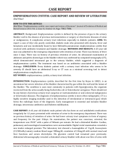
Case Report Original Article Archives of Clinical Experimental Surgery Increased of Langerhans Cells in Smokeless Tobacco-Associated Oral Mucosal Lesions Érica Dorigatti de Ávila1, Rafael Scaf de Molon2, Melaine de Almeida Lawall1, Renata Bianco Consolaro1, Alberto Consolaro1 Bauru Dental School University of São Paulo Bauru–SP, Brazil 1 Abstract Objective: To evaluate the changes in the number of Langerhans Cells (LC) observed in the epithelium of Dog Eea-Like Bladder Herniation smokeless tobacco (SLT-induced) lesions. 2 Methods: Microscopic biopsies carried out in the buccal mucosa of twenty patients, who were Ferhat Cüce1, Onur Sıldıroğlu2sections , Mehmetfrom Incedayı 2 Araraquara Dental Scho São Paulo State Universit Araraquara-SP, Brazil chronic users of smokeless tobacco (SLT), were utilized. For the control group, twenty non-SLT users of SLT Received: February 05, 20 Department of Radiology with normal mucosa were selected. The sections were studied with routine coloring and were immunostained Accepted: February 29, 2 Van Military Hospital Abstract for S-100, CD1a, Ki-67 and p63. These data were statistically analyzed by the Student’s t-test to investigate the Van, Turkey Arch Clin Exp Surg 2012 DOI: 10.5455/aces.2012022 The hernia content involving a bladder segment occurs in 1-4 % of cases of the inguinal hernias. Inguidifferences in the expression of immune markers in normal mucosa and in SLT-induced leukoplakia lesions. Department of Radiology noscrotal hernias are usuallydifference found incidentally on radiological examinations or atbetween the time normal of Gulhane Military Results:bladder There was a significant in the immunolabeling of all markers mucosa Medical AcademyCorresponding author herniorrhaphy. We present an adult case that has a left inguinal bladder hernia detected and evaluated and SLT-induced lesions (p<0.001). The leukoplakia lesions in chronic SLT users demonstrated a significant Érica Dorigatti de Avila Haydarpasa Training Hospital by sonography, intravenous pyelography (IVP), and computerized tomography (CT). İstanbul, Turkey Departamento de Estoma increase in the number of Langerhans cells and in the absence of epithelial dysplasia. da Faculdade de Odontol Key words: Bladder Cystocele, Inguinoscrotal hernia Conclusion: The hernia, increase in the number of these cells represents the initial stage of leukoplakia. Received: June 12,Bauru 2012 Accepted: July 04, 2012 Key words: Smokeless tobacco, leukoplakic lesions, cancer, langerhans cells, chewing tobacco. Universidade de São Pau Arch Clin Exp Surg 2013;X: X-X Avenida Alameda Octávi DOI:10.5455/aces.20120704012529 Introduction Case Report Pinheiro Brizola, 9-75, 17 Scrotal cystocele and urinary bladder A 55-year-old male with inCorresponding contact with thepresented oral mucosa and creates a author: Introduction Bauru–SP, Brasil Onur Sıldıroğlu, MD [email protected] hernia (UBH) are the most common terms termittent left groin swelling and pain for 2 Department more alkaline environment, its products mayof Radiology Among tobacco users, there is a false beused to describe inguinoscrotal herniation years. Concerning the patient’s complaint, Gulhane Military even be more aggressive to tissue [5]. MedicalThe Academy liefthe thatbladder SLT is[1]. safeMost because is not are burned, of of theit UBHs we needed to compress the inguinal canal Haydarpasa Training Hospital percentage of SLT is lower Selimiye M. Tıbbiye C. asymptomatic and are being to complete voiding. The users abdomen was compared which leads many people to diagnosed quit cigarettes Uskudar, İstanbul, Turkey to cigarette users; however, usage is increasing [email protected] during the operation. UBHs had a male presoft on physical examination, but a reducand start using SLT [1]. However, SLT conyoung individuals is therefore a dominance and an increasing prevalence by ible leftamong inguinal hernia extending and into itthe tains higher concentrations of nicotine than age. Early and accurate diagnosis is very im- left hemiscrotum wasdisturbing palpated. danger Urinary[6,7]. significant and cigarettes and, in addition, nearly 30 carciportant to plan the surgery and to avoid the and superficial Ultrasonography (US) were studies on thehyperplasia effects of SLT on the nogenic substances, such as tobacco-specific complications during the surgery. planned forInitial probable prostate 1 2 N-nitrosamines (TSNA), which is formed during the aging process of the tobacco, [2-4] oral mucosa demonstrated the formation of white lesions induced by chronic exposure to X Cuce F et al. and inguinal hernia. US images showed that the left side of the bladder wall was extending into the suprapubic left parietal zone and that there was also an anechoic cystic mass surrounded by a half-thick wall in the inguinal canal. In the latest phase of IVP, contrast mate- Figure 1. Contrast-filled bladder in IVP demonstrates elongation of the bladder base to the left side, and contrast medium is being superposed to left inguinal region (arrow). Figure 2. In the late phase of the contrast-enhanced pelvic CT images, a bladder with lumen-filled contrast medium extending inferiorly to the left inguinal canal (arrow). Figure 3. The 3D CT reconstruction image shows a dog left ear-like, left-sided inguinal bladder hernia (arrow). Arch Clin Exp Surg rial extended beyond the left side of the bladder corpus and a contrast enhancement was superposed at the left obturator foramen, but we could not visualize the relationship between them (Figure 1). The cross-sectional CT images demonstrated elonging of the bladder to the left inguinal canal with contrast-filled lumen; in reconstruction images, this was like a dog ear (Figure 2,3). Discussion UBH is a rare pathology that has been reported in 1 to 4 % of cases of inguinal hernias, whereas digestive or omental hernias are frequently observed [1-6]. The main factor for the herniation mechanism is bladder outlet obstruction (benign prostate hyperplasia, prostatitis, and urethral stricture). The other factors are obesity (especially in elderly males) and loss of bladder tonus with weakness of the surrounding supporting structures [1-3]. The unidentified reason for UBH is much more common in the right side; on the contrary, our patient’s hernia is left-sided [3]. The clinical presentation of UBH is variable, and nonspecific symptoms like dysuria, urinary frequency, urgency, and recurrent urinary tract infection were accompanied by a scrotal mass. The important complaint casting doubt in an elderly patient with scrotal mass is two-phased voiding: after a normal spontaneous urination, the patient evacuates the bladder with manual compression and it is defined as the second phase of urination [1-5]. In patients suffering from groin swelling with twophase urination, US is preferred as the first-line diagnostic choice; it is cost-effective, easy to access, and atraumatic - safety is achieved [2]. In bladder inguinoscrotal herniation, a hipoechoic cystic mass from the bladder through the inguinal canal can be seen. The US can evaluate a bladder hernia according to its relationship with the inferior epigastric artery (generally direct as medial to vessel) and bladder outlet obstruction, such as an enlarged prostate or any subtle sign on IVP [1,2,4]. Being medial to the inferior epigastric artery, our patient’s herniation was an extraperitoneal direct type. In differential diagnosis, US can differentiate the herniated bladder from cystic scrotal mass as hydrocele, spermatocele, and epididymal cyst [4]. The second radiological investigation in exhibiting a bladder hernia is IVP and cystography. Both of them Year 2013 | Volume:X | Issue:X | X-X Bladder herniation have low-level sensitivity [1,2]. During IVP, the patient position is supine and contrast material may not fill the hernia sac because of this position. Post-voiding, prone or erect radiographies must be obtained [1]. The diagnostic triad-suggested bladder hernia in IVP consists of a small bladder, incomplete visualization of the bladder base, and an ipsilateral distal third of ureter displacement to the hernia side [1-3]. Other radiological techniques are cystography and CT. CT has a radiation disadvantage, but it gives more detailed information about a hernia than cystography. Extension of the lesion can be easily revealed by multi-slice CT. This can be important information about scheduling a surgery technique. For example, a bladder diverticulum mimics UBH, which is treated with an abdominal approach to resect [4], whereas surgical repair of bladder herniation is generally done via an inguinal incision, and resection of an uncomplicated UBH gets additional risks in the postoperative period [1,4,7]. Conclusion We have underlined that inguinoscrotal hernia content is rarely vesical. The clinical suspicion is raised if there is a large groin hernia with double-phase voiding symptoms. US should be the first radiological investigation, but CT clearly assesses and distinguishes the bladder from other entities before surgery. Conflict of interest statement We didn’t have any financial support or conflict of interest for this project DOI:10.5455/aces.20120704012529 1. 2. 3. 4. 5. 6. 7. X References Shih-Chieh Huang, Shih-Tsung Huang, Ming-Li Hsieh, Ke-Hung Tsui, Phei-Lang Chang. Inguinoscrotal Bladder Herniation:Report of 2 Cases and Literature Review. J Urol R.O.C. 2001;12:135-138. N. Verbeeck, C. Larrousse, S. Lamy. Diagnosis of inguinal bladder hernias: the current role of sonography. JBR–BTR, 2005;88: 233-236. Osman Raif Karabacak, Alper Dilli, İdil Güneş Tatar, M.Nurettin Sertçelik. Massive Inguino-Scrotal Urinary Bladder Herniation. Ankara Tıp Fakültesi Mecmuası 2009; 62(4):191-193. Kate H. Kraft, Sarah Sweeney, Aaron S. Fink, Chad W.M. Ritenour, Muta M. Issa. Inguinoscrotal bladder hernias: report of a series and reviewof the literature. Can Urol Assoc J 2008;2 (6): 619-23. Shiu-Dong Chung, Hsiao-Chun Chang Pai-Feng Liu, Bin Chiu. Bladder Outlet Obstruction Associated with Inguinal Bladder Hernia. Incont Pelvic Floor Dysfunct 2008; 2 (2): 77-78. Burcu ESEN AKKAŞ, Gülin UÇMAK VURAL, Sait ASLAN, MD,Celaleddin SASANİ, Nur ERÇAKMAK. Bladder Herniation Detected By PET/ CT in A Patient with Thyroid Papillary Carcinoma. Turk J Nucl Med 2009;18(3):98-101. Kishore Kumar, Anil Kumar Sakalecha, Deepak Das,Ponnam Bharath Kumar. Urinary bladder herniation rare preoperative incidental finding radiological features. Int J Biol Med Res. 2012; 3(1): 1459-1460. © GESDAV This is an open access article licensed under the terms of the Creative Commons Attribution Non-Commercial License (http://creativecommons.org/licenses/by-nc/3.0/) which permits unrestricted, non-commercial use, distribution and reproduction in any medium, provided the work is properly cited. www.acesjournal.org
© Copyright 2026 Paperzz