Acute Lymphoblastic Leukemia
Danielle, ALL survivor
This publication was sponsored by
Revised 2014
A Message From Louis J. DeGennaro, Ph.D.
Interim President and CEO of The Leukemia & Lymphoma Society
The Leukemia & Lymphoma Society (LLS) believes we are living
at an extraordinary moment. LLS is committed to bringing you the
most up-to-date blood cancer information. We know how important
it is for you to have an accurate understanding of your diagnosis,
treatment and support options. An important part of our mission
is bringing you the latest information about advances in treatment
for acute lymphoblastic leukemia (ALL), so you can work with your
healthcare team to determine the best options for the best outcomes.
Our vision is that one day the great majority of people who have been
diagnosed with ALL will be cured or will be able to manage their
disease with a good quality of life. We hope that the information in
this booklet will help you along your journey.
LLS is the world’s largest voluntary health organization dedicated to
funding blood cancer research, education and patient services. Since
1954, LLS has been a driving force behind almost every treatment
breakthrough for patients with blood cancers, and we have awarded
almost $1 billion to fund blood cancer research. Our commitment
to pioneering science has contributed to an unprecedented rise in
survival rates for people with many different blood cancers. Until
there is a cure, LLS will continue to invest in research, patient
support programs and services that improve the quality of life for
patients and families.
We wish you well.
Louis J. DeGennaro, Ph.D.
Interim President and CEO
Table of Contents
2
2
6
6
9
13
27
29
31
34
35
47
Introduction
Here to Help
Leukemia
Acute Lymphoblastic Leukemia
Diagnosis and Cell Classification
Treatment
Follow-Up Care
Research and Clinical Trials
Normal Blood and Marrow
The Lymphatic System
Medical Terms
More Information
Acknowledgement
The Leukemia & Lymphoma Society gratefully acknowledges, for her critical
review and important contributions to the material presented in this publication,
Elizabeth Raetz, MD
Professor of Pediatrics
Pediatric Hematology/Oncology
University of Utah
Huntsman Cancer Institute
Primary Children’s Hospital
Salt Lake City, UT
This publication is designed to provide accurate and authoritative information about the subject matter covered.
It is distributed as a public service by LLS, with the understanding that LLS is not engaged in rendering medical
or other professional services.
Acute Lymphoblastic Leukemia
I page 1
Introduction
This booklet provides information about acute lymphoblastic leukemia (ALL) for
patients and their families. Brief descriptions of normal blood and marrow, the
lymphatic system and definitions of medical terms are included.
ALL may be called by other names, including “acute lymphocytic leukemia” and
“acute lymphoid leukemia.”
About 6,070 new cases of ALL were expected to be diagnosed in the United
States in 2013. Based on the most current data, an estimated 66,030 people are
living with, or are in remission from, ALL. Although ALL can occur at any age, it
is the most common type of leukemia in children and young adults younger than
20 years.1
Advances in the treatment of ALL have resulted in improved remission rates.
The number of patients who have gone into remission or have been cured is
increasing. New therapies are under study in clinical trials.
Source: Surveillance, Epidemiology and End Results (SEER) Program (www.seer.cancer.gov).
National Cancer Institute, DCCPS, Surveillance Research Program, Statistical Research and
Applications Branch, updated April, 2013.
1
Here to Help
This booklet will be helpful when you talk to your doctor about your diagnosis
and the tests and treatment you need. We encourage you to take the lead in
asking questions and discussing your fears and concerns. These actions will give
members of your healthcare team the opportunity to answer your questions, extend
emotional support and provide any needed referrals.
A diagnosis of ALL is often a shock to the patient, family members and friends.
Denial, depression, hopelessness and fear are some of the reactions people may
have. Keep in mind that
{{Many
people are better able to cope once their treatment plan is established and
they can look forward to recovery.
{{The
outlook for people with ALL is continuing to improve. New approaches
to therapy are being studied in clinical trials for patients of all ages and at every
stage of treatment.
LLS Has Ways to Help. Treatment for ALL will affect your daily life, at least for a
time. During and after treatment, you may want to have friends, family members
page 2
I 800.955.4572 I www.LLS.org
or caregivers help you get information and support. Making treatment choices;
paying for medical care; communicating with healthcare providers, family members
and friends—these are some of the stressors that go along with a cancer diagnosis.
LLS offers free information and patient services for individuals and families
touched by blood cancers.
Talk with an Information Specialist. Information Specialists are master’s level
oncology nurses, social workers and health educators. They provide accurate
up-to-date disease and treatment information and are available to speak with callers
Monday through Friday, 9 a.m. to 9 p.m. ET at (800) 955-4572. You can email
[email protected] or chat live at www.LLS.org.
Clinical Trials. Our Information Specialists help patients work with their doctors
to find out about specific clinical trials. Information Specialists conduct
clinical-trial searches for patients, family members and healthcare professionals.
You can also use an online clinical-trial search service supported by LLS that offers
patients and caregivers immediate access to listings of blood cancer clinical trials.
Please visit www.LLS.org/clinicaltrials.
Advocacy and Public Policy. The LLS Office of Public Policy (OPP) enlists
volunteers to help advocate for policies and laws to speed the development of new
treatments and improve access to quality medical care. Visit www.LLS.org/advocacy
to find out more or get involved.
Co-Pay Assistance Program. This program offers assistance for financially
eligible patients with certain blood cancer diagnoses to help pay for private or
public health insurance premiums and/or co-pay costs for prescription medications.
Check www.LLS.org/copay or call (877) 557-2672 to speak to a Co-Pay Assistance
Program specialist for eligibility information.
Language Services. Free language services are available when you speak with an
Information Specialist. Let your doctor know if you want a professional healthcare
interpreter who speaks your native language or uses sign language to be present
during your visit. Many times, this is a free service.
InformaciГіn en EspaГ±ol. LLS has a number of resources available in Spanish for
patients, caregivers and healthcare professionals. You can read and download these
resources online at www.LLS.org/espanol or order printed copies by mail or phone.
Free Materials. LLS publishes many free education and support materials for
patients and healthcare professionals. PDF files can be read online or downloaded.
Free print versions can be ordered. Visit www.LLS.org/resourcecenter.
Chapter Programs and Services. LLS chapter offices around the United States
and Canada offer support and education. Your chapter can arrange for
peer-to-peer support through the Patti Robinson Kaufmann First Connection Program.
Acute Lymphoblastic Leukemia
I page 3
The Patient Financial Aid program offers a limited amount of financial aid for
qualified patients. Find your chapter by calling (800) 955-4572 or by visiting
www.LLS.org/chapterfind.
Other Helpful Organizations. Our website, www.LLS.org/resourcedirectory,
offers an extensive list of resources for patients and families about financial
assistance, counseling, transportation, summer camps and other needs.
Telephone/Web Education Programs. LLS provides a number of free, live
telephone and web education programs presented by experts for patients, caregivers
and healthcare professionals. For more information, please visit
www.LLS.org/programs.
Children’s Concerns. Each family that receives a diagnosis of childhood ALL
is thrown into an unfamiliar world of treatment and follow-up care. The child,
parents and siblings need support. Remember that help is available. Don’t
hesitate to ask for assistance for your child, yourself or other family members,
even if you are already working with a psychologist, social worker or child life
specialist. For practical guidance on how to support your child and other family
members, deal with your own concerns, share the news with extended family
and friends and make the transition to life after treatment ends, see the free LLS
publication Coping With Childhood Leukemia and Lymphoma.
The Trish Greene Back to School Program for Children With Cancer. This
program is designed to increase communication among healthcare professionals,
school personnel, parents and patients to assure children with cancer a smooth
transition back to school. For more information about these and other programs,
contact your LLS chapter by visiting www.LLS.org/chapterfind.
Suggestions From Other People Living With Cancer
{{Get
information about choosing a cancer specialist or treatment center.
{{Find out about financial matters: What does your insurance cover?
What financial assistance is available to you?
{{Learn about the most current tests and treatments for ALL.
{{Keep all appointments with the doctor and talk openly about your fears or
concerns or any side effects you experience.
{{Talk with family and friends about how you feel and how they can help.
{{Contact your doctor if you have fatigue, fever, pain or sleep problems so that
any issues can be addressed early on.
{{Get medical advice if you have experienced changes in mood, feelings of
sadness or depression.
page 4
I 800.955.4572 I www.LLS.org
Reach Out. You and your loved ones can reach out for support in several ways.
For example:
{{LLS
offers online Blood Cancer Discussion Boards as well as online chats at
www.LLS.org/getinfo.
{{Local
or Internet support groups and blogs can provide forums for support.
{{Patients
with cancer often become acquainted with one another, and these
friendships provide support.
Information for World Trade Center Survivors. People who were involved
in the aftermath of the attacks of September 11, 2001, may be eligible for help
from the World Trade Center Health Program. These include: responders, workers
and volunteers who helped with rescue, recovery and cleanup at the World Trade
Center and related sites in New York City; survivors who were in the New York
City disaster area, lived, worked, or were in school in the area; and responders to
the Pentagon and the Shanksville, PA crash who have been diagnosed with a blood
cancer. For more information, call the World Trade Center Health Program at
(888) 982-4748 or visit www.cdc.gov/wtc/faq.html.
Depression. Treatment for depression has proven benefits for people living
with cancer. Depression is an illness that should be treated even when a person is
undergoing treatment. Seek medical advice if your mood does not improve over
time—for example, if you feel depressed every day for a two-week period. Contact
LLS or ask your healthcare team for guidance and referrals to other sources of help,
such as counseling services or community programs. For more information you can
contact the National Institute of Mental Health (NIMH) at www.nimh.nih.gov and
enter “depression” in the search box at the top of the web page, or call the NIMH
toll free at (866) 615-6464.
We’d Like to Hear From You. We hope this booklet helps you. Please tell us
what you think at www.LLS.org/publicationfeedback. Click on “LLS Disease &
Treatment Publications—Survey for Patients, Family and Friends.”
Acute Lymphoblastic Leukemia
I page 5
Leukemia
Leukemia is a cancer of the marrow and blood. The four major types of leukemia are
acute myeloid leukemia, chronic myeloid leukemia, acute lymphoblastic leukemia
and chronic lymphocytic leukemia.
Acute leukemia is a rapidly progressing disease that produces cells that are not fully
developed. These cells cannot carry out their normal functions. Chronic leukemia
usually progresses slowly and patients have greater numbers of mature cells. In
general, these more mature cells can carry out some of their normal functions (see
Normal Blood and Marrow on page 31).
With lymphoblastic leukemia, the cancerous change begins in a marrow cell that
normally forms lymphocytes (a type of white blood cell). With myeloid leukemia,
the cancerous change begins in a marrow cell that normally forms red blood cells,
some types of white blood cells and platelets.
The four main types of leukemia are further classified into subtypes. Knowing the
subtype of your disease is important because your treatment plan is based in part on
the subtype (see ALL Subtypes on page 10).
More general information about leukemia is given in the free LLS publications
Understanding Leukemia and The ALL Guide–Information for Patients and Caregivers.
Acute Lymphoblastic Leukemia
How ALL Develops. ALL results from an acquired or a genetic injury to the
DNA of a single cell in the marrow. The effects of ALL include uncontrolled and
exaggerated growth and accumulation of cells called “lymphoblasts” or “leukemic
blasts,” which fail to function as normal blood cells.
The presence of the leukemic blasts blocks the production of normal cells. As a
result, when ALL is diagnosed, the number of healthy blood cells (red blood cells,
white blood cells and platelets) is usually lower than normal.
The medical term for
Is
Low red blood cell count Anemia
Low platelet count Thrombocytopenia (“thrombocyte” is another word for platelet)
Low neutrophil count Neutropenia (a neutrophil is a type of white blood cell)
page 6
I 800.955.4572 I www.LLS.org
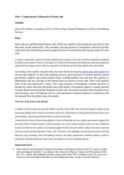
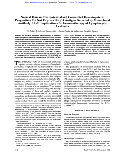
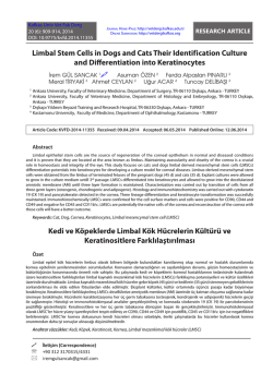
Incidence, Causes and Risk Factors. ALL occurs most often in the first decade
of life but increases in frequency again in older individuals (see Figure 1, below).
11 Lymphoblastic Leukemia: Age-Specific Incidence Rates (2006-2010)
Acute
Incidence (No. per 100,000)
9
7
5
3
1
0
Figure 1. I The horizontal axis shows five-year age intervals. The vertical axis shows the frequency of new cases of
ALL per 100,000 people, by age-group. Note that the risk of ALL is greatest in the first five years of life. An increase
in occurrence is also seen in older individuals (source: Surveillance, Epidemiology and End Results [SEER] Program;
National Cancer Institute; 2013)
The causes of ALL are not clear. A few factors have been associated with an
increased risk of developing the disease. Exposure to high doses of radiation
(carefully studied in the survivors of atomic bomb detonations in Japan) is one such
factor. ALL occurs at different rates in various settings. There are higher leukemia
rates in more developed countries and in higher socioeconomic groups. These and
other findings have led to a hypothesis that reducing children’s exposure to bacterial
infections during the first year of life may have increased the risk of childhood ALL.
Nonetheless, there have been other life-saving benefits from avoidance of bacterial
infections during infancy. A child who has had multiple diagnostic x-rays may be at
a slightly increased risk for ALL; however, more studies need to be done to confirm
these research findings. Previous chemotherapy and radiation treatment may be a
cause of ALL in adults.
Scientists continue to explore possible relationships to lifestyle or environmental
factors. Research supports the view that a number of complex factors may be
involved. One study found that children exposed to agricultural pesticides applied
near their home may experience a significant increased risk of ALL. The findings
from other studies have not been definitive, which is confusing for patients and
their families. They may wonder what they could have done differently to avoid the
disease; unfortunately, at the present time, there is no answer to that question.
Acute Lymphoblastic Leukemia
I page 7
Some cases of ALL relate to a mutation in a lymphocyte that occurs during the
prenatal period (in utero). Usually the leukemia is diagnosed in infancy or in the
first few years after birth. However, in some cases, years may pass before the disease
appears. With ALL, it seems that additional genetic abnormalities can occur after
birth and allow the unregulated cell growth that is needed to trigger the disease,
because there are more mutations found in utero than there are cases of childhood
ALL.
Signs and Symptoms. It is common for a person with ALL to feel a loss of
well-being because of the underproduction of normal cells in the bone marrow.
The person may tire more easily and have shortness of breath during normal
physical activities.
To begin determining the reason for these signs and symptoms, your doctor
will want to examine your blood by doing a blood test called a complete blood
count (CBC). Low numbers of red blood cells, white blood cells and platelets are
common in patients with newly diagnosed ALL.
Other signs and symptoms that a person with ALL may have include
{{Pale
skin coloring from anemia
{{Signs
of bleeding caused by a very low platelet count, including
{{
Black-and-blue
minor injury
{{
The
appearance of pinhead-sized red spots on the skin, called “petechiae”
{{
Prolonged
{{Mild
marks or bruises occurring for no reason or because of a
bleeding from minor cuts
fever
{{Frequent
minor infections
{{Discomfort
{{Enlarged
in bones or joints
spleen, liver or lymph nodes.
Leukemic cells can also collect in the testes in a small number of patients.
Bleeding. A low platelet count predisposes patients to bleeding. Bleeding in the
brain or lung is serious and can be fatal. However, such bleeding usually comes after
minor bleeding, such as nosebleeds, blood in the urine or bruises (see Low Blood Cell
Counts on page 24).
Infection. Severe infection usually does not occur at the time of diagnosis. If the
neutrophil count becomes or remains low because of ALL or its treatment, serious
infection may occur and can be life threatening. However, if proper precautions
are taken during therapy, most patients do not develop life threatening infections
(see Infection on page 25).
page 8
I 800.955.4572 I www.LLS.org
A person who has signs or symptoms that suggest the possibility of leukemia is
usually referred to a specialist. This may be a hematologist/oncologist. The specialist
will order additional tests to make a diagnosis. The signs and symptoms of ALL are
also seen in a number of other, less serious diseases.
Diagnosis and Cell Classification
An accurate diagnosis of the type of leukemia is important. The exact diagnosis
helps the doctor to
{{Estimate
how the disease will progress
{{Determine
the appropriate treatment.
Talk to your doctor about
{{The diagnostic tests that are being done
{{What the results mean
{{Getting copies of the test results.
Blood and Bone Marrow Tests. Blood and bone marrow cells are examined to
diagnose ALL and identify the ALL subtype (see ALL Subtypes on page 10). An
examination of the stained (dyed) blood cells with a light microscope will often
show the presence of leukemic blast cells (immature cells that do not function
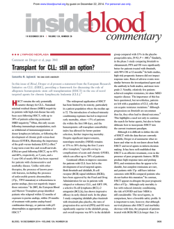
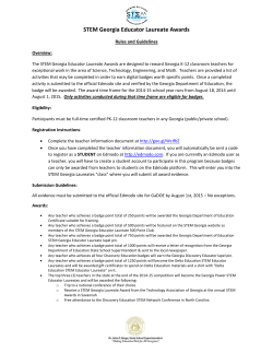
like normal, mature white blood cells). A bone marrow examination is preferred
to diagnose ALL because a proportion of patients do not have leukemic blasts
circulating in the blood at the time of diagnosis (see Figure 2, below).
ALL Blast Cells
Panel A
Panel B
Figure 2. I Panel A shows a photograph of developing cells in healthy marrow. The variation in the
appearance of the cells is characteristic of normal marrow. Panel B shows a photograph of marrow cells from
a patient with acute lymphoblastic leukemia. An unvaried appearance characterizes the leukemic blast cells.
Acute Lymphoblastic Leukemia
I page 9
Blood and Marrow Samples. To do the blood tests, blood samples are generally
taken from a vein in the patient’s arm. Samples of marrow cells are obtained by
bone marrow aspiration and bone marrow biopsy (see page 36). The cells from
the blood and marrow samples are examined under a microscope. Your doctor will
work with a hematopathologist, a specialist who studies blood diseases by looking
at the samples of blood and marrow cells and other tissues.
ALL Subtypes. ALL has many subtypes and can be classified by immunologic,
cytogenetic and molecular genetic tests. Some of these tests may be repeated
during and after therapy to measure the effects of treatment. Depending on the
subtype, the doctor will determine which drugs or drug combinations, drug
dosages, and duration of treatment are most appropriate for the patient, and
whether other types of treatment, such as stem cell transplant, are needed to
achieve the best results.
Immunophenotyping, a process used to identify cells based on the types of proteins
(antigens) on the cell surface, is necessary to establish the diagnosis of either B-cell
ALL, T-cell ALL or acute myeloid leukemia (AML). “Flow cytometry” is the name of
one test that may be used to do immunophenotyping.
ALL is divided into two major subtypes based on the physical characteristics and
the level of development of the leukemia cells. This basic classification helps the
treatment team to start planning the best course of treatment for the patient. The
principal ALL subtypes are
{{B
lymphoblastic leukemia
{{T
lymphoblastic leukemia
The phenotype or physical characteristics of the leukemia cell determine whether
the cells are of B-cell or T-cell origin. The B-cell subtype is identified by finding
cell surface markers on the leukemic blast cells that are the same as those that
develop on normal B lymphocytes. The T-cell subtype is identified by finding
cell surface markers on the leukemic blast cells that are the same as the ones that
develop in normal T lymphocytes.
Not all B lineage disease is treated the same. Mature B-cell leukemia is also
known as “Burkitt leukemia/lymphoma.” It accounts for 2%-3% of ALL patients.
The treatment for Burkitt leukemia is based on therapy for non-Hodgkin
lymphoma and is completely different than the treatment used for ALL. For more
information, see the free LLS publication Non-Hodgkin Lymphoma.
page 10
I 800.955.4572 I www.LLS.org
In some studies, ALL has been subdivided into CD10 (the common acute
lymphoblastic leukemia antigen, abbreviated cALLa) positive and CD10
negative. However, these categories of ALL have not been used in determining
treatment approach.
Genetic classification of ALL cells is also important (See Table 1 on page 12). About
75 percent of adult and childhood cases can be classified into subgroups based on
the chromosome number or DNA analysis, specific chromosomal rearrangements
and molecular genetic changes.
“Karyotyping” and “cytogenetic analysis” are processes used to identify certain
changes in chromosomes and genes. Laboratory tests called “fluorescence in situ
hybridization (FISH)” and “polymerase chain reaction (PCR) assays” may be
done, in which cells in a sample of marrow are studied to look for certain changes
in the structure or function of genes. In some cases, other special tests may be
used. See the free LLS publication Understanding Lab and Imaging Tests for more
comprehensive information about these tests.
Examination of leukemic cells by cytogenetic techniques permits identification
of chromosome or gene abnormalities. Translocations are the most common type
of DNA change that is associated with ALL. In a translocation, the DNA from
one chromosome breaks off and becomes attached to a different chromosome.
Other chromosome changes such as deletions (part of the chromosome is lost)
and inversions (rearrangement of the DNA within part of a chromosome) can
also lead to the development of ALL, but these changes are less common. In many
cases of ALL, the genetic changes are not known. Not all ALL cases exhibit the
same chromosome changes. Some are more common than others and some have a
greater effect on the patient’s prognosis than others.
Other features that are important in guiding treatment approach include the
age of the patient, level of the white blood cell count, involvement of the central
nervous system and involvement of lymph nodes.
Acute Lymphoblastic Leukemia
I page 11
Table 1. ALL Principal Cytogenetic Abnormalities
Abnormality
Associated Prognosis
Hyperdiploidy
More than the normal number of
46 chromosomes
Favorable prognosis
Hypodiploidy
Fewer than the normal number of 46
chromosomes
Poor prognosis
Translocation between chromosomes
12 and 21
Favorable prognosis
“Philadelphia” or
“Ph” chromosome
Translocation between chromosome
22 and chromosome 9
Favorable prognosis with
contemporary therapy
“Ph-like” ALL
(BCR-ABL1-negative)
Poor prognosis
Translocation between chromosome
1 and 19
(associated with CNS leukemia)
Favorable prognosis with
contemporary therapy
Translocation between chromosome
4 and 11
(associated with infant and older
adult age-groups, CNS leukemia)
Poor prognosis
Translocation between chromosome
11 and 19
Poor prognosis for infants
Better prognosis for older children
Translocation between chromosome
8 and 14
Favorable prognosis with short-term
intensive therapy
CRLF2 and Janus kinase gene
mutations
Poor prognosis
NOTCH1 mutations
Favorable prognosis
HOX11 overexpression
Favorable prognosis with
chemotherapy alone
Chromosome 21 amplification
page 12
I 800.955.4572 I www.LLS.org
Requires intensive therapy to avert
poor prognosis
Treatment
A diagnosis of ALL is associated with a wide range of outcomes.
Treatment Planning. A number of factors affect the choice and outcome of
treatment, including
{{The
ALL subtype
{{The
type of leukemic lymphocytes as judged by their appearance
{{Immunophenotype
and chromosome composition
{{Whether
the patient has received chemotherapy in the past to treat another
type of cancer
{{Whether
the ALL is present in the central nervous system or other sites outside
of the bone marrow
{{Whether
the ALL has not responded to treatment or has relapsed
{{The
presence of systemic infection at diagnosis
{{The
patient’s age and general health.
Fast Facts about Treatment Planning
{{A
person who has ALL is usually treated by a hematologist/oncologist.
{{It is essential to seek treatment in a center where doctors are experienced in
the care of patients with acute leukemia.
{{Patients with ALL need treatment as soon as possible after diagnosis. The
approach for treating each patient is based on an individual’s subtype, risk
factors and treatment goals.
Fast Facts About Children and Adolescents
{{For
many children, ALL is curable with current therapies.
{{A number of cancer centers are using pediatric protocols to treat adolescent
and young adult patients.
Acute Lymphoblastic Leukemia
I page 13
Fast Facts About Treatment
{{For
older ALL patients, age alone is not a reason to withhold treatment.
{{Achieving
a remission is important because it is associated with prolonging
survival. The initial goal of treatment is usually to bring about a remission,
in which
{{
There
is no evidence of leukemic blast cells in the blood or marrow
{{
Normal
blood cell production is restored and blood cell counts return
to normal levels.
{{In
most patients, intensive chemotherapy is required to achieve complete
remission. At least two drugs are combined to treat patients initially.
{{The
age of the patient and the type of leukemic lymphocytes based on their
appearance, immunophenotype or chromosome composition can influence
the type of treatment given.
{{More
treatment is needed once a remission is achieved to help prevent a
relapse.
{{Postremission
treatment may consist of maintenance chemotherapy or stem
cell transplantation.
{{If
relapse occurs, treatment options may include different chemotherapy
regimens, allogeneic stem cell transplantation or other investigational therapies.
{{Variations
on standard approaches to treatment are undergoing intensive
study throughout the world. A patient may receive a different number of
drugs, a different sequence of drugs, or drugs different from those described
in this booklet and still be receiving appropriate and effective treatment.
Talk to your doctor about
{{Your treatment options and the results you can expect from treatment
{{The results you might expect with standard therapy
{{The possibility of participating in a clinical trial
Pretreatment Considerations. Adults of childbearing age and parents of children
diagnosed with ALL should ask the doctor for information about addressing the
risk for infertility. See the free LLS publication Fertility for more details.
page 14
I 800.955.4572 I www.LLS.org
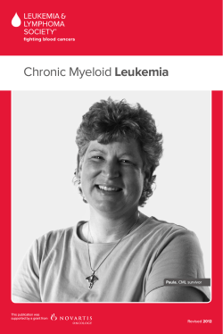
Chemotherapy. There are three parts to the treatment for ALL. These are
induction, consolidation (also called “intensification”) and maintenance. (see
Figure 3 on page 19.) Consolidation and maintenance are postremission therapies.
Induction Therapy. The initial phase of chemotherapy is called “induction.” The
specific drugs, the dosages used, and the timing of their administration, depend on
several factors, including the patient’s age, the specific features of the leukemia and
the overall health of the patient. Several drugs are combined. Typically, the severity
of the disease and the side effects of this initial therapy result in an initial hospital
stay of four to six weeks. Some patients who live with a caregiver and near the
medical facility may be safely discharged sooner. This depends on the policies of the
treatment center and the status of the patient.
A central line (indwelling catheter) is placed surgically in a vein in the upper chest.
The catheter is tunneled under the skin of the chest so that it stays firmly in place.
The external end of the catheter (port) can be used to administer medications,
fluids or blood products or to withdraw blood samples for cell counts and
chemical tests. An alternative is a PICC line (percutaneously inserted central
venous catheter), which can be placed in a vein of the upper arm. See the free LLS
publication Understanding Side Effects of Drug Therapy for additional information
about drug administration.
The goal of induction therapy is to achieve a remission, which means to rid the
blood and marrow of visible leukemic blast cells. A remission is not a cure but it
is a very important part of the process as it allows normal marrow cells to develop
and the patient’s blood counts return to normal levels. Generally, if blast cells are
still evident after the first course of induction chemotherapy, a second course of
chemotherapy, usually using different drugs is given. Table 2, on page 16, gives
examples of the drugs that may be used for induction and postremission treatment
as well as some of the drugs under study in ALL clinical trials. Other drugs may
be added or substituted for higher-risk, refractory or relapsed patients. Allogeneic
stem cell transplantation may be added to the treatment plan for patients with
relapsed ALL or for patients at high risk of relapse after chemotherapy (see pages
21 through 23). Autologous stem cell transplantation is not commonly used to
treat ALL because of the high relapse rate following this type of transplant.
A child with ALL is usually admitted to the hospital, as soon as the diagnosis is
known, to start the induction treatment. Most children enter remission after the
first month of therapy. For some children this is the first time they have stayed away
from home for an extended period of time. Providing age-appropriate information
to your child about the illness and treatment will help him or her to build trust in
both you and the treatment team and to feel comfortable talking about fears and
concerns. For practical guidance about how to support your child and other family
members, deal with your own concerns, share the news with extended family
and friends and make the transition to life after treatment ends, see the free LLS
publication Coping With Childhood Leukemia and Lymphoma.
Acute Lymphoblastic Leukemia
I page 15
Table 2. Some Drugs Used for Treatment and/or in Clinical Trials for ALL
Most antileukemic drugs interact with the cell’s genetic
material (the DNA).
Antitumor Antibiotics
{{daunorubicin (CerubidineВ®)
{{doxorubicin (AdriamycinВ®)
{{mitoxantrone (NovantroneВ®)
{{idarubicin (IdamycinВ®)
DNA-Repair Enzyme Inhibitors
{{etoposide (VP-16; VePesidВ®,
EtopophosВ®)
{{teniposide (VM-26; VumonВ®)
{{topotecan (HycamtinВ®)
DNA Synthesis Inhibitor
{{carboplatin (ParaplatinВ®)
DNA-Damaging Agents
{{cyclophosphamide (CytoxanВ®)
{{ifosfamide (IfexВ®)
Enzymes That Prevent Cells
From Surviving
{{Asparaginase Erwinia chrysanthemi
(ErwinazeВ®)
{{pegaspargase (PEG-L-asparaginase;
OncasparВ®)
Tyrosine Kinase Inhibitors
{{imatinib mesylate (GleevecВ®)
{{dasatinib (SprycelВ®)
{{nilotinib (TasignaВ®)
{{ponatinib (IclusigВ®)
Antimetabolites
{{azacitidine (VidazaВ®)
{{cladribine (2-CdA; LeustatinВ®)
{{clofarabine (ClolarВ®)
{{cytarabine (cytosine arabinoside,
ara-C; Cytosar-UВ®)
{{fludarabine (FludaraВ®)
{{hydroxyurea (HydreaВ®)
{{6-mercaptopurine (PurinetholВ®)
{{methotrexate
{{nelarabine (ArranonВ®)
{{6-thioguanine (thioguanine;
TabloidВ®)
Drug That Prevents Cells
From Dividing
{{vincristine (OncovinВ®)
{{liposomal vincristine (MarqiboВ®)
Synthetic Hormones
{{prednisone
{{prednisolone
{{dexamethasone
Monoclonal Antibodies
{{alemtuzumab (CampathВ®)
{{rituximab (RituxanВ®)
I Lists some of the standard drugs and some of the drugs currently being studied to treat ALL patients.
Various approaches to ALL treatment are undergoing study in clinical trials. A patient may be treated with drugs
that are not listed in this table and still be receiving appropriate and effective treatment. However, it is essential to
seek treatment in a center where doctors are experienced in the care of patients with acute leukemia.
Table 2.
page 16
I 800.955.4572 I www.LLS.org
Table 3. Examples of Therapy Used in the Treatment of ALL
Induction therapy given in the first month may include
{{Doxorubicin
or daunorubicin by vein
{{Asparaginase by injection into a muscle or by vein
{{Vincristine by vein
{{Corticosteroid (dexamethasone or prednisone) by mouth
{{Methotrexate by injection into the spinal fluid
{{6-Mercaptopurine by mouth
{{Cytarabine by injection into the spinal fluid.
Postremission therapy given in cycles for two to three years
may include
{{Vincristine
by vein
{{Cyclophosphamide by vein
{{Daunorubicin or doxorubicin by vein
{{Thioguanine by mouth
{{Prednisone or dexamethasone by mouth
{{Mercaptopurine by mouth
{{Methotrexate by mouth, by vein, or into a muscle
{{Methotrexate by injection into the spinal fluid
{{Cytarabine by injection into the spinal fluid
{{Hydrocortisone by injection into the spinal fluid
{{Radiation therapy to the head.
Postremission Therapy (Consolidation and Maintenance Therapy). Since
residual leukemia cells that are undetectable by blood or marrow examination
remain after remission, the optimal treatment for patients who have ALL requires
additional intensive postremission therapy. As in the induction phase, individual
factors such as the age of the patient, the ability to tolerate intensive treatment,
cytogenetic findings, the availability of a stem cell donor and other considerations
may influence the treatment approach.
Consolidation therapy is usually given in cycles for four to six months. The goal of
this phase of treatment is to reduce the number of leukemic cells still remaining.
Generally, several chemotherapy drugs are combined to help prevent the leukemia
cells from developing drug resistance. When necessary, intrathecal therapy (drugs
that are administered directly into the spinal canal) is continued.
Acute Lymphoblastic Leukemia
I page 17
Maintenance therapy is usually given for about two years. During the first months
of maintenance, treatment protocols may include one or two intensified treatments
similar to the ones used during induction. These intensified treatments are also
known as “re-induction” or “delayed intensification” treatments. In most cases,
postremission chemotherapy also includes drugs not used during induction
treatment (see Table 3 on page 17).
Some types of high-risk ALL—such as T-cell ALL or ALL in the very young
(infants) or in adults—are usually treated with higher doses of drugs during
induction, consolidation and maintenance therapy.
Central Nervous System (CNS) Prophylaxis. ALL cells often collect in
the lining of the spinal cord and brain, called the “meninges.” If not treated, the
meninges can harbor leukemia cells, and relapse can occur in these sites (meningeal
leukemia). For this reason, treatment called “central nervous system prophylaxis” is
directed to those sites. The treatment involves injecting drugs, such as methotrexate,
into the spinal column. Areas of the body that are less accessible to chemotherapy
given by mouth or in the vein are sometimes referred to as “sanctuary sites.” Cranial
radiation for pediatric patients, except in cases of T-cell ALL and patients who have
a CNS relapse, is not being used in some doctors’ practices. Treatment without
radiation decreases the chance of long-term and late effects for the patient, such as
organ damage, the development of second cancers and neurocognitive impairment.
page 18
I 800.955.4572 I www.LLS.org
ALL Treatment Overview
Treatment Phase
Features
Goal
INDUCTION
(4-6 weeks)
-Multiagent chemotherapy
(may include vincristine,
anthracyclines, corticosteroids,
asparaginase,
cyclophosphamide,
other agents)
-CNS prophylaxis (intrathecal
therapy, cranial irradiation)
Achieve
remission
CONSOLIDATION/
INTENSIFICATION
(4-6 months)
-Given in cycles
-Drug combinations used may
be similar to induction
-Intrathecal therapy may be
continued
-Consider HSCT* for certain
high-risk patients
-Eliminate
remaining
leukemic cells
after induction
therapy
-Presymptomatic
CNS treatment
MAINTENANCE
(2 years—adults)
(2-3 years—children)
- Drug combinations given in 1
or 2 intensified treatments
-May include daily
mercaptopurine, weekly
methotrexate, periodic
vincristine, corticosteroids
and intrathecal therapy
-Prevent disease
relapse
*Hematopoietic stem cell
transplantation (HSCT)
Based on NCCN Guidelines.
Figure 3. I The figure above provides general information. There are many different ALL treatment approaches.
Speak to your doctor to develop a treatment plan specific to you.
Ph-Positive ALL. About one out of four to five adults with ALL and a small
number of children (about 2 to 4 percent) with ALL have a subtype called
“Ph-positive (Philadelphia-positive) ALL.” Patients with this subtype of ALL have
a chromosome alteration that results in a specific gene mutation referred to as
“BCR-ABL.” These patients are treated with the tyrosine kinase inhibitor drugs
(TKIs) imatinib mesylate (GleevecВ®), dasatinib (SprycelВ®), nilotinib (TasignaВ®)
or ponatinib (IculsigВ®), in addition to other multidrug chemotherapy. Gleevec
treatment with chemotherapy is effective for some Ph-positive ALL patients.
Sprycel and Tasigna are used to treat Ph-positive ALL patients who do not
tolerate or respond to Gleevec or those who develop resistance to it. Iclusig is
FDA approved to treat adult patients who have T315I-positive Philadelphia
chromosome positive (Ph+) ALL or Ph+ ALL for whom no other tyrosine-kinase
inhibitor therapy is indicated. TKIs specifically block the leukemia-causing effects
of the BCR-ABL gene mutation in many patients. TKIs given alone would not
result in cures for Ph-positive ALL patients, so these drugs are combined with
chemotherapy. Studies are ongoing to learn the usefulness of this approach for
Ph-positive ALL, and many results have been promising. New combinations of
drugs are being studied in clinical trials for the treatment of Ph-positive ALL.
For more information about clinical trials, see page 29.
Acute Lymphoblastic Leukemia
I page 19
Young Adults. Older adolescents and adults younger than 40 years are often called
“young adults.” Traditionally, treatment for this group has been similar to adult
treatment protocols. However, clinical trials are looking into using a variety of
pediatric protocol options. Some of these treatment options include combination
chemotherapy using different dosing amounts; combination chemotherapy
including rituximab (RituxanВ®) and intensified doses of nonmyelotoxic drugs, such
as prednisone, vincristine (OncovinВ®) or PEG-asparaginase. Asparaginase Erwinia
chrysanthemi (ErwinazeВ®) is offered as an alternative when the patient is allergic to
PEG-asparaginase. Speak to your doctor or call an Information Specialist to learn
about the different clinical trials that may be available to you.
Childhood Versus Adult Forms of ALL. ALL has an unusual pattern of age
distribution (see Figure 1, page 7). The risk of developing ALL peaks between ages
1 to 4 years and then decreases until about age 50. At age 50, the incidence increases
again, especially among men. As with other types of leukemia, incidence increases
again as a person gets older.
The adult form of ALL is more resistant to treatment than the childhood form;
however over the last few years several factors have contributed to longer remissions
and prolonged survival for adult patients with ALL. These include
{{Improved
outcomes with allogeneic stem cell transplantation
{{Use
of tyrosine kinase inhibitors for Philadelphia chromosome (Ph+) ALL
{{Use
of intensified pediatric-like therapy for adolescents and young adults.
For patients with ALL that is resistant to treatment or who have relapsed, allogeneic
stem cell transplantation may be the best option, if they are able to achieve
complete remission before transplantation. Likewise, patients with high-risk
disease are recommended for transplantation if it is unlikely that they will achieve
remission with chemotherapy alone.
For ALL patients older than 60 years, patient performance status, other health issues
and ALL risk features are all considered in developing a treatment plan. Age alone is
not a reason to withhold treatment. Standardized measures of strength and reaction
time are used to determine physiological age, which is a better indicator of tolerance
for therapy. However, older patients may have a poorer response to therapy because
{{The
leukemic cells of older ALL patients have a higher occurrence of unfavorable
cytogenetic and molecular abnormalities.
{{Older
patients may have other medical problems (called “comorbidities”),
including heart, lung or kidney disease or diabetes mellitus. The doctor may
have to select less toxic drugs or decrease the dosage and frequency of treatment.
page 20
I 800.955.4572 I www.LLS.org
It is important to know that even in otherwise healthy patients aged 75 years or older,
the principal cause of treatment failure is not toxicity, but failure of the treatment to
eliminate the ALL cells. Occasionally, very elderly patients refuse treatment or are so ill
from unrelated illnesses that treatment may be unreasonable.
There are new treatments under study for all ages and stages of disease.
Talk to your doctor about
{{Whether treatment in a clinical trial is right for you.
Minimal Residual Disease (MRD). Sensitive molecular testing techniques permit
the identification of small amounts of residual leukemia cells, known as minimal
residual disease (MRD), at times when blood and marrow appear normal. This
approach can be used if the leukemia cells have a detectable molecular abnormality
or immunophenotype. It can also permit more sensitive follow-up of patients in
remission and can help determine whether additional treatment is necessary. Studies
in both children and adults with ALL have shown that there is a strong correlation
between MRD and the risk or relapse. There is also a prognostic value to measuring
MRD during and immediately after the initial induction therapy. The detection of
MRD on day 29 of treatment (end of induction) may be useful in determining the
need for additional induction therapy. In some pediatric institutions, doctors are
checking for MRD on day eight as an indicator of slow early-responders.
Stem Cell Transplantation. Some patients may benefit from intensive chemotherapy
alone followed by standard or reduced-intensity stem cell transplantation.
The decision to undergo a transplant should be discussed with your doctor. About
75 to 80 percent of children treated for ALL will not need a transplant. For an adult,
the decision depends on the features of the leukemia and the patient’s general health
and age.
Which patients are likely to benefit from transplantation after their first complete
remission is a question under study in clinical trials. Some of the main factors that
influence the approach used are
{{Patient
age
{{Ability
to tolerate intensive treatment
{{Cytogenetic
{{Availability
and molecular characteristics of the ALL cells
of an HLA-matched related or unrelated stem cell donor.
See the free LLS publications Blood and Marrow Stem Cell Transplantation and
Cord Blood Stem Cell Transplantation for comprehensive information about
stem cell transplantation.
Acute Lymphoblastic Leukemia
I page 21
Allogeneic Stem Cell Transplantation. This treatment uses donor stem cells
to restore a patient’s marrow and blood cells. For standard-risk patients in first
remission, the choice between a transplant (standard or reduced-intensity) and
continued chemotherapy is not clear.
For high-risk patients, an allogeneic transplant is an option for those patients in first
remission who have a matched related or matched unrelated donor. Cord blood
stem cells may be an alternative source for donor stem cells if an appropriate sibling
or unrelated donor is not available. Allogeneic stem cell transplantation is a curative
treatment option for some high-risk ALL patients in first remission.
Children who relapse less than six months following initial treatment or while in
chemotherapy have a lower chance of a second remission. For these children and
for children with refractory disease, transplantation with a matched related or
matched unrelated donor may be considered. Cord blood stem cells may also be a
source for the transplant. For children who do undergo transplantation, the use of
unrelated human leukocyte antigen (HLA)-matched donors appears to be just as
successful as it is for related HLA-matched donors (for example, siblings), making
more donors available through stem cell transplantation registries.
Reduced-Intensity Allogeneic Stem Cell Transplantation. The benefits
and risks of reduced-intensity allogeneic stem cell transplantation have not yet
been clearly established for ALL patients. Patients who are too old or too ill to
have a standard allogeneic stem cell transplant may be candidates for a reducedintensity transplant if a suitable donor is available. The conditioning therapy used
for a reduced-intensity transplant is of lower intensity than that for a standard
allogeneic stem cell transplant; it does not completely inactivate the patient’s
immune system or treat the ALL as aggressively.
Reduced-intensity allogeneic stem cell transplantation is based on two considerations:
{{Much-improved
immunosuppressive therapy prevents the patient from rejecting
the donor’s stem cells, even though the patient’s immune system has not been
fully suppressed by the lower-intensity conditioning therapy.
{{The anticipated attack of the donor’s immune cells successfully suppresses the
patient’s leukemia cells. This attack is referred to as a “graft-versus-leukemia effect”
or “GVL.” Over time, if the transplant is successful, the donor’s stem cells replace
the patient’s immune cells. The engrafted donor immune cells recognize minor
tissue antigens on the patient’s leukemia cells and continue to suppress their growth.
The risk of graft-versus-host disease (GVHD) is an important consideration and
a potentially disabling side effect.
Talk to your doctor about
{{Whether a stem cell transplant is an option for you.
page 22
I 800.955.4572 I www.LLS.org
Autologous Stem Cell Transplantation. This procedure uses the patient’s
own stem cells to restore blood cell production. This type of transplant is not
commonly used to treat ALL.
Refractory Leukemia or Relapsed Leukemia. Most patients achieve an initial
remission. However, some patients have residual leukemic cells in their marrow
even after intensive treatment. This is referred to as “refractory leukemia.” Other
patients achieve remission but then have a decrease in normal blood cells and a
return of leukemia cells in the marrow. This situation is referred to as a “relapse.”
With refractory leukemia, different drugs from those used in the first course
of treatment may be administered in an effort to induce remission. Stem cell
transplantation may be an option following remission that may result in a more
durable remission. In patients who relapse, the duration of the remission, the
patient’s age and the cytogenetic findings in the leukemia cells influence the
approach to therapy. Drugs similar to those administered initially, different drugs
or stem cell transplantation may be used to treat the leukemia.
There are several drugs approved by the Food and Drug Administration (FDA) to
treat relapsed or refractory ALL patients.
Nelarabine (ArranonВ®) is approved for patients with relapsed T-cell ALL.
Clofarabine (ClolarВ®) is approved for patients who are between 1 and 21 years with
relapsed or refractory ALL after they have received at least two prior chemotherapy
regimens. Although treatment with clofarabine alone is not curative, it may lead
to a temporary remission for the patient that is then followed by allogeneic stem
cell transplantation, which may result in a cure. Clofarabine is also being studied
in combination with other drugs in clinical trials for the treatment of children,
adolescents and adults with relapsed or refractory ALL.
Liposomal vincristine (MarqiboВ®) is approved for adult patients with Ph
chromosome-negative ALL who have relapsed two or more times, or whose
leukemia has progressed following two or more regimens of therapy.
The following factors may increase the risk for relapse after initial treatments:
{{Microscopic
of therapy
{{Age
{{A
evidence of leukemia (minimal residual disease) after 20 weeks
30 years and older
high white blood cell count at the time of diagnosis
{{Disease
that has spread beyond the bone marrow to other parts of the lymphatic
system, such as the spleen
Acute Lymphoblastic Leukemia
I page 23
{{Certain
genetic abnormalities, such as the presence of the Philadelphia
chromosome or MLL (mixed-lineage leukemia) gene translocations
{{The
need for four or more weeks of induction chemotherapy in order to achieve
a first complete remission.
Patients with one or more of these risk factors may be candidates for stem cell
transplantation once they are in first remission. Talk to your doctor for more
information.
Several drugs and drug combinations that can be used to treat ALL are being
studied in clinical trials. LLS Information Specialists offer guidance on how
patients can work with their doctors to find out if a specific clinical trial is an
appropriate treatment option. Information Specialists conduct clinical-trial
searches for patients, family members and healthcare professionals. You can use
an LLS-supported online clinical trial search service that offers patients and
caregivers immediate access to listings of blood cancer clinical trials, by visiting
www.LLS.org/clinicaltrials.
Talk to your doctor about
{{Therapies under study in clinical trials for refractory or relapsed ALL.
Disease and Treatment Side Effects. Most ALL treatment side effects are
temporary and subside once the body adjusts to therapy or when therapy is
completed. During the course of treatment and at the end of therapy, healthy
new cells will begin to grow and develop. Severe side effects are treated on an
inpatient basis.
Low Blood Cell Counts. ALL decreases the production of normal blood cells.
In addition, chemotherapy is toxic to both normal blood cells and ALL cells. The
normal blood cells are eliminated from the marrow along with ALL cells. For the
patient, this results in a severe deficiency in the number of
{{Red
blood cells (anemia)
{{Platelets
(thrombocytopenia)
{{White
blood cells called “neutrophils” (neutropenia) and “monocytes”
(monocytopenia).
Transfusion of red blood cells and platelets is almost always needed for a period
of several weeks during treatment. After that, the blood cell counts usually return
toward normal.
page 24
I 800.955.4572 I www.LLS.org
Infection. During treatment for ALL, the deficiency of neutrophils and
monocytes (types of white blood cells) can lead to infection from bacteria and
fungi normally present in the environment, on the skin, in the nose and mouth,
on the gums, or in the colon. The risk of infection may be increased because
chemotherapy damages the lining of the mouth and intestines, making it easier
for bacteria to enter the blood. When the white blood cell count is low and
infection risk is increased, antibiotics are given to prevent or treat infection.
Transfusion is not generally used for patients with a low neutrophil count,
but it can be used in patients with high fever, infection that is unresponsive to
antibiotics, blood fungal infections or septic shock.
Growth factors may be given to the patient to stimulate the marrow to make
new white blood cells. The growth factors used most frequently are G-CSF
(granulocyte colony-stimulating factor; filgrastim [NeupogenВ®] and pegfilgrastim
[NeulastaВ®]) and GM-CSF (granulocyte-macrophage colony-stimulating factor;
sargramostim [LeukineВ®]). These agents are used in children only in special
circumstances.
Because the patient has an increased risk of developing an infection, the medical
staff, family and friends need to practice frequent and vigorous hand washing and
take other precautions to avoid exposing patients to bacteria, viruses and other
infection-causing agents. Caregivers for patients with central lines or ports need
to be meticulous in the cleaning of catheters.
Patients at home should not delay in seeking medical attention if any signs of
infection develop. A rise in temperature to 101В°F or higher, or the onset of chills,
may be the only sign of infection in a patient with a very low white blood cell
count. Other signs of infection may include persistent coughing; tenderness at a
site prone to infection, such as the area surrounding the anus or the facial sinuses;
sore throat; pain during urination; or frequent loose stools.
ALL patients are advised to receive certain vaccinations. It is recommended
that children receive an annual influenza vaccine. Adult patients are advised
to receive vaccinations for pneumococcal pneumonia and influenza. There
are two types of pneumococcal vaccines available for adults: a pneumococcal
polysaccharide vaccine (PPSV23) and a pneumococcal conjugate vaccine
(PCV13). Immunizations using live organisms or with high viral loads, such as
the herpes zoster or shingles vaccine, should not be administered. Your doctor
can give you more information.
Other Side Effects. Chemotherapy affects tissues that normally have a high rate
of cell turnover. Thus, the lining of the mouth, the lining of the intestines, the
skin and the hair follicles may be affected. Common side effects may include
Acute Lymphoblastic Leukemia
I page 25
{{Mouth
ulcers
{{Diarrhea
{{Temporary
hair loss
{{Rashes
{{Nausea
{{Loss
and vomiting
of appetite
{{Fatigue.
Fortunately, drugs that counteract nausea and vomiting can be given to prevent
or relieve these distressing side effects. Some ALL patients find that acupuncture
treatments relieve chemotherapy-associated nausea and vomiting.
Some ALL patients may build up the concentration of uric acid in their blood as
a result of a very high white blood cell count. The use of chemotherapy may also
increase uric acid levels. Uric acid is a chemical in the cell. It enters the blood and is
excreted in the urine. If many cells are killed simultaneously by therapy, the amount
of uric acid in the urine can be so high that kidney stones can form. This may
seriously interfere with the flow of urine. Drugs such as allopurinol (ZyloprimВ®) or
rasburicase (ElitekВ®) can be given to minimize the buildup of uric acid in the blood.
There are drugs and other supportive therapies to prevent or manage many side
effects. For more information see the free LLS publications Blood Transfusion,
Cancer-Related Fatigue Facts and Understanding Side Effects of Drug Therapy.
Sometimes, a drug or a drug combination causes effects that continue for a period
of time after treatment ends. Some effects may be long-lasting (see Long-Term and
Late Effects of Treatment on page 27).
Talk to your doctor about
{{Possible side effects and follow-up care.
page 26
I 800.955.4572 I www.LLS.org
Follow-up Care
Some of the tests that were done to diagnose ALL may be repeated to
{{Follow the effects of treatment
{{Make
decisions about whether to continue, intensify, change or stop treatment.
After treatment, a patient who is in remission and has completed therapy continues
to be examined regularly by his or her doctors. Careful periodic assessment of the
patient’s health, blood cell counts and, if indicated, marrow is required. As time
progresses, assessments may be less frequent, but should continue indefinitely.
It is important to keep a record of your cancer treatment so that your doctor can
follow up on specific late effects that may be associated with those treatments.
This information would include your diagnosis, the names of chemotherapy drugs
taken, radiation treatment information, surgery information, transplantation
information, information about any other treatments, and the names and dates of
any significant complications and the treatment received for those complications.
This can help your doctor develop a follow-up schedule for you.
To find a follow-up clinic and other resources for child and adult survivors, contact
our Information Specialists.
Both adults and children may experience difficulties when they return to their daily
routines after such a long period of treatment. Getting support throughout this
time, and for as long as needed, is important and will be helpful as you return to
your “normal” life.
Long-Term and Late Effects of Treatment. Children and young adults who
have been treated for ALL may be at increased risk for heart damage, other cancers
and neurologic or cognitive problems. Patients should be seen by a primary care
doctor for a general health examination at least once a year. They should also be
examined regularly by an oncologist.
It is important to know about the potential for long-term effects of treatment so
that any problems can be identified early and managed. Treatment for individuals
who have ALL sometimes causes effects that continue after treatment ends (longterm effects) or develop much later in life (late effects). Various factors can influence
the risk of developing long-term or late effects, including
{{Type
{{Age
and duration of treatment
at the time of treatment
{{Gender
and overall health.
Acute Lymphoblastic Leukemia
I page 27
Most ALL patients are treated with an anthracycline, such as daunorubicin
(CerubidineВ®). Anthracyclines have been associated with increased risk for heart
muscle injury or chronic heart failure. Heart disease may not become apparent
until many years after therapy ends.
Current prevention strategies for reducing heart damage include: limiting the
cumulative dose of the anthracycline, altering drug schedules, using anthracycline
structural analogs (chemical structure of the analog drug is modified to be less toxic
but equally effective as the original drug) and liposomal encapsulated anthracyclines
(the therapeutic agent has a special coating to reduce side effects), offering
cardioprotective drugs and nutritional supplements.
Avascular necrosis and pain in the hip bones or shoulders may occur in some young
patients after chemotherapy. Patients with these conditions may eventually require
joint replacement surgery.
Sometimes cranial irradiation is used for patients with T-cell ALL or those who
experience a relapse. Doctors are limiting the use of this treatment to avoid
the risk of long-term or late effects such as neurocognitive impairment and the
development of second cancers.
Stem cell transplantation is used to treat some patients with ALL. This treatment
has been associated with long-term or late effects, including infertility, thyroid
dysfunction, chronic fatigue and risk for developing a second cancer (lymphoma,
melanoma of the skin, or cancer of the tongue and salivary glands, central nervous
system, bone, soft tissue and thyroid gland). The number of patients who develop
second cancers is small.
Children may experience side effects of treatment, both in the short- and
long-term, that can affect learning, including effects on growth, cognitive
development and psychosocial development. Going back to school also brings new
challenges to families whose main focus has been getting through treatment. By
being aware of possible effects, parents can work with the school to help their child.
See the free LLS publications Coping With Childhood Leukemia and Lymphoma and
Learning & Living With Cancer: Advocating for your child’s educational needs, which
provide information about the challenges children may face and what can be done,
the laws that protect your child and ways that schools can help.
Fertility. Recent studies show that both males and females treated for ALL as
children or adolescents were not generally at increased risk for major complications
during pregnancy or for infant malformation or death. Certain childhood cancers
and treatments can increase the risk for preterm birth and low birth weight. Talk to
your doctor for additional information.
Long-term and late effects can be managed. For more information see the free LLS
publications Long-Term and Late Effects of Treatment for Childhood Leukemia or
Lymphoma, Long-Term and Late Effects of Treatment in Adults and Understanding
Side Effects of Drug Therapy.
page 28
I 800.955.4572 I www.LLS.org
Talk to your doctor about
{{Possible long-term and late effects and follow-up care.
Treatment Outcomes. A few decades ago there were very low cure rates in both
children and adults diagnosed with ALL. Today, nearly 90 percent of children and
40 percent of adults can expect long-term, leukemia-free survival—and probable
cure—with contemporary treatment. Currently, emphasis is placed not only on
improving the cure rate but also on improving quality of life by preventing acute
and late treatment-related complications, such as second cancers, cardiotoxicity
and endocrinopathy.
“Relative survival” compares the survival rate of a person diagnosed with a disease
to that of a person without the disease. In children under 15 years of age, the
five-year relative survival rate has increased from 3 percent in 1964 to 89.2 percent
in 2006 as a result of successful treatments made possible by clinical trials.
In adults, the probability of remission has increased dramatically in the last 10
years, and extended remissions are also more frequent. Several areas of research are
likely to lead to further progress.
Research and Clinical Trials
New approaches under study in clinical trials for ALL treatment, many of which are
being supported by LLS research programs, hold the promise of increasing the rate
of remission and finding a cure for ALL.
Clinical Trials. Every new drug or treatment regimen goes through a series of
clinical trials before it becomes part of standard therapy. Clinical trials are carefully
designed and rigorously reviewed by expert clinicians and researchers to ensure
as much safety and scientific accuracy as possible. Participation in a carefully
conducted clinical trial may be the best available therapy. Patient participation in
past clinical trials has resulted in the therapies we have today.
LLS Information Specialists, at (800) 955-4572, can offer guidance on how patients
can work with their doctors to determine if a specific clinical trial is an appropriate
treatment option. Information Specialists will conduct individualized clinical-trial
searches for patients, family members and healthcare professionals. This service is
also available at www.LLS.org/clinicaltrials.
Acute Lymphoblastic Leukemia
I page 29
Research Approaches. There are clinical trials for newly diagnosed patients and for
patients with relapsed or refractory disease. A number of approaches are under study
in clinical trials for the treatment of patients with ALL. Some of the objectives are
{{To
achieve a greater understanding of ALL cytogenetic abnormalities and how
they affect prognosis
{{To
refine techniques to assess the high risk of relapse in individual patients to
ensure that intensive treatment is given primarily to high-risk cases
{{To
find most effective combinations of chemotherapy drugs while reducing
undesired side effects
{{To
develop treatment strategies to prevent or reverse chemotherapy resistance
{{To
refine stem cell transplants to increase effectiveness, reduce complications
and determine which patients are most likely to benefit by this treatment
{{To
develop new and/or refine existing immunotherapy agents so that they can
be used in frontline treatment
{{To
refine techniques for faster detection of minimal residual disease
after induction therapy so that the patient’s treatment plan can be more
individualized.
Agents Under Study. The following are examples of specific agents under study
in clinical trials for ALL.
Proteasome Inhibitor
{{Bortezomib (Velcade®)—This drug, approved to treat myeloma and some types
of lymphoma, is now being studied for the treatment of relapsed pediatric-ALL
patients and patients with T-cell ALL.
Antimetabolite
{{Clofarabine (Clolar®)—Already approved to treat pediatric ALL, it is now
showing promising results in studies of adults with ALL. It is also being studied
in combination with other drugs in clinical trials for the treatment of children,
adolescents and adults with relapsed or refractory ALL.
Janus kinase (JAK) Inhibitor
{{Ruxolitinib (Jakafi®)—Already approved to treat myelofibrosis patients, it
is being studied in clinical trials in the treatment of pediatric refractory and
relapsed ALL.
Immunotherapies
{{Monoclonal antibodies rituximab (Rituxan®) and alemtuzumab (Campath®)—
These drugs are already approved in the treatment of other blood cancers. They
are currently being studied in clinical trials for ALL.
{{Monoclonal
antibody blinatumomab (AMG 103)—This new drug has shown
promising results in early studies for adult ALL patients who have already
received chemotherapy.
page 30
I 800.955.4572 I www.LLS.org
{{Combination
chemotherapy with or without Rituxan—This is being studied for
the treatment of younger patients with B-cell ALL.
{{Chimeric
antigen receptor (CAR) therapy—This is another type of
immunotherapy. The patient cells are removed through apheresis and modified
in a laboratory so they can be reprogrammed to target tumor cells through a
gene modification technique. The cells are then returned to the patient following
chemotherapy. This technique is being studied in trials for pediatric ALL.
To learn more about clinical trials, you can read the free LLS publication
Understanding Clinical Trials for Blood Cancers. We also encourage you to contact an
Information Specialist and visit www.LLS.org for more information about specific
treatments for ALL under study in clinical trials.
Normal Blood and Marrow
Blood and Marrow. Blood is composed of plasma and cells suspended in plasma.
Plasma is largely made up of water in which many chemicals are dissolved. These
chemicals include
{{Proteins
{{
Albumin,
the most common protein in blood
{{
Blood-clotting
proteins, made by the liver
{{Erythropoietin, a protein made by the kidneys that stimulates red cell production
{{
Immunoglobulins,
antibodies made by plasma cells in response to infections
including those we develop from our vaccinations (such as poliovirus
antibodies, which are made by normal plasma cells in the bone marrow)
{{Hormones
(such as thyroid hormone and cortisol)
{{Minerals
(such as iron and magnesium)
{{Vitamins
(such as folate and vitamin B12)
{{Electrolytes
(such as calcium, potassium and sodium).
The cells suspended in plasma include red cells, platelets and white cells
(neutrophils, monocytes, eosinophils, basophils and lymphocytes).
{{The
red cells make up a little less than half the volume of the blood. They are
filled with hemoglobin, the protein that picks up oxygen in the lungs and
delivers it to the cells all around the body; hemoglobin then picks up carbon
dioxide from the body’s cells and delivers it back to the lungs, where it is
discharged when we exhale.
Acute Lymphoblastic Leukemia
I page 31
{{The
platelets are small cells (one-tenth the size of red cells) that help stop
bleeding at the site of an injury in the body. For example, when a person has a
cut, the vessels that carry blood are torn open. Platelets stick to the torn surface
of the vessel, clump together and plug up the bleeding site with the help of
blood-clotting proteins such as fibrin and electrolytes such as calcium. Later, a
firm clot forms. The vessel wall then heals at the site of the clot and returns to its
normal state.
{{The
neutrophils and monocytes are white cells known as “phagocytes” (eating
cells) because they can ingest bacteria or fungi and kill them. Unlike the red cells
and platelets, the monocytes can leave the blood and enter the tissues, where
they can attack invading organisms and help combat infection. Eosinophils and
basophils are white cells that respond to allergens or parasites.
{{Most
lymphocytes, another type of white cell, are found in the lymph nodes,
the spleen and the lymphatic channels, but some enter the blood. There are
three major types of lymphocytes: T lymphocytes (T cells), B lymphocytes
(B cells) and natural killer (NK) cells. Each of these cells is a key part of the
immune system.
Blood Cell & Lymphocyte Development
Stem Cells
Multipotential
Hematopoietic Cells
Multipotential
Lymphoid Cells
Differentiate & mature into
six types of blood cells
Differentiate & mature into
three types of lymphocytes
Red cells
Neutrophils
Eosinophils
Figure 4.
page 32
Basophils
Monocytes
Platelets
T Lymphocytes
B Lymphocytes
Natural Killer Cells
I Stem cells develop into blood cells (hematopoiesis) and lymphocytic cells.
I 800.955.4572 I www.LLS.org
Marrow is a spongy tissue where blood cell development takes place. It occupies
the central cavity of bones. In newborns, all bones have active marrow. By the time
a person reaches young adulthood, the bones of the hands, feet, arms and legs no
longer have functioning marrow. The spine (vertebrae), hip and shoulder bones,
ribs, breastbone and skull contain the marrow that makes blood cells in adults.
The process of blood cell formation is called “hematopoiesis.” A small group of
cells, the stem cells, develop into all the blood cells in the marrow by the process of
differentiation (see Figure 4, on page 32).
In healthy individuals, there are enough stem cells to keep producing new blood
cells continuously. Blood passes through the marrow where it picks up the fully
developed and functional red and white cells and platelets that will circulate in the
blood stream.
Some stem cells also enter the blood and circulate. They are present in such small
numbers that they cannot be counted or identified by standard blood count tests.
Their presence in the blood is important because they can be collected by a special
technique. There are also methods to induce more stem cells to leave their home in
the marrow and circulate in the blood, allowing a greater number of stem cells to be
collected. If enough stem cells are harvested from a compatible donor, they can be
transplanted into a recipient.
Stem cell circulation, from marrow to blood and back, also occurs in the fetus. After
birth, placental and umbilical cord blood can be collected, stored and used as a
source of stem cells for transplantation.
Acute Lymphoblastic Leukemia
I page 33
The Lymphatic System
The Lymphatic System. The marrow is really two organs in one. The first is the
blood cell-forming organ. The second is the lymphocyte-forming organ and is a part
of the immune system.
The marrow produces three main types of lymphocytes:
{{B
lymphocytes (B cells), which make antibodies in response to foreign antigens,
especially microbes
{{T
lymphocytes (T cells), which mature in the thymus. The T lymphocytes
have several functions, including assisting B lymphocytes to make antibodies
against invading bacteria, viruses or other microbes. The antibody attaches to the
microbe, making it possible for other white blood cells to recognize the antibody
and pull it into the cell (ingest it) along with its attached microbe. The white
blood cell then kills and digests the microbe
{{Natural
killer (NK) cells, which attack virus-infected cells without requiring
antibody or other mediation. T cells and NK cells have other functions as well
and are important elements in research efforts to design immunotherapies to
treat lymphoma and other cancers.
The lymphocytes circulate through channels called “lymphatics,” which connect the
lymph nodes to each other throughout the body. The lymphatic channels collect
into large ducts that empty into blood vessels. Lymphocytes enter the blood via
these ducts. Most lymphocytes are found in the lymph nodes and other parts of
the lymphatic system such as the skin; spleen; tonsils and adenoids (special lymph
nodes); intestinal lining; and, in young people, the thymus.
page 34
I 800.955.4572 I www.LLS.org
Medical Terms
For longer definitions of words or for definitions of words you do not see in this
section, visit www.LLS.org/glossary.
Allogeneic Stem Cell Transplantation. A treatment that uses donor stem cells
to restore a patient’s marrow and blood cells. The patient is given conditioning
therapy (high-dose chemotherapy with or without total body radiation) to treat
the blood cancer and “turn off” the immune system so that the donor cells are not
rejected. For more information, see the free LLS publication Blood and Marrow
Stem Cell Transplantation.
Anemia. A decrease in the number of red blood cells and, therefore, in the
hemoglobin concentration of the blood. If severe, anemia can cause a pale
complexion, weakness, fatigue and shortness of breath on exertion.
Anthracyclines (Antitumor Antibiotics). Chemotherapy agents that interact
directly with the DNA in the nucleus of cells, thus interfering with cell survival.
Antibodies. Proteins released by plasma cells (derived from B lymphocytes) that
recognize and bind to the specific foreign substances called “antigens.” Antibodies
coat, mark for destruction or inactivate foreign particles such as bacteria, viruses or
harmful toxins. They can be used to identify and classify types of blood cancers or
be altered to make them useful in antibody-mediated immunotherapy.
Antigen. A foreign substance, usually a protein, that stimulates an immune
response when it is ingested, inhaled or comes into contact with the skin or mucous
membranes. Examples of antigens are bacteria, viruses or allergens.
Autologous Stem Cell Transplantation. A treatment that uses a patient’s
own stem cells to delay the progression of certain blood cancers. The autologous
transplantation process takes place after the patient achieves a complete response
(remission), or a good partial response, to induction drug therapy. The stem cells are
collected, then frozen for later use. After the patient receives conditioning therapy
(intensive chemotherapy and/or radiation), the cells are thawed and infused back
into the patient. For more information, see the free LLS publication Blood and
Marrow Stem Cell Transplantation.
Basophil. A type of white blood cell that participates in certain allergic reactions.
Biopsy. A procedure to obtain tissue for diagnosis. In many cases, a special needle
can be used to obtain the tissue. In some cases, a larger piece of tissue may be
surgically removed.
Acute Lymphoblastic Leukemia
I page 35
Blast Cells. The earliest marrow cells identified by the light microscope. Blasts
represent about one percent of normally developing marrow cells. In acute
leukemias, abnormal blast cells (similar in appearance to normal blast cells)
accumulate in large numbers, constituting up to 80 percent of all marrow cells.
Abnormal blast cells interfere with the production of normal red blood cells, white
blood cells and blood cells in the marrow.
Blood Cell Count. A laboratory test that requires a small blood sample to provide
information about the types and numbers of cells circulating in the blood. The term
“complete blood count” or “CBC” is often used to refer to this test.
Blood Cells. Any of the three main types of cells in the blood: red blood cells,
which carry oxygen; white blood cells, which principally prevent or combat
infections; and platelets, which help prevent bleeding.
Bone Marrow. A spongy tissue in the hollow central cavity of the bones that is the
site of blood cell formation. See Normal Blood and Marrow on pages 31 through 33.
Bone Marrow Aspiration. A test to examine marrow cells to detect cell
abnormalities. A marrow sample is usually taken from the patient’s hip bone. After
medication is given to numb the skin, the liquid sample is removed using a special
needle inserted through the bone and into the bone marrow.
Bone Marrow Biopsy. A test to examine marrow cells to detect cell
abnormalities. This test differs from a bone marrow aspiration in that a small
amount of bone filled with marrow is removed, usually from the hip (pelvic) bone.
After medication is given to numb the skin, a special hollow biopsy needle is used
to remove a core of bone containing marrow. Bone marrow aspiration and bone
marrow biopsy may be done in the doctor’s office or in a hospital. The two tests
are almost always done together.
CBC. See Blood Cell Count.
Central Line (Indwelling Catheter). A special tube inserted into a large vein in
the upper chest. The central line, sometimes referred to as an “indwelling catheter,”
is tunneled under the skin of the chest to keep it firmly in place. The external end
of the catheter can be used to administer medications, fluids or blood products
or to withdraw blood samples. With meticulous care, central lines can remain in
place for long periods of time (many months) if necessary. They can be capped and
remain in place in patients after they leave the hospital, and be used for outpatient
chemotherapy or blood product administration. See Port.
page 36
I 800.955.4572 I www.LLS.org
Central Nervous System (CNS) Prophylaxis. In certain types of leukemia,
particularly acute lymphoblastic leukemia and acute monocytic leukemia with
high blood cell counts, leukemic cells have a propensity for entering the covering of
the spinal cord and brain (the meninges). This process is often not apparent until
months or years after remission when the leukemia returns, first in the coverings of
the CNS, then in the marrow and blood. To prevent this type of relapse (meningeal
leukemia), virtually all children and adults with acute lymphoblastic leukemia
who enter remission are treated by placing appropriate chemotherapy in the fluid
that bathes the spinal cord and brain to prevent the leukemia from returning in
these sites. In some cases, x-ray therapy is administered to the head as well. These
approaches are very effective in eliminating leukemia cells in the coverings of the
brain and spinal cord.
Chemotherapy. The use of chemicals (drugs or medications) to kill malignant
cells. Numerous chemicals have been developed for this purpose, and most act to
injure the DNA of the cancer cells. When the DNA is injured, the cells cannot
grow or survive.
Chromosome. Any of the 46 structures in the nucleus of all cells in the human
body (except the red blood cells) that contain a strand of DNA. This strand is made
up principally of genes, which are specific stretches of the DNA. Each chromosome
has a long arm (called “q”) and a short arm (called “p”). The number or size of
chromosomes may be altered in blood cancer cells due to chromosome breakage
and rearrangement. See Translocation.
Clinical Trials. Carefully planned and monitored research studies, conducted by
doctors. The goal of clinical trials for blood cancers is to improve treatment and
quality of life and to increase survival.
Clonal. The designation for a population of cells derived from a single transformed
parent cell. Virtually all cancers are derived from a single cell with an injury
(mutation) to its DNA and thus are monoclonal. Leukemia, lymphoma and
myeloma are examples of clonal cancers; that is, cancers derived from a single
abnormal cell.
Colony-Stimulating Factor. See Growth Factor.
Complete Blood Count (CBC). See Blood Cell Count.
Computed Tomography (CT) Scan. A technique for imaging body tissues and
organs. X-ray transmissions are converted to detailed images using a computer to
synthesize x-ray data. The images are displayed as a cross-section of the body at any
level from the head to the feet.
Conditioning Treatment. Intensive therapy given to a patient in preparation for stem
cell transplant. It typically includes cytotoxic drugs with or without total body radiation.
Acute Lymphoblastic Leukemia
I page 37
Cord Blood Stem Cells. Stem cells that are present in the blood drained from the
placenta and umbilical cord after a baby is born. These stem cells have the capability
to repopulate the marrow of a compatible recipient and produce blood cells. Frozen
cord blood is a source of donor stem cells for transplantation to HLA-matched
recipients. Most cord blood transplants are given by matched or nearly matched
unrelated donors.
Cytogenetic Analysis. The process of analyzing the number and size of the
chromosomes of cells. In addition to detecting chromosome alterations, in some
cases it is possible to identify the actual genes that have been affected.
Cytopenia. A reduction in the number of cells circulating in the blood.
Cytotoxic Drugs. Anticancer drugs that act by killing cells or preventing them
from dividing. See Chemotherapy.
Differentiation. The process by which stem cells give rise to functional cells
of a single blood cell line. Differentiation of stem cells forms red blood cells,
platelets and white blood cells (neutrophils, monocytes, eosinophils, basophils and
lymphocytes). See Hematopoiesis.
DNA. The genetic material in the cell.
DNA Repair Enzyme Inhibitors. Chemotherapy drugs that prevent certain cell
proteins from working and make the DNA more susceptible to injury.
DNA Synthesis Inhibitors. Chemotherapy drugs that react with DNA to alter it
chemically and keep it from permitting cell growth.
Eosinophil. A type of white blood cell that participates in allergic reactions and
helps fight certain parasitic infections.
Erythrocytes. See Red Blood Cells.
Erythropoietin (EPO). A hormone required for the normal production of red
blood cells. It is produced mainly by the kidneys and is released into the blood
in response to decreased levels of oxygen in the blood. Epoetin alfa (ProcritВ® or
EpogenВ®) and darbepoetin alfa (AranespВ®) are laboratory-made forms of the human
hormone erythropoietin that can be used to treat anemia.
FDA. The short name for the United States Food and Drug Administration. Part of
the FDA’s job is to assure the safety and security of drugs, medical devices and the
US food supply.
FISH. See Fluorescence In Situ Hybridization (FISH).
page 38
I 800.955.4572 I www.LLS.org
Flow Cytometry. A test that permits the identification of specific cell types within
a sample of cells. The test may be used to examine blood cells, marrow cells or
cells from a biopsy. One use of flow cytometry is to determine whether a sample
of cells is composed of T cells or B cells. This permits the doctor to determine if
the leukemia or lymphoma is of the B- or T-cell type. Flow cytometry is also used
to select stem cells from a mixed-cell population so that they can be used later in a
stem cell transplant.
Fluorescence In Situ Hybridization (FISH). A technique for studying
chromosomes in tissue using DNA probes tagged with fluorescent molecules that
emit light of different wavelengths (and different colors). The probes match to the
chromosomes within the cells, and the chromosomes fluoresce in color.
G-CSF (Granulocyte Colony-Stimulating Factor). See Growth Factor.
GM-CSF (Granulocyte-Macrophage Colony-Stimulating Factor). See
Growth Factor.
Graft-Versus-Host Disease (GVHD). The immune attack by lymphocytes in
the donor’s marrow or blood cell suspension (the graft) against the tissues of the
recipient (the host).The principal sites affected by GVHD are the skin, the liver and
the gastrointestinal tract.
Granulocyte. A type of white blood cell that has a large number of granules in the
cell body. Neutrophils, eosinophils and basophils are types of granulocytes.
Growth Factor. A chemical used to stimulate the production of neutrophils and
shorten the period of low neutrophil counts in the blood after chemotherapy.
Granulocyte colony-stimulating factor (G-CSF) and granulocyte-macrophage
colony-stimulating factor (GM-CSF) are examples of growth factors that are made
commercially. GM-CSF can also stimulate monocytes.
Hematocrit. The proportion of the blood occupied by the red blood cells.
Normal values are 40 to 54 percent in males and 35 to 47 percent in females. If the
hematocrit is below normal, the condition is called “anemia.” If the hematocrit is
above normal, the condition is called “erythrocytosis.”
Hematologist. A doctor who specializes in the treatment of blood cell diseases.
This person is either an internist who treats adults or a pediatrician who treats
children.
Hematopathologist. A type of pathologist who studies diseases of blood cells by
looking at peripheral blood smears, bone marrow aspirates and biopsies, and lymph
nodes and other tissues. The hematopathologist uses his or her expertise to identify
diseases such as blood cancers.
Acute Lymphoblastic Leukemia
I page 39
Hematopoiesis. The process of blood cell development in the marrow. The most
undeveloped cells in the marrow are stem cells. They start the process of blood
cell development. The stem cells begin to develop into young or immature blood
cells such as red blood cells or white blood cells of various types. This is called
“differentiation.” The young or immature blood cells then further develop into
fully functional blood cells. This is called “maturation.” The mature cells leave the
marrow, enter the blood and circulate throughout the body. Hematopoiesis is a
process that is active normally throughout life since most blood cells live for short
periods and must be replaced continuously.
Hemoglobin. The iron-containing pigment in red blood cells that carries oxygen
to the tissue cells. A reduction in the number of red blood cells decreases the
amount of hemoglobin in the blood. A decreased blood hemoglobin concentration
is called “anemia.”
HLA. The abbreviation for human leukocyte antigen(s). These antigens are proteins
on the surface of most tissue cells, and they give an individual his or her unique
tissue type. HLA factors are inherited from mother and father, and the greatest
chance of having the same HLA type is between siblings. On average, one in four
siblings is expected to share the same HLA type. The testing for HLA factors is
referred to as “tissue typing.” Before transplantation takes place, tissue typing is
performed in order to determine if the donor and recipient are compatible.
Immune System. Cells and proteins that defend the body against infection.
Lymphocytes, lymph nodes and the spleen are parts of the body’s immune system.
Immunophenotyping. A method that uses the reaction of antibodies with cell
antigens to determine a specific type of cell in a sample of blood cells, marrow cells
or lymph node cells. The antibodies react with specific antigens on the cell. A tag
is attached to an antibody so that it can be detected. The tag can be identified by
the laboratory detector used for the test. As cells carrying their array of antigens
are tagged with specific antibodies, they can be identified; for example, myeloid
leukemic cells can be distinguished from lymphoblastic leukemic cells.
Immunosuppression. A state in which the immune system does not function
properly and its protective functions are inadequate. The patient is more susceptible
to infections, including those from microbes that are usually not highly infectious.
This can occur as a result of intensive chemotherapy and radiation therapy,
especially when used in high doses to condition a patient for transplantation. See
Graft-Versus-Host Disease (GVHD).
Indwelling Catheter. See Central Line (Indwelling Catheter).
page 40
I 800.955.4572 I www.LLS.org
Intrathecal. The designation for the space between the covering or lining of the
central nervous system (CNS) and the brain or spinal cord. This lining is called
the “meninges.” In some situations, drugs have to be administered directly into the
spinal canal when cancer cells are present in the meninges. This procedure is called
“intrathecal therapy.”
Karyotype. The systematic arrangement, using images, of the 46 chromosomes in
the human cell in 22 matched pairs (maternal and paternal member of each pair) by
length from longest to shortest and other features, with the sex chromosomes shown
as a separate pair (either XX or XY). The 22 pairs are referred to as “autosomes.”
Leukocytes. See White Blood Cells.
Leukocytosis. An increase above normal in the concentration of blood leukocytes
(white blood cells).
Leukopenia. A decrease below normal in the concentration of blood leukocytes
(white blood cells).
Lumbar Puncture. A procedure to remove spinal fluid from the space surrounding
the spinal cord or to administer anticancer drugs to either prevent or treat leukemia
or lymphoma of the coverings (meninges) of the central nervous system (CNS).
Another term for lumbar puncture is “spinal tap.”
Lymphatic System. The system comprising the lymph nodes, the thymus (in
the first several decades of life), the lymphatic channels, the lymphatic tissue of the
marrow, the gastrointestinal tract, the skin, the spleen, and the T, B and natural
killer cells contained in those sites.
Lymph Nodes. Small structures (the size of beans) that contain large numbers
of lymphocytes and are connected with each other by small channels called
“lymphatics.” These nodes are distributed throughout the body.
Lymphoblast. The leukemic cell that replaces the normal marrow cell.
Uncontrolled and exaggerated growth and accumulation of these leukemic cells
means that they fail to function as normal blood cells.
Lymphocyte. A type of white blood cell that is the essential cell type in the body’s
immune system. There are three major types of lymphocytes: B lymphocytes, which
produce antibodies to help combat infectious agents like bacteria, viruses and fungi;
T lymphocytes, which have several functions, including assisting B lymphocytes to
make antibodies; and natural killer (NK) cells, which can attack virus-infected cells
or tumor cells.
Macrophage. See Monocyte/Macrophage.
Acute Lymphoblastic Leukemia
I page 41
Magnetic Resonance Imaging (MRI). A technology that provides detailed
images of body structures. It differs from the CT scan in that the patient is not
exposed to x-rays. The signals generated in the tissues in response to a magnetic
field produced by the instrument are converted by computer into images of body
structures. Thus, the size, or a change in size, of organs—such as the lymph nodes,
liver and spleen—or tumor masses can be measured.
Marrow. See Bone Marrow.
Minimal Residual Disease (MRD). The small amounts of cancer cells that may
remain after treatment, even when blood and marrow may appear to be normal.
These residual cells can only be identified by sensitive molecular or flow
cytometry-based techniques.
Monoclonal. See Clonal.
Monoclonal Antibodies. Antibodies made by cells belonging to a single clone.
These highly specific antibodies can be produced in the laboratory. In cancer
therapy, they can be used for the targeted delivery of drugs or radioactive substances
to cancer cells.
Monoclonal Antibody Therapy. Therapy using proteins made in the laboratory
that either react with or attach to antigens on the cancer cells to which they
are targeted. The antibodies are used therapeutically in three ways: as “naked”
antibodies (monoclonal antibodies); as antibodies to which radioactive isotopes are
attached (radioimmunotherapies); and as antibodies to which toxins are attached
(immunotoxins).
Monocyte/Macrophage. A type of white blood cell that represents about 5 to
10 percent of the cells in normal human blood. The monocyte and the neutrophil
are the two major microbe-eating and microbe-killing cells in the blood. When a
monocyte leaves the blood and enters the tissue, it becomes a macrophage.
Multidrug Resistance (MDR). A characteristic of cells that makes them resistant
to the effects of several different classes of drugs.
Mutation. An alteration in the structure of a gene that results from a change to the
part of the DNA that represents the gene. See Oncogene.
Myelocyte. A cell of the marrow that is a precursor of the mature granulocytes of
the blood. Myelocytes are not present in the blood of healthy individuals.
Neutropenia. A decrease below normal in the concentration of neutrophils, a type
of white blood cell.
page 42
I 800.955.4572 I www.LLS.org
Neutrophil. The principal phagocyte (microbe-eating cell) in the blood. This blood
cell is the main cell that combats infections. A severe deficiency of neutrophils
increases the patient’s susceptibility to infection.
Oncogene. A mutated gene that is the cause of a cancer. Several subtypes of acute
myeloid leukemia, acute lymphoblastic leukemia and lymphoma, and nearly all
cases of chronic myeloid leukemia are associated with an oncogene.
Oncologist. A doctor who diagnoses and treats patients with cancer. Oncologists
are usually internists who treat adults or pediatricians who treat children. Radiation
oncologists specialize in the use of radiation to treat cancer, and surgical oncologists
specialize in the use of surgical procedures to diagnose and treat cancer. These
doctors cooperate and collaborate to provide the best treatment plan (surgery,
radiation therapy, chemotherapy, or immunotherapy) for the patient.
Pancytopenia. A decrease below normal in the concentration of the three major
blood cell types: red blood cells, white blood cells and platelets.
Pathologist. A doctor who identifies disease by studying tissues under a
microscope. See Hematologist; Hematopathologist.
Percutaneously Inserted Central Venous Catheter (PICC or PIC Line). A
long, thin, flexible tube that is inserted into the body and can be left in place for
weeks or even months for administration of medications, fluids and nutrition. It can
also be used to obtain blood samples. The PICC eliminates the need for standard
intravenous (IV) administration.
Peripheral Blood Smear. A sample of blood placed on a slide and stained (dyed)
so that the cells can be examined under a microscope.
Petechiae. Pinhead-sized sites of bleeding in the skin. This type of bleeding results
from a very low platelet count and is typically seen on the legs, feet, trunk and arms.
Phagocytes. Cells that readily eat (ingest) microorganisms such as bacteria or
fungi and kill them as a means of protecting the body against infection. The two
principal phagocytes are neutrophils and monocytes. They leave the blood and enter
the tissues in which an infection has developed.
Philadelphia Chromosome (Ph Chromosome). An abnormality of
chromosome 22 found in the marrow and blood cells of patients with chronic
myeloid leukemia and of some patients with acute lymphoblastic leukemia.
The abnormality, a shortening of the long arm of this chromosome, was first
observed and reported by doctors at the University of Pennsylvania; thus the name
“Philadelphia chromosome.” Since this discovery, the lost piece of chromosome 22
has been shown to stick (translocate) to chromosome 9 in most cases. Indeed, some
of chromosome 9 also sticks (translocates) to chromosome 22. This is known as
Acute Lymphoblastic Leukemia
I page 43
a “balanced translocation,” because virtually equal lengths of partial chromosome
arms exchange position. Because chromosome 22 is a very short chromosome and
chromosome 9 a very long one, the lengthening of chromosome 9 was less apparent
than the shortening of 22 until more sensitive detection techniques became
available. The abnormality of chromosome 22 is now usually abbreviated as “Ph
chromosome.”
PIC/PICC Line. See Percutaneously Inserted Central Venous Catheter
(PIC/PICC Line).
Plasma. The liquid portion of the blood, in which the blood cells, platelets,
proteins and various other components are suspended. It is also referred to as “blood
plasma.”
Platelets. Small blood cells (about one-tenth the volume of red blood cells) that
stick to the site of blood vessel injury, aggregate and seal off the injured blood vessel
to stop bleeding. “Thrombocyte” is a synonym for platelet and is often used as the
prefix in terms describing platelet disorders, such as thrombocytopenia (too few) or
thrombocythemia (too many).
Platelet Transfusion. Transfusion of donor platelets, which may be needed to
support some patients treated for blood cancer. The platelets can be collected from
several unrelated donors and given as pooled, random-donor platelets. Sometimes
the platelets are collected from a single donor using a special machine that separates
the platelets from the blood.
Polymerase Chain Reaction (PCR). A technique to expand trace amounts of
DNA or RNA so that the specific type of the DNA or RNA can be studied or
determined. This technique has become useful in detecting a very low concentration
of residual blood cancer cells, too few to be seen using a microscope. PCR can
detect the presence of one blood cancer cell among 500,000 to 1 million blood
cells. PCR requires a specific DNA (or RNA) abnormality or marker, like an
oncogene, in the leukemia or lymphoma cells in order to be used for identifying
residual abnormal cells.
Port. A small device used with a central line (catheter) that allows access to a vein.
The port is placed under the skin of the chest. To give medicines or nutrition or
to take blood samples, the doctor or nurse puts a needle through the skin into the
port. A numbing cream can be put on the skin before the port is used.
Promyelocyte. A cell of the marrow that is very early in development along the
pathway to myeloid cells. It represents the next stage after the blast cell stage.
Radiation Therapy. The use of x-rays and other forms of radiation in treatment.
Radiation therapy may be useful in the treatment of some localized blood cancers.
page 44
I 800.955.4572 I www.LLS.org
Recurrence/Relapse. The return of a disease after it has been in remission
following treatment.
Red Blood Cells. Blood cells that carry hemoglobin, which binds oxygen and
carries it to the tissues of the body. Red blood cells are also called “erythrocytes.”
Reduced-Intensity Stem Cell Transplantation. A form of allogeneic
transplantation, now in clinical trials. In reduced-intensity transplantation (also
called “nonmyeloblative stem cell transplantation”) patients receive lower doses
of chemotherapy drugs and/or radiation in preparation for the transplant. For
more information, see the free LLS publication Blood and Marrow Stem Cell
Transplantation.
Refractory Disease. Disease that does not go into remission or improve
substantially after treatment with initial standard therapy for the disease. Newly
diagnosed patients or relapsed patients may have refractory disease. In refractory
leukemia, a proportion of malignant cells resist the damaging effects of one or
several drugs. See Resistance to Treatment.
Relapsed Disease. Disease that initially responded to therapy but has begun to
progress.
Remission. The disappearance of evidence of a disease, usually as a result of
treatment. The terms “complete” and “partial” are sometimes used to modify the
term “remission.” Complete remission means that all evidence of the disease is gone.
Partial remission means the disease is markedly improved by treatment, but residual
evidence of the disease is present. Long-term benefit usually requires a complete
remission, especially in acute leukemia or progressive lymphomas.
Resistance to Treatment. The ability of cancer cells to grow despite exposure to a
drug that ordinarily kills cells or inhibits their growth. Cells develop drug resistance
in several different ways. See Multidrug Resistance (MDR).
Risk Factor. A factor that is scientifically established to increase a person’s chance of
getting a disease. Risk factors can be classified as either genetic (inherited), lifestylerelated, or environmental.
RNA. Abbreviation for ribonucleic acid, a molecule in cells that carries out DNA’s
instructions for making proteins.
Sanctuary Sites. Areas in which it is difficult to get a sufficient concentration
of chemotherapy to destroy leukemia cells. For example, in acute lymphoblastic
leukemia, the coverings (meninges) of the brain and spinal cord and the testes are
notable sanctuary sites.
Spinal Tap. See Lumbar Puncture.
Acute Lymphoblastic Leukemia
I page 45
Spleen. An organ located in the left upper portion of the abdomen just under the
left side of the diaphragm. It contains clusters of lymphocytes and also filters old or
worn-out cells from the blood. Enlargement of the spleen is called “splenomegaly.”
Surgical removal of the spleen is known as “splenectomy.”
Stem Cells. Primitive cells in marrow that are essential to the formation of red
blood cells, white blood cells and platelets. Stem cells are largely found in the
marrow, but some leave the marrow and circulate in the blood. Using special
techniques, the stem cells in blood can be collected, preserved by freezing and later
thawed and used for stem cell therapy. See Hematopoiesis.
Stem Cell Transplantation. See Allogeneic Stem Cell Transplantation;
Autologous Stem Cell Transplantation.
Thrombocythemia. An above-normal concentration of platelets in the blood.
Thrombocytopenia. A decrease below normal in the concentration of platelets in
the blood.
Toxin. A naturally derived substance that is poisonous to cells. A toxin can be
attached to antibodies that then attach to cancer cells. The toxin may kill the cancer
cells.
Translocation. An abnormality of chromosomes in marrow or lymph node cells
that occurs when a piece of one chromosome breaks off and attaches to the end of
another chromosome. See Mutation.
Transplantation. See Allogeneic Stem Cell Transplantation; Autologous Stem Cell
Transplantation.
White Blood Cells. Any of the five major types of infection-fighting cells in the
blood: neutrophils, eosinophils, basophils, monocytes and lymphocytes. White
blood cells are also called “leukocytes.”
page 46
I 800.955.4572 I www.LLS.org
More Information
Free LLS publications include
Blood Transfusion
Cancer-Related Fatigue Facts
Choosing a Blood Cancer Specialist or Treatment Center
Coping With Childhood Leukemia and Lymphoma
Learning & Living With Cancer. Advocating for your child’s educational needs
Long-Term and Late Effects of Treatment for Childhood Leukemia or Lymphoma Facts
Long-Term and Late Effects of Treatment in Adults Facts
Pictures of My Journey: Activities for kids with cancer
The ALL Guide—Information for Patients and Caregivers
Understanding Clinical Trials for Blood Cancers
Understanding Side Effects of Drug Therapy
Understanding Lab and Imaging Tests
Visit “Suggested Reading” at www.LLS.org/resourcecenter to see helpful books on a
wide range of topics.
References
Chow EJ, Kamineni A, Daling JR, et al. Reproductive outcomes in male childhood
cancer survivors: a linked cancer-birth registry analysis. Archives of Pediatrics & Adolescent
Medicine. 2009;163(10):887-894.
FDA approves Erwinaze to treat a form of leukemia. [news release]. Updated November
18, 2011. Accessed December 18 2013. FDA; 2011.
http://www.fda.gov/NewsEvents/Newsroom/PressAnnouncements/ucm280525.htm.
Accessed December 18, 2013.
FDA. (2012). FDA approves Marqibo to treat rare type of leukemia. [News Release].
Retrieved from http://www.fda.gov/newsevents/newsroom/pressannouncements/
ucm315027.htm. Updated August 9, 2012. Accessed December 18, 2013.
Hoelzer D. Novel antibody-based therapies for acute lymphoblastic leukemia. Hematology.
American Society of Hematology Education Program 2011; 2011:243-249.
Howlader N, Noone AM, Krapcho M, et al. (eds). SEER Cancer Statistics Review,
1975-2010, National Cancer Institute. Bethesda, MD. From http://seer.cancer.gov/
csr/1975_2010/, based on November 2012 SEER data submission, posted to the SEER
web site, April 2013.
Hunger SP, Xiaomin L., Devidas M, et al. Improved survival for children and adolescents
with acute lymphoblastic leukemia between 1990 and 2005: A report from the Children’s
Oncology Group. Journal of Clinical Oncology. 2012; 30(14): 1663-1669.
Khaled S, Thomas S, Forman S. Allogeneic hematopoietic cell transplantation for acute
lymphoblastic leukemia (ALL) in adults. Current Opinion in Oncology. 2012; 24(2):
182-190.
Acute Lymphoblastic Leukemia
I page 47
Ching-Hon Pui. Acute lymphoblastic leukemia. In: Lichtman MA, Beutler E,
Kaushansky K, et al. eds. Acute lymphoblastic leukemia. Williams Hematology. 8th ed.
New York, NY: McGraw Hill Professional; 2010: chap 93.
Mueller BA, Chow EJ, Kamineni A, et al. Pregnancy outcomes in female childhood and
adolescent cancer survivors: a linked cancer-birth registry analysis. Archives of Pediatrics &
Adolescent Medicine. 2009;163(10):879-886.
National Cancer Institute PDQВ®. Childhood Acute Lymphoblastic Leukemia Treatment.
Bethesda, MD: National Cancer Institute. Date last modified November 26, 2013.
http://www.cancer.gov/cancertopics/pdq/treatment/childALL/HealthProfessional.
Accessed December 18, 2013.
National Comprehensive Cancer Network. Practice Guidelines in Oncology—v.2.2013.
Acute Lymphoblastic Leukemia. http://www.nccn.org/professionals/physician_gls/pdf/all.
Accessed December 18, 2013.
Leukemia patients remain in remission more than two years after receiving genetically
engineered T cell therapy. [news release]. Penn Medicine News; 2012.
http://www.uphs.upenn.edu/news/News_Releases/2012/12/tcell/. Accessed December
18, 2013.
Pui CH, Carroll WL, Meshinchi S, et al. Biology, risk stratification, and therapy of pediatric
acute leukemias: an update. Journal of Clinical Oncology. 2011;29(5): 551-565.
Pui CH, Mullighan CG, Evans W, et al. Pediatric acute lymphoblastic leukemia: where
are we going and how do we get there? Blood. 2012;120(6): 1165-1174. Epub 2012
June 22.
Rheingold, S. Pediatric ALL: Update on Treatment and Follow up Care. Teleconference of
the Leukemia & Lymphoma Society, Leukemia Education Series; October 23, 2013.
http://www.lls.org/?gclid=CPiXlqjl8LoCFUYaOgodKigAxA#/resourcecenter/
pastprograms/leukemia/102313_peds_all/. Accessed December 18, 2013.
Rull RP, Gunier R, Von Behren J, et al. Residual proximity to agricultural pesticide
applications and childhood acute lymphoblastic leukemia. Environmental Research. 2009
Oct;109(7):891-899. Epub 2009 Aug 22.
Vardiman JW, Thiele J, Arber DA, et al. The 2008 revision of the World Health
Organization (WHO) classification of myeloid neoplasms and acute leukemia: rationale
and important changes. Blood. 2009; 114(5):937-951.
Zerra P, Cochran TR, Franco VI, et al. An expert opinion on pharmacological
approaches to reducing the cardiotoxicity of childhood acute lymphoblastic leukemia
therapies. Expert Opinion on Pharmacotherapy. 2013; 14(11):1497-513. doi:
10.1517/14656566.2013.804911. Epub 2013 May 27.
page 48
I 800.955.4572 I www.LLS.org
REACH OUT TO OUR
INFORMATION SPECIALISTS
The Leukemia & Lymphoma
Society’s (LLS) Information
Specialists provide patients,
families and healthcare
professionals with the latest
information on leukemia,
lymphoma and myeloma.
Our team consists of master’s
level oncology professionals who
are available by phone Monday
through Friday, 9 am to 6 pm (ET).
Co-Pay Assistance
LLS’s Co-Pay Assistance Program
helps blood cancer patients cover
the costs of private and public
health insurance premiums,
including Medicare and Medicaid,
and co-pay obligations. Support
for this program is based on the
availability of funds by disease.
For more information,
call 877.557.2672 or
visit www.LLS.org/copay.
For a complete directory of our patient services programs, contact us at
800.955.4572 or www.LLS.org
(Callers may request a language interpreter.)
For more information, please contact:
or:
National Office
1311 Mamaroneck Avenue, Suite 310, White Plains, NY 10605
Contact our Information Specialists 800.955.4572 (Language interpreters available upon request)
www.LLS.org
Our Mission:
Cure leukemia, lymphoma, Hodgkin’s disease and myeloma, and improve the
quality of life of patients and their families.
LLS is a nonprofit organization that relies on the generosity of individual,
foundation and corporate contributions to advance its mission.
PS33 40M 2/14
© Copyright 2026 Paperzz