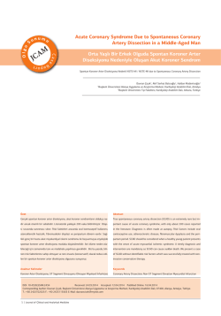
414
Türk Kardiyol Dern Arş - Arch Turk Soc Cardiol 2014;42(4):414 doi: 10.5543/tkda.2014.26925
Cardiogenic shock caused by huge para-aortic hematoma
and pseudoaneurysm after Bentall operation
Bentall ameliyatı sonrası kardiyojenik şoka neden olan
dev para-aortik hematom ve psödoanevrizma
Para-aortic hematoma
due to leaking aortic
conduit is an uncommon complication after Bentall operation.
Especially in the early
Department of Cardiology,
postoperative period,
Dışkapı Yıldırım Beyazıt
patients may have very
Training and Research Hospital,
subtle or no symptoms.
Ankara
Compression of the
coronary arteries and
ascending aorta by the hematoma may compromise
myocardial vascularization, causing clinical signs of
ischemic heart disease and heart failure. In this setting,
our case emphasizes that early noninvasive evaluation
may provide early detection of that complication. A
21-year-old male patient was referred to our department with a possible diagnosis of cardiogenic shock.
Bentall operation had been performed one month before in another hospital because of an ascending aortic aneurysm and aortic valve insufficiency. His systolic blood pressure was lower than 80 mmHg. The
physical examination revealed altered mental status,
Hamza Sunman
Mehmet Erat
Mehmet Doğan
Ekrem Yeter
A
B
weak peripheral pulse, cool skin, and the
metallic sound of the heart valve. ECG
was normal except for sinus tachycardia. Transthoracic and transesophageal
echocardiography revealed decreased left ventricular function, normally functioning metallic valve in
the aortic position, and huge para-aortic hematoma
(Figure A). Additionally, this hematoma was seen to
cause moderate aortic graft compression (Video 1*).
Detailed examination showed blood flow through the
junction between the aortic graft and right coronary
ostium, and this flow caused a whirlpool-like echocontrast in a large pseudoaneurysm (Figure B, Videos
2, 3*). Computed tomographic angiography showed
the leakage of contrast material from the right coronary artery anastomosis into the pseudoaneurysm
(37x38x40 mm) and a huge para-aortic hematoma
(92x106x120 mm) (Figure C). Cardiothoracic surgery
was consulted, and the patient was taken to the operating room. An exploratory thoracotomy was performed
together with hematoma evacuation. Unfortunately,
he died due to severe hemorrhagic stroke within the
first postoperative week.
C
Figures– (A) Transesophageal echocardiography (TEE) showed a large para-aortic hematoma. Ao: Ascending aorta; Asterisk: Paraaortic hematoma; LV: Left ventricle. (B) Spontaneous echo contrast within the large pseudoaneurysm (star) was seen in the upper
esophageal TEE view. (C) Computed tomographic evaluation. Ao: Ascending aortic graft; White star: Pseudoaneurysm; Arrows: Paraaortic hematoma; White arrow: Proximal part of right coronary artery; Black star: Leakage of contrast from the right coronary artery
anastomosis. *Supplementary video files associated with this presentation can be found in the online version of the journal.
© Copyright 2026 Paperzz
![Full Text PDF [399K] - 日本歯科理工学会japanese soc dental](http://s3.paperzz.com/store/data/005363974_1-ec5f0d213f984bbc958ca66dcd196891-250x500.png)