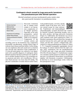
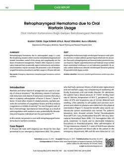
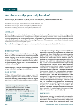
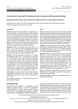
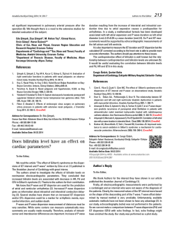
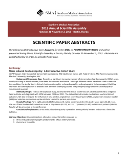
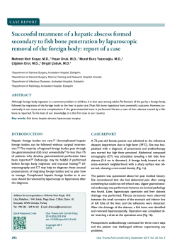
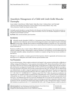
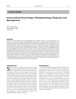
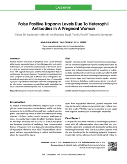
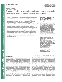
380
Türk Kardiyol Dern Arş - Arch Turk Soc Cardiol 2014;42(4):380-383 doi: 10.5543/tkda.2014.43896
Rapid retraction of a post-infarction
intramyocardial dissecting hematoma
Hızla gerileyen bir miyokart içi hematom olgusu
Ebru Özpelit, M.D., Özer Badak, M.D., Mehmet Emre Özpelit, M.D.,# Ömer Kozan, M.D.
Department of Cardiology, Dokuz Eylul University Faculty of Medicine, Izmir;
#
Department of Cardiology, Izmır University Medicalpark Hospital, Izmir
Summary– A 60-year-old male with a recent anterior myocardial infarction (MI) was referred to our hospital for implantable cardioverter defibrillator (ICD) implantation. He
was on the 42nd day of MI and clinically stable on admission. Electrocardiography showed right bundle branch block
with QS pattern on anterior leads. Transthoracic echocardiographic examination revealed an ejection fraction of
25% with akinesis of the apex and mid-apical segments of
anterior and septal walls. In the apical-septal region, a pulsatile cavity with systolic expansion surrounded by a thin
endomyocardial border was visualized. Color-Doppler interrogation did not demonstrate any flow within that structure.
These findings suggested an intramyocardial dissecting
hemorrhage formed after MI. Cardiac magnetic resonance
imaging also confirmed an intramyocardial hematoma in the
mid-apical anteroseptal region. A conservative approach
was assumed as the patient was hemodynamically stable.
The planned ICD implantation was postponed due to the
high risk of perforation. Subsequently, oral anticoagulant
therapy with warfarin was initiated against risk of intracardiac thrombus formation. The existing dual antiplatelet
therapy was also continued. One week after hospital discharge, he was rehospitalized due to a very high INR of 6.3.
The repeated transthoracic echocardiography revealed an
almost complete resolution of the intramyocardial dissecting
hematoma and adhesion of the surrounding myocardial layers. Oral anticoagulant therapy was discontinued. Echocardiographic examinations showed no change compared to
the last examination during hospitalization. This case illustrates a conservatively managed intramyocardial dissecting
hematoma case, in which anticoagulant and antiaggregant
therapy yielded a rapid retraction without any complication.
Özet– Bir buçuk ay önce ön duvar miyokart enfarktüsü nedeniyle başka bir merkezde revaskülarizasyon uygulanan
ve 42. günde klinik stabilizasyon sonrası kalp içi defibrilatör (ICD) takılması amacıyla kliniğimize gönderilen 60
yaşında erkek hastanın başvuru sırasında yakınması yoktu. Elektrokardiyografide sağ dal bloğu zemininde önyüzü
gören derivasyonlarda QS paterni olan hastanın transtorasik ekokardiyografisinde sol ventrikül apeksi, ön duvar
ve septumun orta-apikal segmentleri akinetikti. Ejeksiyon
fraksiyonu %25’ti. Ekokardiyografik incelemede, sol ventrikülün apikoseptal bölgesinde sistolde genişleyen, diyastolde küçülen pulsatil bir boşluk gözlendi. Renkli Doppler
incelemesinde, bu boşluk içerisinde akım saptanmadı. Bu
bulgularla, hastada akut miyokart enfarktüsüne sekonder
miyokart içi hematom geliştiği düşünüldü. Kalbin manyetik
rezonans görüntülemesi ile tanısı doğrulanan hasta, klinik
olarak stabil olduğu için miyokart içi hematom yönünden
konservatif olarak izlendi. Planlanan ICD implantasyonu
yüksek perforasyon riski nedeniyle ertelendi. Daha sonra, hematom olan bölgede trombus gelişiminin önlenmesi amacıyla varfarin ile oral antikoagülan tedavi başlandı.
Hastanın almakta olduğu ikili antitrombosit tedaviye de
devam edildi. Taburcu edilme sonrası birinci hafta kontrolünde hastanın INR’si 6,3’tü. Aktif kanaması olmayan hastanın yapılan ekokardiyografisinde daha önce gözlenen miyokart içi hematomun tamamen yok olduğu gözlendi. Oral
antikoagülan tedavisi kesilen hasta takipte stabil seyretti.
Ekokardiyografik kontrollerinde hematomun tekrarlamadığı
görüldü. Bu olguda konservatif olarak izlenen miyokart içi
hematomun, antiagregan ve antikoagülan tedavi ile hızla
gerilediği bildirilmiştir.
Received: October 25, 2013 Accepted: December 25, 2013
Correspondence: Dr. Ebru Özpelit. Dokuz Eylül Üniversitesi Tıp Fakültesi, Kardiyoloji Anabilim Dalı, İzmir.
Tel: +90 232 - 412 24 13 e-mail: [email protected]
© 2014 Turkish Society of Cardiology
Rapid retraction of a post-infarction intramyocardial dissecting hematoma
381
I
n t r a m y o c a r d i a l Abbreviations:
dissecting hema- ICD Implantable cardioverter defibrillator
toma (IDH) is a rare IDH Intramyocardial dissecting hematoma
complication of acute INR International normalized ratio
MI Myocardial infarction
myocardial infarction (MI). It consists
of a cystic cavity filled with blood, which is enclosed
by sheets of myocardium.[1] Little is known about
the optimal management of these patients. Although
there is a certain predominance of surgical therapy,
conservatively treated cases have also been reported.
[2-4]
The appropriate medication is also an issue of controversy, with respect to the usage of antiaggregants
and anticoagulants. We describe a case of IDH with
an unexpectedly rapid resolution under conservative
treatment with a high level of anticoagulation.
CASE REPORT
A 60-year-old man with a recent anterior MI was
referred to our hospital for implantable cardioverter
defibrillator (ICD) implantation. He was at the 42nd
day of MI and clinically stable on admission. Troponin values were in normal range. Electrocardiography
showed right bundle branch block with QS pattern
with inverted T waves and QT prolongation (Figure
1). Transthoracic echocardiographic examination revealed an ejection fraction of 25% with akinesis of the
apex and mid-apical segments of the anterior and septal walls. At the apical-septal region, a pulsatile cavity with systolic expansion surrounded by a thin endomyocardial border was visualized. Color-Doppler
interrogation did not demonstrate any flow within that
A
Figure 1. ECG of the patient showing pathological Q waves
in anterior-inferior leads and QT prolongation.
structure (Figure 2a, b, Video 1*). These findings suggested an IDH formed after MI. For a definite diagnosis, cardiac magnetic resonance imaging (MRI) was
performed, which also confirmed an intramyocardial
hematoma in the mid-apical anteroseptal region (Figure 3). A conservative approach was assumed as the
patient was hemodynamically stable. The plan of ICD
implantation was postponed due to the high risk of
perforation. Subsequently, oral anticoagulant therapy
with warfarin was initiated against the risk of intracardiac thrombus formation. The existing dual antiplatelet therapy was also continued. The patient was
discharged one week later in stable condition with an
international normalized ratio (INR) of 2.1, and warfarin dose of 5 mg/day. In the following week, he was
rehospitalized due to a very high INR value.[3,5] The
repeated transthoracic echocardiography revealed an
B
Figure 2. (A) Apical intramyocardial dissecting hematoma in four-chamber view. (B) Apical intramyocardial dissecting hematoma in two-chamber view.
Türk Kardiyol Dern Arş
382
form of cardiac rupture of the left ventricular wall.
The underlying mechanism is a hemorrhage dissecting among the spiral myocardial fibers, creating a
neocavitation limited by the myocardium.[6] The hematoma may expand, rupturing into adjacent structures, spontaneously resolve, or transform into an
organized thrombus. Because of the potential risk of
fatal myocardial rupture, surgical correction was the
preferred method of management for the initial cases
in the literature. However, today, the high operative
mortality and good clinical course of conservatively
treated patients have changed the approach. At present, surgical correction is mostly preferred in hemodynamically unstable patients. In the largest published
series of IDH, Vargas-Barrón et al.[5] reported complete reabsorption in five of 15 cases with a mean 12
months of follow-up. In these five cases, IDHs were
limited to the apical segments of the left ventricle. In
addition, four spontaneously resolved cases of IDH
were reported by different authors, all of which were
also apical in location.[7-10] Based on these observations, conservative management seems a reasonable
option in patients with apical IDH. The present case
also confirms this statement, in which complete resolution was evident in a relatively very short period of
time compared to the previous cases. In most of the
reported cases, spontaneous resolution occurred in
several months. However, in this case, it took only
two weeks for complete retraction. The mechanism of
retraction was interpreted as spontaneous drainage of
the dissecting hematoma through the endomyocardial
Figure 3. Hyperintense apical intramyocardial hematoma in
cardiac MRI.
almost complete resolution of IDH and adhesion of
the surrounding myocardial layers. Neither thrombus
formation nor pericardial effusion was present in the
echocardiographic examination (Figure 4a, b, Video
2*). Oral anticoagulant therapy was discontinued,
and the patient was discharged on dual antiplatelet
therapy after normalization of INR. In the six months
following hospital discharge, serial echocardiographic examinations showed no change compared to the
last examination during hospitalization. The clinical
follow-up was uneventful during this period.
DISCUSSION
Intramyocardial dissecting hematoma is an unusual
A
B
Figure 4. (A) Complete resolution of intramyocardial hematoma in apical four-chamber view. (B) Complete resolution
of intramyocardial hematoma in apical two-chamber view.
Rapid retraction of a post-infarction intramyocardial dissecting hematoma
layer into the left ventricular cavity. It is difficult to
explain definitively this relatively shorter time course;
however, the high level of anticoagulation seems to be
a reasonable cause. There is no clear recommendation
about the use of anticoagulants and antiaggregants in
the treatment of IDH. In most of the reported cases,
anticoagulants and antiaggregants were used because
of other coexisting indications such as embolic stroke
or visible intracardiac thrombus. Although there was
no such clinical condition in this case, the use of oral
warfarin and dual antiplatelet therapy resulted in a
rapid retraction of the IDH. An argument could be
made as to whether it is safe to use this medication
solely for this entity because of the risk of rupture.
Even in the presence of such a risk, further conservatively treated cases of IDH will help to clarify the
issue. Another issue of discussion in conservatively
treated IDH patients is the implantation of ICD and/
or cardiac resynchronization therapy (CRT) device, as
in this case. Hematomas in the apicoseptum of the left
ventricle and right ventricle increase the risk of myocardial rupture during implantation of the leads. In
our opinion, each case should be evaluated separately,
weighing the advantages of such devices and risks
of the procedure. As long as our case was stable on
amiodarone therapy, it was preferred not to implant
an ICD temporarily.
In conclusion, despite the growing awareness of
IDH, many issues remain unknown, especially about
its management and prognosis. This report illustrates
a conservatively managed case in which anticoagulant
and antiaggregant therapy yielded a rapid retraction
of the hematoma without any complication. Further
data are needed to make definitive recommendations
regarding the follow-up of these patients.
Conflict-of-interest issues regarding the authorship or
article: None declared.
*Supplementary video file associated with this article
can be found in the online version of the journal.
383
right ventricle. Am Heart J 1992;124:1641-2. CrossRef
2. Bapat VN, Naik AM, Lokhandwala Y, Tendolkar AG. Images
in cardiovascular medicine. Intramyocardial dissecting hematoma. Circulation 1998;97:2470-2. CrossRef
3. Nakata A, Hirota S, Tsuji H, Takazakura E. Interventricular
septal dissection in a patient with an old myocardial infarction. Intern Med 1996;35:33-5. CrossRef
4. Jiménez J, Almería C, Zamorano JL, Alfonso F, Ribera JM,
Sánchez-Harguindey L. Intramyocardial dissection of the
posterior wall of the left ventricle with shunt to coronary sinus after myocardial infarction. [Article in Spanish] Rev Esp
Cardiol 2001;54:247-9. [Abstract] CrossRef
5. Vargas-Barrón J, Romero-Cárdenas A, Roldán FJ, MolinaCarrión M, Avila-Casado C, Villavicencio R, et al. Long-term
follow-up of intramyocardial dissecting hematomas complicating acute myocardial infarction. J Am Soc Echocardiogr
2005;18:1422. CrossRef
6. Harpaz D, Kriwisky M, Cohen AJ, Medalion B, Rozenman
Y. Unusual form of cardiac rupture: sealed subacute left ventricular free wall rupture, evolving to intramyocardial dissecting hematoma and to pseudoaneurysm formation-a case
report and review of the literature. J Am Soc Echocardiogr
2001;14:219-27. CrossRef
7. Bahlmann E, Schneider C, Vitali Serdoz L, Hoffmann-Riem
M, Broemel T, Kuck KH. Spontaneous retraction of an intramyocardial dissecting hemorrhage and multiple left ventricular thrombus formations in subacute myocardial infarction
and antiphospholipid syndrome: a case report with long-term
follow-up. J Am Soc Echocardiogr 2006;19:578.e5-8.
8. Stewart S, Huddle R, Stuard I, Schreiner BF, DeWeese JA.
False aneurysm and pseudo-false aneurysm of the left ventricle: etiology, pathology, diagnosis, and operative management. Ann Thorac Surg 1981;31:259-65. CrossRef
9. Drozdz J, Kasprzak JD, Krzeminska-Pakula M. Spontaneous closure (thrombosis) of the intramyocardial dissection:
40-month follow-up. J Am Soc Echocardiogr 2002;15:10234. CrossRef
10.Galache Osuna JG, Marquina Barcos A, Cay Diarte E, Sánchez-Rubio Lezcano J, Salazar González JJ, Placer Peralta LJ.
Conservative management of a post infarction intramyocardial dissecting hematoma. [Article in Spanish] Rev Esp Cardiol
2003;56:735-7. [Abstract] CrossRef
REFERENCES
Key words: Cardiomyopathies/diagnosis/ultrasonography; heart rupture; hematoma; interventricular septal rupture; myocardial infarction
1. Mohan JC, Agarwala R, Khanna SK. Dissecting intramyocardial hematoma presenting as a massive pseudotumor of the
Anahtar sözcükler: Kardiyomiyopatiler/tanı/ultrasonografi; kalp yırtığı; hematom; interventrküler septal yırtık; miyokart enfarktüsü.
© Copyright 2026 Paperzz