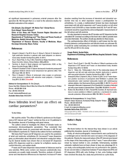
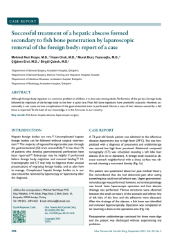
CASE REPORT
142
A Case of Ramsay Hunt Syndrome with
Atypical Presentation
Atipik Prezentasyonlu Bir Ramsay Hunt Sendromu Olgusu
Kamil KAYAYURT, Ozcan YAVASI, Ozlem BILIR, Gokhan ERSUNAN, Baris GIAKOUP
Department of Emergency, Recep Tayyip Erdogan University Faculty of Medicine, Rize
SUMMARY
ÖZET
Ramsay Hunt syndrome is a rare complication of herpes zoster
which results from the reactivation of the latent varicella-zoster virus in the geniculate ganglion. Although facial nerve is the most
common affected nerve in Ramsay Hunt syndrome, other cranial
and cervical nerves can also be affected. We present an atypical
case of Ramsay Hunt syndrome in a 42-year-old male, with cervical
nerve involvement. As spontaneous recovery rate in Ramsay Hunt
syndrome is low, early diagnosis and treatment plays a key role in
full recovery of paralysis.
Ramsay Hunt sendromu, varisella-zoster virüsün latent olarak kaldığı
genikulat ganglionda aktifleşmesiyle oluşan herpes zosterin nadir bir
komplikasyonudur. Ramsay Hunt sendromunda fasiyal sinir en sık etkilenen sinir olmasına rağmen diğer kraniyal sinirler ve servikal sinirler
de tutulabilir. Bu yazıda, 42 yaşındaki erkek hastada servikal tutulumun
da eşlik ettiği atipik bir Ramsay Hunt sedromu olgusu sunuldu. Ramsay
Hunt sedromunda spontan iyileşme oranları düşük olduğundan bu hastaların tanılarının erken dönemde konması ve tedavilerinin hemen başlanması paralizinin tam olarak iyileşmesinde kilit role sahiptir.
Key words: Facial palsy; Ramsay Hunt syndrome; varicella-zoster virus.
Anahtar sözcükler: Fasiyal paralizi; Ramsay Hunt sendromu; varisella-zoster
virüs.
Introduction
Here, we report a case of RHS, in which C2-C4 cervical nerves
are affected together with the facial nerve.
Ramsay Hunt Syndrome (RHS), also known as Herpes Zoster Oticus, was first defined by James Ramsay Hunt in 1907.
[1]
The disease is a rare complication of the primary varicella
zoster virus infection (VZV). Activated many years after inoculation, VZV is followed by a latency period in the geniculate
ganglion and spreads along the sensory tract of the facial
nerve. Ipsilateral facial paralysis, otalgia, and vesicular rash
of the skin of external auditory canal compose the classical
triad of the disease. In contrary to Bell’s palsy, the patients
who are untreated or delayed in their treatment have poor
prognosis and the full recovery rate is as low as 10-30% in
these patients.[2,3] RHS is also known as cranial polyneuropathy and besides facial nerve, cranial nerves 5, 8, 9, 10, 11, 12,
and C2-C4 cervical nerves may also be affected. The eighth
cranial nerve is the most common involved nerve together
with facial nerve, while the others are rarely involved.[4]
Case Report
A 42-year-old man presented to our emergency department
with the complaints of redness, numbness, and pain around
left eye and face following an episode of otalgia, otorrhea,
decreased hearing, and swelling of the left ear lasting three
days. He did not have any chronic disease and medication
history. His vital signs were as follows: blood pressure, 140/90
mmHg; heart rate, 76 bpm; respiratory rate, 16/minute; and
temperature, 36.7 ºC. Physical examination revealed edema
of the external auditory canal and auricula of the left ear with
a vesicular rash and serous discharge, periorbital and facial
erythema and edema, stage 2 peripheric facial paralysis on
the left side and vesicular rash of the left anterior cervical
region (Figure 1a-c). Ophtalmic examination revealed no
Submitted: March 14, 2014 Accepted: April 29, 2014 Published online: June 24, 2014
Correspondence: Dr. Kamil Kayayurt. Recep Tayyip Erdogan Universitesi Tip Fakultesi, Acil Tip Anabilim Dalı, Rize, Turkey.
e-mail: [email protected]
Turk J Emerg Med 2014;14(3):142-145
doi: 10.5505/1304.7361.2014.82788
Kayayurt K et al.
(a)
143
A Case of Ramsay Hunt Syndrome with Atypical Presentation
(b)
(c)
Figure 1. (a) Left facial palsy and periorbital edema. (b) Vesicular rash on the left face and periauricular region with serous
auricular discharge. (c) Vesicular rash on the left anterior cervical region.
pathological findings. Hematological or biochemical findings were not substantial except a white blood cell count of
14.6 K/mL. The patient was hospitalized with the diagnosis
of RHS and acyclovir 10 mg/kg/day and prednisolone 1 mg/
kg/day were initiated. Following the regression of his clinical
symptoms and physical findings, he was discharged at the
third day of his admittance for ambulatory treatment and
follow-up.
Discussion
In United States of America, it is estimated that one out of
every three people are infected by VZV.[5] However, clinical findings of herpes zoster do not develop in all infected
subjects. The population based herpes zoster incidence was
reported as 22.4 / 10,000 in a prospective study conducted
by Paul and Thiel.[6] RHS develops in 0.2% of primary herpes
zoster infections.[5] The incidence of the disease demonstrated an increase after fifth decade and peaked at eighth
decade, parallel to the decreasing cellular immunity with the
aging process. It is 20% more common in females compared
to males.[7]
The present case developed facial paralysis and vesicles
around the left ear and neck following an episode of otalgia, otorrhea, decreased hearing, and swelling of the left ear,
which lasted three days. Vesicular rash may manifest prior
to or following facial paralysis or may not be observed at
any time. When present, these vesicles may be observed at
the external auditory canal, auricule, skin of the cheeks, the
anterior two thirds of the tongue, hard palate, or cervical
region.[4] Our patient had vesicles at the external auditory
canal, auricule, and anterior cervical region with no presentation in other regions. The key clinical finding is facial paralysis and RHS, which covers 12% of cases with non-traumatic
facial paralysis.[7] Otalgia is the second most common symptom and 73% of the patients manifest otalgia as a complaint.
[2]
Involvement of the vestibulocochlear nerve together with
facial nerve is observed in 50% of the cases, causing hearing
loss, vertigo, nausea, vomiting and nystagmus.[4] Dysphagia,
disturbances of gaze and taste, double vision suggest involvement of other cranial nerves. Furthermore, cardiac arrhythmias may develop when vagal nerve is involved.[2,4] Our
patient did not have any cranial nerve involvement other
than facial nerve.
The cervical involvement observed in our case is not a typical finding in RHS.[5] Three different theories are proposed
for this clinical situation. The first theory is spreading of VZV
through cerebrospinal fluid (CSF) or by hematogenous way.
In 38% of the cases with cutaneous herpes zoster, pleocytosis is detected in CSF.[8] Another study reported presence of
21% of the VZV in CSF of the patients manifesting cutaneous lesions.[9] The second proposed theory is the presence of
anastomoses between cervical nerves and branches of the
facial nerve as an anatomical variation, leading to the spread
of inflammation to more than one dermatome along these
anastomoses.[5,10] The last theory is the simultaneous activation of the virus in more than one ganglion, causing clinical
symptoms.[5]
The case presented here was diagnosed clinically. There was
no need for ancillary laboratory or imaging studies. RHS is
a clinical diagnosis with the unilateral facial paresis, otalgia
144
Turk J Emerg Med 2014;14(3):142-145
and vesicular rash in ipsilateral ear, hard palate, and the anterior two third of the tongue being sufficient for diagnosis.
It is hard to differentiate between RHS and Bell’s palsy when
vesicles are not present in patients. Complement fixation
tests and increased antibody levels may support the diagnosis. The detection of the virus by polymerase chain reaction
in mononuclear cells of external auditory canal fluid, tear,
CSF, and blood is accepted as the gold-standard in diagnosis of VZV.[11] Jonsson et al. evaluated the cases with facial
paralysis and reported that there were no extra benefits of
contrast enhanced magnetic resonance imaging for either
diagnosis or prognosis of the patients.[12]
There are not so many randomized, prospective controlled
studies for RHS in the medical literature. The data is mostly based on the case reports and retrospective case series.
The actual management is starting with a combination of
an antiviral agent and steroids.[13] The most commonly used
antiviral agent is acyclovir, however; a new generation antiviral drugs such as valacyclovir, famciclovir, penciclovir, and
brivudine are being preferred due to increasing resistance.
[14]
Prednisolone is suggested as the drug of choice for steroid therapy. In a retrospective study, Murakami et al. compared the effectiveness of the combination therapy of acyclovir and prednisolone with prednisolone therapy alone.
They concluded that the full neurological recovery rate of
the combination therapy group was significantly higher
than the group treated merely with prednisolone. Moreover, when patients were grouped according to the time of
initiation of drug therapy, the group in which therapy was
initiated in the first three days showed a full neurological recovery rate of 75%. The rate decreased to 48% in the second
group in which therapy was initiated in 4 to 7 days similar
to the decrease of 30% in the third group in which therapy
was initiated in later than 7 days. There was no clinically significant difference between therapeutic effects of parenteral
and oral administration of acyclovir.[3] Kinishi et al. compared
the groups of patients who started acyclovir alone and prednisolene alone therapy in the first seven days. They reported
significantly better nerve stimulation test results in the acyclovir alone group.[15]
Since patients suffer from severe pain, pain management
must also be taken into consideration in addition to the
treatments mentioned above. Cellulitis may develop following infection of the vesicles, necessitating antibiotic therapy.
Following discharge from the hospital, most of the patients
need rehabilitation and long term follow-up.[5] Our patient
was administered a combination therapy of acyclovir and
prednisolone after the clinical diagnosis was made. He was
discharged at the third day for ambulatory treatment and
follow-up.
The most important aspect of RHS when compared to Bell’s
palsy is the significantly worse prognosis when left untreated. In non-treated patients or patients in whom treatment
was started later than seven days, the full neurological recovery rate is significantly as low as 10 to 30%. Advanced
age, involvement of more than one cranial nerve, and a
higher stage of facial paralysis during first admittance are
well-defined prognostic indicators of RHS.[2,3] The absence of
the prognostic factors as well as the early initiation of therapy yielded full neurological recovery in the present case,
consistent with the literature. The most common complication of RHS is postherpetic neuralgia that increases with age.
Meningoencephalitis, myelitis, cerebrovascular problems
associated with vasculitis, ventriculitis, and cerebral venous
thrombosis may also develop.[4] Examination of our patient
during follow-up visits indicated no development of any of
these listed complications.
Conclusion
RHS is a medical emergent condition necessitating early
diagnosis and treatment in order to have a good prognosis. In patients with facial paralysis but no external vesicular
findings, otoscopic and audiometric examinations must be
performed. The combination therapy with antivirals and steroids must be initiated as soon as possible in order to minimize the risk of permanent neuronal damage.
Conflict of Interest
The authors declare that there is no potential conflicts of interest.
References
1. Hunt JR. On herpetic inflammation of the geniculate ganglion. A new syndrome and its complications. J Nerv Ment Dis
1907;34:73-96. CrossRef
2. Ko JY, Sheen TS, Hsu MM. Herpes zoster oticus treated with
acyclovir and prednisolone: clinical manifestations and
analysis of prognostic factors. Clin Otolaryngol Allied Sci
2000;25:139-42. CrossRef
3. Murakami S, Hato N, Horiuchi J, Honda N, Gyo K, Yanagihara
N. Treatment of Ramsay Hunt syndrome with acyclovir-prednisone: significance of early diagnosis and treatment. Ann
Neurol 1997;41:353-7. CrossRef
4. Wagner G, Klinge H, Sachse MM. Ramsay Hunt syndrome. [Article in English, German] J Dtsch Dermatol Ges 2012;10:23844. [Abstract] CrossRef
5. Worme M, Chada R, Lavallee L. An unexpected case of Ramsay Hunt syndrome: case report and literature review. BMC
Res Notes 2013;6:337. CrossRef
6. Paul E, Thiel T. Epidemiology of varicella zoster infection. Results of a prospective study in the Ansbach area. [Article in
German] Hautarzt 1996;47:604-9. [Abstract] CrossRef
Kayayurt K et al.
A Case of Ramsay Hunt Syndrome with Atypical Presentation
7. Robillard RB, Hilsinger RL Jr, Adour KK. Ramsay Hunt facial
paralysis: clinical analyses of 185 patients. Otolaryngol Head
Neck Surg 1986;95:292-7.
8. Gold E. Serologic and virus-isolation studies of patients
with varicella or herpes-zoster infection. N Engl J Med
1966;274:181-5. CrossRef
9. Haanpää M, Dastidar P, Weinberg A, Levin M, Miettinen A,
Lapinlampi A, et al. CSF and MRI findings in patients with
acute herpes zoster. Neurology 1998;51:1405-11. CrossRef
10.Brown H, Burns S, Kaiser CW. The spinal accessory nerve plexus, the trapezius muscle, and shoulder stabilization after radical neck cancer surgery. Ann Surg 1988;208:654-61. CrossRef
11.Sweeney CJ, Gilden DH. Ramsay Hunt syndrome. J Neurol
Neurosurg Psychiatry 2001;71:149-54. CrossRef
12.Jonsson L, Tien R, Engström M, Thuomas KA. Gd-DPTA enhanced MRI in Bell’s palsy and herpes zoster oticus: an overview and implications for future studies. Acta Otolaryngol
1995;115:577-84. CrossRef
13.Hato N, Murakami S, Gyo K. Steroid and antiviral treatment
for Bell’s palsy. Lancet 2008;371:1818-20. CrossRef
14. Dworkin RH, Johnson RW, Breuer J, Gnann JW, Levin MJ, Backonja M, et al. Recommendations for the management of herpes zoster. Clin Infect Dis 2007;44 Suppl 1:S1-26. CrossRef
15.Kinishi M, Amatsu M, Mohri M, Saito M, Hasegawa T, Hasegawa S. Acyclovir improves recovery rate of facial nerve palsy in
Ramsay Hunt syndrome. Auris Nasus Larynx 2001;28:223-6.
145
© Copyright 2026 Paperzz