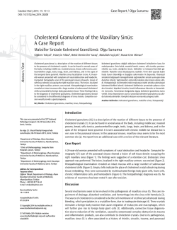
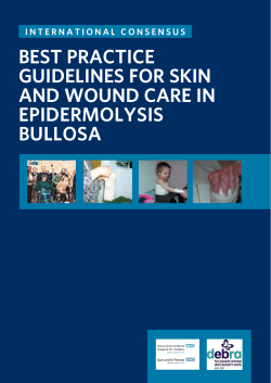
OLGU SUNUMU/CASE REPORT
J Turgut Ozal Med Cent 2014;21(2):145-7
Journal Of Turgut Ozal Medical Center www.jtomc.org A Rare Cause of Nasal Obstruction: Concha Bullosa of the Inferior
Turbinate
Emrah Sapmaz1, Hilal Irmak Sapmaz2, Yüksel Toplu3, Ramazan Öçalan1, Işıl Çakmak Karaer1
1
State Hospital, Otorhinolaryngology Clinic, Malatya, Turkey
İnönü University, Faculty of Medicine, Department of Anatomy, Malatya, Turkey
3
İnönü University, Faculty of Medicine, Department of Otorhinolaryngology, Malatya, Turkey
2
Abstract
Concha bullosa of the inferior turbinate is an extremely rare anatomic malformation. Inferior concha bullosa is the structure with air sacs on
the inferior turbinate body and may be associated with maxillary sinus. Inferior concha bullosa is generally asymptomatic and diagnosed
incidentally by computed tomography. Its main clinical symptoms are nasal obstruction, headache, and postnasal drip. Several techniques
have been suggested as treatment options. With this study, we intend to report the case of a 23 year old woman who has nasal
obstruction, headache, and postnasal drip was diagnosed with inferior, middle and superior concha bullosa and we have performed
laterization through radiofrequency ablation on inferior turbinate and partial resection on the middle turbinate.
Key Words: Inferior Concha Bullosa; Nasal Congestion; Computed Tomography.
Burun Tıkanıklığının Nadir Bir Sebebi: İnferior Konka Bülloza
Özet
İnferior konka bülloza oldukça nadir görülen bir anatomik varyasyondur. İnferior konka bülloza, inferior konkanın kemik lamelinin içerisinde
hava keseciğinin olmasıdır ve bazen bu hava kesesi maksiller sinüslede alakalı olabilir. Genellikle asemptomatiktir ve görüntüleme
yöntemleriyle tesadüfen tespit edildir. Bazen burun tıkanıklığı, baş ağrısı ve geniz akıntısı gibi semptomlarla neden olabilir. Tedavisinde
çeşitli teknikler tarif edilmiştir. Biz bu makalede 23 yaşında burun tıkanıklığı, baş ağrısı ve geniz akıntısı nedeniyle kliniğimize baş vuran ve
üst orta ve alt konklarda konka bülloza tespit ettiğimiz,,inferior konka büllozanın tedavisinde lateralizasyon ve radyofrekansla ablasyon
yaptığımız aynı zamanda orta konka büllozaya parsiyel rezeksiyon uyguladığımız bir bayan vakayı sunduk.
Anahtar Kelimeler: Alt Konka Bülloza; Nazal Tıkanıklık; Bilgisayarlı Tomografi.
did not help and observing no allergens in the skin prick
test, the patient was asked for a paranasal sinus
tomography scan. The coronal paranasal sinus
tomography showed pneumatization in the left inferior,
middle and upper turbinates (Figure 1 and 2).Since the
pneumatization in the inferior turbinate was connected
to the maxillary sinus, we have performed laterization
through radiofrequency ablation on inferior turbinate
and partial resection on the middle turbinate. The
patient had no intraoperative and postoperative
problems. After 3 postoperative months, there was a
significant reduction in symptoms.
INTRODUCTION
Stretching from the lateral nasal wall extending into the
nose, nasal turbinates are crucial structures. Inferior
turbinal ethmoid and maxillary turbinate develop
through the 8th to 10th weeks of fetal life (1). Concha
bullosa (CB) is the pneumatization of the middle
turbinate, and less commonly, of the top turbinate (2).
Inferior concha bullosa (ICB) is the structure with air sacs
on the inferior turbinate body and may be associated
with maxillary sinus. Although middle turbinate
pneumatization is fairly common, the pneumatization of
the inferior turbinate is rare (3). It can either be unilateral
or bilaterally (3,4).
CASE REPORT
A 23 year old female patient with continuous headache
in the last year and complaints of breathlessness was
admitted to our clinic. The patient did not have any
history of trauma. In the anterior rhinoscopy and
endoscopic examination, we have observed inferior
turbinate on the left with a minimal septal deviation
along with hypertrophy in the middle turbinates. Upon
learning that previous medical treatment she was given
Figure 1. ICB (white arrow)
(white arrow)
145
Figure
2.
MCB
ve
UCB
Journal of Turgut Ozal Medical Center
commonly used but in cases in which ventilation is
connected to the maxillary sinus, this method is
contraindicated (4).
DISCUSSION
Nasal turbinates are important structures in maintaining
normal nasal functions. They are the most important
substructures in moistening, directing and cleaning the
air taken in through the nose (5). Generally, the lower
turbinates become hypertrophied due to reasons such
as allergic causes, being exposed to irritants, chronic
sinus infections, and vasomotor rhinitis. In the presence
of septal deviation, compensatory hypertrophy may
occur in the turbinate as a response to the deviation. It is
very rare for the hypertrophy of the inferior turbinate to
stem from the pneumatized inferior turbinate. Unlike
other turbinates, the embryological development of the
inferior turbinates springs from what is called
maxsilloturbinal structures (6).
It has been suggested that applying the resection to
such patients may lead to iatrogenic sub-meatal
antrostomy. It has been argued that, in these cases, the
resection may disrupt the physiology of the maxillary
sinus and initiate mucous recirculation phenomena (2). In
cases with small pneumatization, it may even be enough
to break the lateral wall by just pushing it towards
medial (15). Because it may result in atrophic rhinitis, the
removal of the whole inferior turbinate or damaging the
mucosa is contraindicated (16).
As a result of the relationship between the inferior
turbinate and the maxillary sinus as observed in the
computerized tomography, and the insufficient
improvement in the turbinate hypertrophy after the
administration of mucosal decongestants and steroids,
we preferred radiofrequency ablation and lateralization
in our patient's case.
Among the major hypotheses concerning the formation
of the pneumatized inferior turbinate, we can mention
the double lamellar ossification of inferior concha
between the 5th and 9th weeks followed by the
invagination of the mucous membrane within the
concha, and the extension of maxillary sinus aeration
into the inferior turbinate. That the hypertrophy of the
inferior turbinates causes nasal obstruction and that it is
affected by surgical intervention have both clinical and
surgical importance (7).
Consequently, ICB should be considered in the
differential diagnosis of patients with nasal obstruction
and hypertrophied inferior turbinate.
REFERENCES
However, because the more important area is the
middle meatus in endoscopic sinus surgery, middle
turbinate variations attract more attention. This is the
reason why variations of inferior turbinate are usually in
the form of case reports. It was Zinreich et al. who
evaluated IDB as a variation of the turbinates for first
time in 1988 (8). Yang et al. (9) among 59,238
tomographic scans of the paranasal sinuses have
identified ICB in a total of 16 patients, two of them being
bilateral ICB. Similarly, Christmas et al. have reported
three ICB cases that caused nasal blockage (10).
1.
ICB is usually asymptomatic and is usually diagnosed by
imaging methods (2, 11). If there is a wide range of
ventilation, it can cause nasal congestion. In addition to
congestion, it may also lead to nasal discharge, coinfections, headaches and epiphora (12, 13). Clinically, it
may not be easy to differentiate it from hypertrophied
turbinates. At this point, applying decongestant can be
useful for the differential diagnosis.
6.
ICB should be treated when it is symptomatic. Maximum
air passages and mucosal functions should be
maintained during the treatment. Nasal steroids may be
administered as a medical treatment but often the
problem does not respond well and surgical intervention
may be required. Turbinoplasty with microdebrider,
submucosal
diathermy,
radiofrequency
ablation,
resection of the free wall, breaking the wall with forceps
are among the surgical techniques offered for the
treatment (2,14).
10.
2.
3.
4.
5.
7.
8.
9.
11.
12.
13.
14.
Due to the fact that it has been tried many times with
positive results, the resection of the lateral wall is
146
Bolger WE, Butzin CA, Parsons DS. Paranasal sinus bony
anatomic variations and mucosal abnormalities: CT analysis
for endoscopic sinus surgery. Laryngoscope 1991;101:5664.
Unlu HH, Altuntas A, Aslan A, Eskiizmir G, Yucel A. Inferior
concha bullosa. J Otolaryngol 2002;31:62-4.
Dawlaty EE. Inferior concha bullosa--a radiological and
clinical rarity. Rhinology 1999;37:133-5.
Dogru H, Doner F, Uygur K, Gedikli O, Cetin M.
Pneumatized inferior turbinate. Am J Otolaryngol
1999;20:139-41.
Pittore B, Al Safi W, Jarvis SJ. Concha bullosa of the inferior
turbinate: an unusual cause of nasal obstruction. Acta
Otorhinolaryngol Ital;31:47-9.
Braun H, Stammberger H. Pneumatization of turbinates.
Laryngoscope 2003;113:668-72.
Spear SA, Brietzke SE, Winslow C. Bilateral bifid inferior
turbinates. Ann Otol Rhinol Laryngol 2003;112:195-6.
Zinreich SJ, Mattox DE, Kennedy DW, Chisholm HL, Diffley
DM, Rosenbaum AE. Concha bullosa: CT evaluation. J
Comput Assist Tomogr 1988;12:778-84.
Yang BT, Chong VF, Wang ZC, Xian JF, Chen QH. CT
appearance of pneumatized inferior turbinate. Clin Radiol
2008;63:901-5.
Christmas DA, Jr., Merrell RA, Jr., Mirante JP, Yanagisawa
E. Pneumatized inferior turbinate: report of three cases. Ear
Nose Throat J 2004;83:152-3.
Aydin O, Ustundag E, Ciftci E, Keskin IG. Pneumatization of
the inferior turbinate. Auris Nasus Larynx 2001;28:361-3.
Kiroglu AF, Cankaya H, Yuca K, Kara T, Kiris M. Isolated
turbinitis and pneumatization of the concha inferior in a
child. Am J Otolaryngol 2007;28:67-8.
Gocmen H, Oguz H, Ceylan K, Samim E. Infected inferior
turbinate pneumatization. Eur Arch Otorhinolaryngol
2005;262:979-81.
Ozcan KM, Gedikli Y, Ozcan I, Pasaoglu L, Dere H.
Microdebrider for reduction of inferior turbinate: evaluation
of effectiveness by computed tomography. J Otolaryngol
Head Neck Surg 2008;37:463-8.
www.totmdergisi.org
15. Cannon CR. Endoscopic management of concha bullosa.
Otolaryngol Head Neck Surg 1994;110:449-54.
16. Clement WA, White PS. Trends in turbinate surgery
literature: a 35-year review. Clin Otolaryngol Allied Sci
2001;26:124-8.
Received/Başvuru: 29.08.2013, Accepted/Kabul: 04.10.2013 For citing/Atıf için
Correspondence/İletişim
Emrah SAPMAZ
State Hospital, Otorhinolaryngology
TURKEY
E-mail: [email protected]
Clinic,
Sapmaz E, Irmak Sapmaz H, Toplu Y, Ocalan R, Cakmak
Karaer I. A rare cause of nasal stuffines: Inferior concha
bullosa. J Turgut Ozal Med Cent 2014;21:145-7 DOI:
10.7247/jtomc.2013.1221
MALATYA,
147
© Copyright 2026 Paperzz