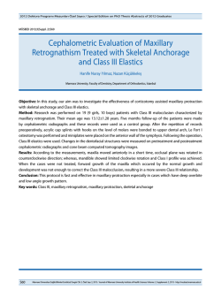
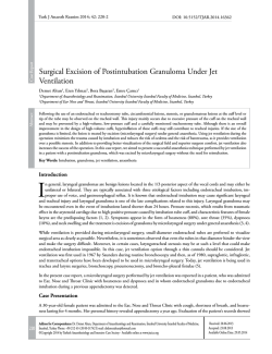
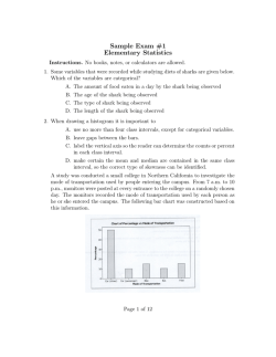
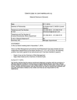
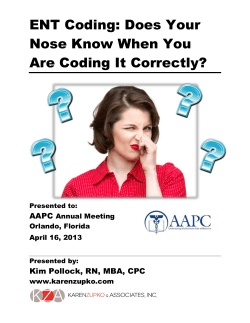
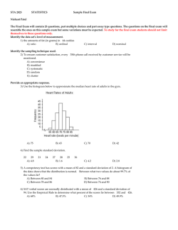
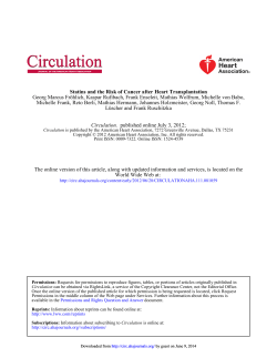
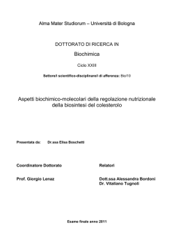
Case Report / Olgu Sunumu
İstanbul Med J 2014; 15: 131-3
DOI: 10.5152/imj.2014.28208
Cholesterol Granuloma of the Maxillary Sinüs:
A Case Report
Maksiller Sinüsde Kolesterol Granülümü: Olgu Sunumu
Abstract / Özet
Çiğdem Tokyol1, Hüseyin Yıldız2, Betül Demirciler Yavaş1, Abdullah Ayçiçek3, Murat Cem Miman4
Cholesterol granuloma is a description of the reaction of different tissues
to the presence of cholesterol crystals. It can be found in several areas of
the body, including middle ear, mastoid process, breast, sella turcica, pontocerebelline angle, testis, lungs, brain, and kidneys, and in the apex of
the temporal bone pyramid. Maxillary sinus localization is rare. A 24-yearold woman presented with symptoms of nasal obstruction and headache.
Computed tomography scan of the paranasal sinuses showed a lesion of
soft tissue density occupying the right maxillary sinus. The lesion, localized
in the right maxillary antrum, was excised. Histopathological examination
revealed an intact mucosa with a large number of submucosal cholesterol
clefts surrounded by foreign body granulation tissue. These findings led us
to the diagnosis of cholesterol granuloma. Cholesterol granuloma should
be considered in the differential diagnosis of sinus lesions. Complete excision would provide a good prognosis.
Kolesterol granülomu değişik dokuların kolesterol kristallerine karşı bir
reaksiyonudur. Orta kulak, mastoid kemik, meme, sella tursika, pontoserebellar açı, testis, akciğerler, beyin, böbrekler ve temporal kemikde görülebilir. Maksiller sinüs lokalizasyonu nadirdir. Yirmi dört yaşında kadın
hasta burun tıkanıklığı ve başağrısı yakınmaları ile başvurdu. Paranazal
sinüslerin bilgisayarlı tomografisinde sağ maksiller sinüste yumuşak doku
dansitesi izlendi. Sağ maksiller antrumda lokalize olan lezyon eksize edildi. Histopatolojik incelemede intakt respiratuar epitel altında submukozal
kolesterol kleftleri izlendi. Kolesterol kleftlerinin çevresinde yabancı cisim
dev hücreleri, köpüksü hücreler, kronik inflamatuar hücreler ve hemosiderin mevcuttu. Tanımlanan bulgularla olguya kolesterol granülomu tanısı
verildi. Sinüs lezyonlarının ayırıcı tanısında kolesterol granulomu da akılda bulundurulmalıdır. Komplet eksizyon sonrasında prognoz iyidir.
Anahtar Kelimeler: Kolesterol granülomu, maksiller sinüs, histopatoloji
Key Words: Cholesterol granuloma, maxillary sinus, histopathology
Introduction
This case was presented at the“23th National
Pathology Congress”, 06-10 November 2013,
İzmir, Türkiye.
Bu olgu 23. Ulusal Patoloji Kongresi’nde
sunulmuştur, 06-10 Kasım 2013, İzmir, Türkiye
Department of Patology, Afyon Kocatepe
University Faculty of Medicine, Afyonkarahisar,
Türkiye
2
Clinic of Otolaryngology, Sungurlu State
Hospital, Çorum, Türkiye
3
Department of Otolaryngology, Afyon Kocatepe
University Faculty of Medicine, Afyonkarahisar,
Türkiye
4
Department of Otolaryngology, İzmir Kocatepe
University Faculty of Medicine, İzmir, Türkiye
1
Address for Correspondence
Yazışma Adresi:
Çiğdem Tokyol, Department of Patology, Afyon
Kocatepe University Faculty of Medicine,
Afyonkarahisar, Türkiye
Phone: +90 505 817 56 63
E-mail: [email protected]
Received/Geliş Tarihi:
19.11.2013
Accepted/Kabul Tarihi:
07.03.2014
© Copyright 2014 by Available online at
www.istanbulmedicaljournal.org
© Telif Hakkı 2014 Makale metnine
www.istanbultipdergisi.org web sayfasından
ulaşılabilir.
Cholesterol granuloma (CG) is a description of the reaction of different tissues to the presence of
cholesterol crystals (1). It can be found in several areas of the body, including middle ear, mastoid
process, breast, sella turcica, pontocerebelline angle, testis, lungs, brain, and kidneys, and in the
apex of the temporal bone pyramid. It is seen associated with chronic middle ear disease but is
very rare in the paranasal sinuses. In the paranasal sinuses, maxillary sinus seems to be the most
common site (2). We report here an additional case with a review of the relevant literature.
Case Report
A 24-year-old woman presented with symptoms of nasal obstruction and headache. Computed tomography (CT) scan of the paranasal sinuses showed a lesion of soft tissue density occupying the
right maxillary sinus (Figure 1). The findings were suggestive of a retention cyst. Endoscopic sinus
approach was performed. The lesion, localized in the right maxillary antrum, was excised (Figure 2).
Histopathological examination revealed an intact mucosa with a large number of submucosal
cholesterol clefts (Figure 3). Empty clefts indicate the place of cholesterol crystals dissolving during
tissue embedding. They were surrounded by multinucleated foreign body giant cells, foam cells,
chronic inflammatory cells, and hemosiderin (Figure 4). The histopathologic diagnosis was CG. No
evidence of recurrence has been noted 1 year after excision.
Discussion
Several mechanisms seem to be involved in the pathogenesis of maxillary sinus CG. They are impairment of drainage, disturbed ventilation, and hemorrhage into the sinus with hemolysis (3).
The source of cholesterol is considered to be the cell membrane of erythrocytes destroyed during
bleeding, which precipitates in a crystalline form, due to inadequate drainage (4). These crystals
stimulate a foreign body reaction that causes migration of leukocytes and macrophages, which
will further give rise to foreign body giant cells (3). Additionally, connective tissue degeneration due to reduction of the ventilation, caused by osteomeatal complex obstruction by trauma
and inflammatory products, can also contribute to cholesterol crystals. Due to its pathogenesis,
maxillary sinus CG is often associated to a history of rhinitis, sinusitis, trauma, and paranasal
İstanbul Med J 2014; 15: 131-3
Figure 1. CT image of the paranasal sinuses showing a lesion of soft
tissue density occupying the right maxillary sinus
CT: computed tomography
Figure 3. Large number of submucosal empty clefts and overlying
intact respiratory epithelium (HEx40)
Figure 2. Intraoperative endoscopic photography of the soft tissue
mass localized in the right maxillary antrum
sinus surgery, especially because they can cause local bleeding
focuses (4-8).
132
Since first reported by Graham and Michaels in 1978, there have
been 47 cases of maxillary sinus cholesterol granuloma in the
literature (3-6, 9-14). CG of the maxillary sinus affects men more
than women, particularly those in their 40s (10). It develops more
frequently in Caucasians and Turkish (2). The left antrum is more
commonly affected than the right one. Bilateral involvement is
rare (2, 11). About half of the patients present with non-specific
symptoms. Nasal obstruction, postnasal drip, or rhinorrhea can be
noted. An episode of clear golden yellow rhinorrhea is the only
specific symptom of maxillary sinus CG (2, 9). As with the clinical features, CT scanning usually reveals nonspecific findings. The
most common changes are antrum opacification and a cystic appearance, while other less common features include bone expansion and erosion (10). Magnetic resonance imaging was reported
to have more specific findings. There is increased signal intensity
in T1- and T2-weighted images. It can be attributed to the effect of
Figure 4. Chronic inflammatory cells and hemosiderin surrounding
the empty clefts (HEx100)
hemoglobin breakdown products derived from microhemorrhages
around cholesterol crystals (8). However, histopathological examination is necessary for final diagnosis. The treatment of choice is
surgical excision. The prognosis is good after operation. Complete
excision is important to avoid recurrence (2).
Conclusion
We present here a case of maxillary sinus CG located in the right
maxillary antrum of a 24-year-old woman. Cholesterol granuloma
should be considered in the differential diagnosis of sinus lesions.
Complete excision would provide a good prognosis.
Tokyol et al. Cholesterol Granuloma
Informed Consent: Written informed consent was not obtained
due to the retrospective nature of this case.
References
1.
Peer-review: Externally peer-reviewed.
Author Contributions: Concept - Ç.T., A.A.; Design - Ç.T., A.A.; Supervision - M.C.M.; Funding - A.A., H.Y.; Materials - H.Y., B.D.Y.;
Data Collection and/or Processing - H.Y., B.D.Y.; Analysis and/or
Interpretation - H.Y., B.D.Y;Literature Review - M.C.M., H.Y.; Writing - Ç.T., H.Y.; Critical Review - M.C.M., A.A.; Other - M.C.M., B.D.Y.
Conflict of Interest: No conflict of interest was declared by the
authors.
Financial Disclosure: The authors declared that this case has received no financial support.
Hasta Onamı: Olgunun retrospektif tasarımından dolayı hasta
onamı alınmamıştır.
2.
3.
4.
5.
6.
7.
8.
9.
Hakem değerlendirmesi: Dış bağımsız.
Yazar Katkıları: Fikir - Ç.T., A.A.; Tasarım - Ç.T., A.A.; Denetleme
- M.C.M.; Kaynaklar - A.A., H.Y.; Malzemeler - H.Y., B.D.Y.; Veri
toplanması ve/veya işlemesi - H.Y., B.D.Y.; Analiz ve/veya yorum
- H.Y., B.D.Y; Literatür taraması - M.C.M., H.Y.; Yazıyı yazan - Ç.T.,
H.Y.; Eleştirel İnceleme - M.C.M., A.A.; Diğer - M.C.M., B.D.Y.
10.
11.
12.
Çıkar Çatışması: Yazarlar çıkar çatışması bildirmemişlerdir.
13.
Finansal Destek: Yazarlar bu olgu için finansal destek almadıklarını
beyan etmişlerdir.
14.
Barnes L, Peel RL, editors. Head and neck pathology: a text/atlas
of differential diagnosis. Tokyo: Igaku-Shoin Medical Publications,
1990.p.471-2.
Chao TK. Cholesterol granuloma of the maxillary sinus. Eur Arch Otorhinolaryngol 2006; 263: 592-7. [CrossRef]
Milton CM, Bickerton RC. A review of maxillary sinus cholesterol granuloma. Br J Oral Maxillofac Surg 1986; 24: 293-9. [CrossRef]
Bella Z, Torkos A, Tiszlavicz L, Iván L, Jóri J. Cholesterol granuloma
of the maxillary sinus resembling an invasive, destructive tumor. Eur
Arch Otorhinolaryngol 2005; 262: 531-3. [CrossRef]
Ko MT, Hwang CF, Kao YF, Lui CC, Huang CC, Peng JP. Cholesterol granuloma of the maxillary sinus presenting as sinonasal polyp. Am J
Otolaryngol 2006; 27: 370-2. [CrossRef]
Ramani P, Murugesan K, Chandrasekar T, Anuja N. Cholesterol granuloma of maxillary sinus. Int J Oral Maxillofac Surg 2006; 35: 1063-5.
Sarioglu S, Pabuççuoglu U, Arzu Topal N. Cholesterol granuloma and
aspergilloma of the maxillary sinus. Eur Arch Otorhinolaryngol 2001;
258: 74-6. [CrossRef]
Leon ME, Chavez C, Fyfe B, Nagorsky MJ, Garcia FU. Cholesterol granuloma of the maxillary sinus. Arch Pathol Lab Med 2002; 126: 217-9.
Graham J, Michaels L. Cholesterol granuloma of the maxillary antrum. Clin Otolaryngol Allied Sci 1978; 3: 155-60. [CrossRef]
Karaky AA, Sawair FA, Baqain ZH, Hassona Y, Khraisat A. Cholesterol granuloma of the maxillary sinus encountered during floor augmentation
procedure: A case report. Clin Implant Dent Relat Res 2010; 12: 249-53.
Xu W, Jin X. Cholesterol granuloma of the maxillary sinus: 2 cases
report. Lin Chuang Er Bi Yan Hou Ke Za Zhi 1998; 12: 549-51.
Astarci HM, Sungu N, Samim EE, Ustun H. Presence of cholesterol granuloma in the maxillary and ethmoid sinuses. Oral Maxillofac Surg
2008; 12: 101-3. [CrossRef]
Nguyen CV, Hudacko R, Theise ND, Tabaee A. Endoscopic management of cholesterol granuloma of the maxillary sinus. J Otolaryngol
Head Neck Surg 2009; 38: E69-72.
Alzahrani M, Morinière S, Duprez R, Beutter P, Bakhos D. Cholesterol
granuloma of the maxillary sinus. Rev Laryngol Otol Rhinol (Bord)
2010; 131: 309-11.
133
© Copyright 2026 Paperzz