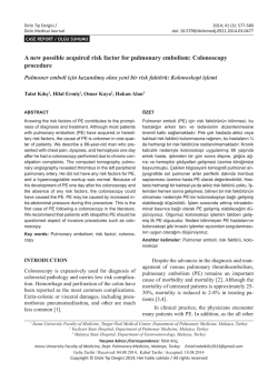
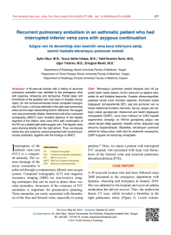
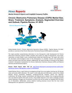
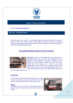
Türk Kardiyol Dern Arş - Arch Turk Soc Cardiol 2014;42(6):587 doi: 10.5543/tkda.2014.47195
587
Pulmonary artery sling and azygos lobe in an asymptomatic 11-year-old boy
On bir yaşında semptomsuz erkek çocukta pulmoner arter sling ve azigos lobu
Savaş Demirpençe
Barış Güven#
Vedide Tavlı*
Department of Pediatric
Cardiology, Sanliurfa Children
Hospital, Sanliurfa;
#
Department of Pediatric
Cardiology, Izmir University,
Medical Park Hospital, Izmir;
*Department of Pediatric
Cardiology, Sifa University Faculty
of Medicine, Izmir
A
C
A 11-year-old boy was referred to our unit for the evaluation of a murmur. His
past medical history was unremarkable. A soft systolic murmur was heard at the
left lower sternal border. Electrocardiography was normal. Chest radiography
was normal. Left pulmonary artery (PA) was absent from its usual location,
and we did not see main and pulmonary arteries in their regular orientations in
parasternal short axis view of transthoracic two-dimensional and color Doppler
echocardiography (Fig. A and B). Computerized tomography angiography revealed anomalous origin of left PA from right PA, which was consistent with a
diagnosis of PA sling and normal variant of azygos lobe and fissure in the right
lung. There was no external compression to the right or left main bronchus as
left pulmonary artery courses retrotracheally to left pulmonary hilum (Fig. C).
Ventilation perfusion scintigraphy and pulmonary function tests were normal.
Barium esophagogram was performed, which demonstrated indentation in the
left anterolateral part of esophagus (Fig. D). Since our patient had no history
of cough, recurrent wheezing/bronchitis attacks, vomiting and dysphagia, we
decided to follow-up clinically.
B
D
Figures– (A, B) Transthoracic two-dimensional and color Doppler echocardiography in parasternal short
axis view showing location of the LPA is not normal. (C) Axial contrast enhanced chest tomography reveals anomalous origin of LPA from the posterior aspect of RPA. LPA is curving
behind trachea as it courses to left pulmonary hilum. (D) Barium esophagogram is demonstrating indentation of esophagus by the surround LPA (Arrow) (LPA: Left pulmonary artery,
MPA: Main pulmonary artery, RPA: Right pulmonary artery, T: Trachea, Ao: Aorta).
© Copyright 2026 Paperzz