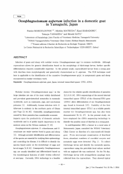
ERCİYES ÜNİVERSİTESİ VETERİNER FAKÜLTESİ DERGİSİ
Journal of Faculty of Veterinary Medicine, Erciyes University
Derleme / Review Article
11(2), 121-126, 2014
Pregnancy Diagnosis in Goats By Ultrasonography
Sinem Özlem ENGİNLER1, Kutlay GÜRBULAK2
Department of Obstetrics and Gynaecology, Faculty of Veterinary Medicine, Istanbul University,
Avcilar Campus, 34320, Avcılar, Istanbul-TURKEY
2
Department of Obstetrics and Gynaecology, Faculty of Veterinary Medicine, Erciyes University, Kayseri-TURKEY
1
Summary: Ultrasonography is a technique commonly used in veterinary reproduction in species including goats. Pregnancy in
goats can be detected as early as the 16th day post-breeding via transrectal way. However, because of the early embryonic losses,
it is recommended to delay the transrectal examination until days 32-34 of gestation. Transabdominal (TA) ultrasonography for
pregnancy diagnosis in goats can be preferred from the second trimester on because after this period the gravid uterus starts to
descend into the cranio-ventral direction. The transrectal (TR) ultrasonography in this period becomes inadequate to distinguish
the parts of the foetus. But it can still detect the pregnancy. By using the TA ultrasonography with 3.5mHz, fluid filled vesicles were
observed approximately from 25 day onwards in goats. Transvaginal ultrasound scanning can be an early pregnancy diagnosis
in goats approximately 7 weeks post mating. In conclusion, the TR can be considered superior to TA in small ruminants to detect
the pregnancy as it leads 4-5 days earlier diagnosis in the TR way than the TA way. Pregnancy can be detected, using the TR
ultrasonography after second trimester; but, as the gravid uterus descends cranio-ventral direction, it is difficult to distinguish the
parts of the foetus. In this review, the ways of ultrasonography for pregnancy determination in goats were discussed.
Key Words: Goat, pregnancy determination, ultrasonography
Keçilerde Ultrasonografi ile Gebelik Teşhisi
Özet: Ultrasonografi veteriner reprodüksiyonunda keçileri de içeren türlerde sıklıkla kullanılan bir tekniktir. Transrektal yolla
keçilerde gebelik çiftleşmeden sonra en erken 16.günde saptanabilir. Ama keçilerde erken embriyonik ölümlerden transrektal
muayene için dolayı gebeliğin 32-34. günlerine kadar beklenilmesi önerilir. Keçilerde transabdominal ultrasonografi (TA) ile gebelik
teşhisi gebeliğin 2. yarısından sonra tercih edilebilir çünkü bu dönemden sonra gebe uterus kranio-ventrale doğru inmeye başlar
ve transrektal ultrasonografi bu dönemde yavru kısımlarının ayırt edilmesinde yetersiz kalırken gebelik yine bu yolla da teşhis
edilebilir. Keçilerde 3.5 mHz TA ultrasonografi ile içi sıvı dolu keseler yaklaşık 25 gün ve sonrasında rahatlıkla gözlemlenmiştir.
Transvaginal ultrasonografi taraması keçilerde çiftleşme sonrasında yaklaşık 7.haftada erken gebeliğin teşhisine izin verebilir.
Sonuç olarak, TR, TA’ya göre gebeliği 4-5 gün önceden teşhis etmeye izin verdiği için daha iyi olarak düşünülebilir. Gebelik,
TR muayenesinde de teşhis edilebilir fakat gebe uterus kranio-ventrale indiğinden fötusun kısımlarının ayırt edilmesi güçtür. Bu
derlemede, keçilerde gebeliğin saptanmasında ultrasonografinin yolları tartışıldı.
Anahtar Kelimeler: Gebelik teşhisi, keçi, ultrasonografi
Introduction
Pregnancy in a goat herd would require the farmer
to consider some special animal expenses like feed,
labour and labour management because pregnant
animals may require special feed supplementation,
extra care and special areas to shelter. Thus, with
the special feed supplemantation, mortality due to
metabolic diseases like pregnancy toxemia incidence
can decrease (16).
Considerably, costs associated with diseases can be
reduced. Moreover, pregnancy detection aids culling
or rebreeding of barren does and provides a valuable
tool for controlled breeding programs. In addition, the
exact information about the stage of gestation could
be effective on drying off the lactating animals for
adequate period and to monitor females near birth
(13). Inability to detect early pregnancy can result in
economic losses in milk and offspring production due
to longer kidding intervals. Ultrasonography has been
used in small ruminants to detect pregnancy. The
Doppler system (42) based on the fetal circulation
or fetal movements and A mode system (44) which
detects tissue interfaces with different acoustic
impedance can detect pregnancy during the second
half of the gestation accurately.
Real-time (B-mode) ultrasonography has been
available since the early 1980s. It is rapid, safe
and practical to diagnose pregnancy in livestock.
Improvement in small ruminants’ ultrasonography
techniques for detection of pregnancy is the priority
for accurately managing reproduction. The present
paper was aimed to review the best way for pregnancy
diagnosis in goats by ultrasonography.
Geliş Tarihi / Submission Date : 29.11.2013
Kabul Tarihi / Accepted Date : 05.03.2014
121
Keçilerde gebelik teşhisi…
Erciyes Üniv. Vet. Fak. Derg. 11(2) 121-126, 2014
Ultrasound waves are reflected between different
tissues to the transducers which converts the
electrical energy in the form of audible or visual
signals and sounds from tissues are shown as vertical
deviations on a baseline on the screen (7, 23). Units
with an oscilloscope display reflections as peaks or
blips on the screen (11). These units are sensitive at a
depth of 10 to 20 cm. The transducer can be placed in
front of the udder while animal is at standing position
(23). A-mode ultrasound has been reported to be
reliable from 50 to 120 days of gestation in sheep and
goats (43,44). Although A-mode is a quick, practical
technique, it is not possible to evaluate the foetal
number or viability with A-mode ultrasonography
(15). Non-pregnancy is considered when the peaks
are present only in the left half of the screen. When
fluid-filled structure is observed, peaks can be able
to visualise on the right half of the screen too (11).
Accuracy was detected in a study (11) as 80-85%
between 60-120 days of gestation.
2)TM mode Ultrasonic Techniques
Figure 1. Foetus of a Saanen goat during pregnancy
(A) (Enginler 2013). Placentome during pregnancy in
a Saanen goat (B) (Enginler 2013). Foetal biparietal
diameter in a Saanen goat foetus during gestation (C)
(Enginler 2013). Postpartum uterine involution in a
Saanen goat (D) (Enginler 2013).
Ultrasonography
Ultrasound is defined as any sound frequency above
the human ear range greater than 20,000 Hz. Sound
waves in ultrasound devices are produced from the
vibration of special crystals named “piezoelectric
crystals” located in an ultrasound transducer. Vibration
of these crystals was formed with the electric current
pulses. A proportion of the sound waves reflected
back to the probe is converted to electric current and
leads to display an echo on the ultrasound screen.
The transducer acts like both sender and receiver of
the echoes.
These echoes shade like the tones of gray on the
screen (31). Yet, ultrasound is a preferable tool
as it is simple, reliable and invasive technique in
small ruminants. Early pregnancy and embryo
determination by ultrasonography in goats can be
distinguished as echogenic areas in anechogenic
regions. Besides, foetal vesicles, foetus, foetal organs
and foetal movements, skull, foetal bones, ribs can
be seen in advanced pregnancy (8). Ultrasonography
can be applied via transrectally, transabdominally
(trancutaneously) and transvaginally in goats. Linear,
sector and convex probes can be used during
ultrasonography with the frequency of 3.5-5-7.5 mHz
as probe options in small ruminants.
1)A-mode Ultrasonic Techniques
122
In this technique, horizontal scans are continuously
analyzed with respect to time on a screen in which
data can be obtained with an A-mode unit. Mobile
structures can be displayed as oblique lines (graphs)
and immobile structures can be displayed as direct
lines on the screen (7). This tool includes an A-mode
unit which is completed with an electronic system
which allows to scan by time. During scanning,
obtained images can be seen as a photo or can be
printed on a paper strip in the form of perpetual motion
type electrocardiography. The TM-mode recently has
been used in cardiology, it can also be useful to obtain
heart activities and to observe of the fetal movements
in obstetrical ecography (7).
3)Real-Time B-mode Ultrasonic Techniques
Real-time B mode ultrasonography was developed
in Australia. In this method, the lines in A-mode can
be displayed as points. The brightness of the points
shows the severity of reflected sounds. Topographic
acoustic cross section of the area can be obtained
when the transducer was moved at intended plans
(23). It produces two dimensional moving images
of the uterus, foetal fluids, foetus, foetal heart
beat and placentomes on the screen which can be
photographed by a camera (7,23). Real-time B mode
ultrasonography allows effective diagnosis, especially
in obstetrics and gynaecology (12,34).
a)Transrectal Ultrasonography: The goat’s rectum
can be manually emptied before the examination
and ultrasonic coupling gel can be applied to the
transducer face covered with a lubricated plastic
sleeve before its introduction into the rectum. The
transducer is then progressed cranially along the
rectal floor to overlie the repoductive tract. Similar to
Erciyes Üniv. Vet. Fak. Derg. 11(2) 121-126, 2014 this transrectal technique, Gürbulak et al. (19) have
used the extention rod with prob to detect postpartum
uterine involution in Tuj Breed sheep. The transducer
face must be pressed firmly against the rectal mucosa
in order to facilitate the visualisation of the gestational
sac properly. A straw bale can be located under the
animal or abdomen can be pushed upwards manually
to allow the foetus seen on the equipment’s monitor.
Santiago-Moreno et al. (38,39) have reported the
earliest pregnancy period as 16th day of the gestation
in goats via transrectal way. Singh et al. (41) have
used 5mHz B-mode transrectal transducer for early
pregnancy diagnosis and their accuracy was detected
as 66% (4/6) from 17 to 19th days; 83% (5/6) from
21 to 23th days and 100% (6/6) from 24 to 26th days,
respectively.
Amer (3) reported a small non-echogenic vesicle
about 1 cm in diameter in the uterine lumen from
19.5±0.3 day onwards and the foetal heart beat was
distinguished on day 22.9±0.7 by the same author
using 6 mHz TR ultrasonography. Padilla-Rivas et al.
(32) have reported that they predicted the pregnancy
from day 21.9 onwards, using 7.5mHz linear array
transducer transrectally in Boer goats. Four days later
on day 26 the foetal heart beat was recognizable.
Baronet and Vaillancourt (8) have reported that
pregnancy could be diagnosed at the 20-30th gestation
day by TR ultrasonography. Besides, Arthur et al. (6)
have reported accurate detection of pregnancy after
the 25th day of gestation transrectally in goats. In the
TR ultrasonography, 3.5-5-7.5mHz transducers can
be used properly and in early pregnancy detection,
7.5mHz transducer can be preferred. GonzalesBulnes et al. (18) recommended not to perform
ultrasonography until the 32-34th days of gestation
because of the early embryonic losses in goats.
Garcia et al. (17) reported the accurate fetal number
detection as the 45-50th gestational day. Placentomes
can be detected by the TR ultrasonography, using a
5mHz linear probe between days 28 to 30 of gestation.
They firstly appear as small echogenic areas on the
surface of the endometrium, later as the gestational
age increases the placentomes appear as cupshaped, hyperechogenic structures with the concave
surfaces (8-10,21). Authors (8,10,20,27,35,37) have
reported that 3-5mHz transducers could be used to
detect pregnancy in goats for either the TA and the TR
ultrasonography. Some fluid filled compartments can
be an evidence for pregnancy that can be observed
a few days earlier by the TR ultrasonography than
the TA ultrasonography (24). Hesselink and Taverne
(22) have reported that foetus could not be observed
using the TR ultrasonography in goats at day 30 of
the gestation. Doize et al. (13) have reported that
one main advantage of the TR approach in small
ruminants was that no other special transducer was
needed, a regular linear transducer could be used for
S. Ö. ENGİNLER, K. GÜRBULAK
examination.
b)Transabdominal Ultrasonography: Transabdominal
ultrasonography can be preferred from the second
trimester (>50 days) for pregnancy diagnosis in goats
because after this period the gravid uterus started
to descend into the cranio-ventral direction and TR
ultrasonography becomes inadequate in this period
(15). The TA ultrasonography can be used on animals
when they are in standing position or layed on their
backs (dorsal recumbency) and well restrained. Area
of scanning can be chosen as ventral abdominal wall
just in front of the udder and may be necessary to
be hair-shaved before the examination. Sufficient
ultrasonic gel can be applied to the ventral abdomen
to improve the image quality. In a study by Amer (4),
the TA ultrasonography with a 3.5-5mHz sector-array
transducer was used for early pregnancy detection
in goats. Amer (3) has reported that with 3.5mHz
TA ultrasonography, fluid filled vesicles were reliably
spotted from 24.7±0.4 day onwards and fetal heart
beats were first identified as late as 27.0±0.6 date of
gestation. Padilla-Rivas et al. (32) have reported that
they predicted the pregnancy from day 26.4 onwards,
using 3.5mHz linear array transducer transabdominally
in Boer goats, and the foetal heart beat was firstly
observed on day 33.1 in their study. Buckrell (10) has
reported that pregnancy could be diagnosed between
the 25-30th gestation days transabdominally from
inguinal region in Saanen goats. Küplülü et al. (26)
have used 5mHz B-mode real time ultrasonography
transabdominally in their study between the 15th and
the 38th gestation days in Saanen goats and they found
that the 25th gestation day is the most accurate period
for pregnancy diagnosis transabdominally. They also
found that early periods (the 15, 19 and 20th gestation
days) were inaccurate for pregnancy diagnosis
transabdominally because the uterus was still located
in the pelvic canal. Enginler et al. (14) reported that
transabdominal ultrasonography was more difficult
to perform than transrectal ultrasonography when
used to detect the biparietal diameter and fetal sex on
weekly examinations from the 10th to the 14th weeks
of gestation in Saanen goat foetuses.
c)Transvaginal Ultrasonography: Transvaginal probe
is not often a preferable tool in field conditions as it
is expensive. As it is well known, transvaginal probe
can also be used in large animals for ovum pickup procedures. Koker et al. (25) have preferred
transvaginal ultrasonography because of the risk
of haemorrhage and luminal wall injury during
TR ultrasonography in Saanen goats’ pregnancy
detection. B-mode real time scanner attached to an
annular array sector of 5-7.5 mHz multi-frequency
transvaginal probe to predict the pregnancy in
Saanen goats have reported by them. Transvaginal
examinations were performed with the animal in the
normal standing position, with gel lubricated probe
123
Keçilerde gebelik teşhisi…
covered by a disposable sheath and cleaned with a
chlorhexidine solution prior each examination. They
have introduced the probe into the vagina firstly at a
45 degree angle upward and then forward straight
cranially. Pregnancy could be diagnosed when the
gestational sac was observed as a circular or an
elongated anechoic structure (≥ 2cm in diameter)
within the uterine cavity which was lining cranial to
the urinary bladder. They found that transvaginal
ultrasound scanning can diagnose early pregnancy
in Saanen goats 7 week post mating. Aria et al. (5)
have reported that transvaginal ultrasonography is
easier than TA ultrasonography. They have found
transvaginal ultrasonography was hygenic and safer
than transrectal technique in small ruminants in their
study conducted on 48 Massese sheep and on 3
goats.
4)Doppler Ultrasonography
In Doppler ultrasonography, transducer emits
ultrasound waves; motionless structures reflect the
sounds at the same frequency, whereas moving
organ like foetal heart and pulsatile arteries reflects
the sound in different frequency and then determined
frequency variations converted into the audible
sounds (2,11). When an ultrasound beam encounters
a moving object towards the transducer this is known
as positive Doppler shift (31). An object travelling
away from the transducer, it leads reduced frequency
and named as negative Doppler shift (31). Pregnancy
diagnosis with doppler ultrasonography can include
movements as an indication of pregnancy such
as foetal heart beat, foetal circulation and foetal
movements (42-44). Foetal blood flow is the most
diagnostic feature. Foetal pulse which is faster than
maternal pulse or foetal movements are all positive
criteria for pregnancy (28,29). Colour Doppler, Power
Doppler and Pulsed Doppler Systems have been used
in small ruminant practices. Umbilical cord vessels,
caudal cava vein and ductus venosus of the foetus
could be investigated with doppler ultrasonography
(1,33, 36,40).
a)Transabdominal Doppler Ultrasonography: After
shaving of the abdomen, the transducer can be
applied in front of the udder. The examination can
be performed on the standing doe. The lubricated
probe can be applied to the skin in the inguinal region
across the abdomen cranial to pelvic brim. The ideal
time has been reported as between 40 and 75 days
of gestation (23).
b)Transrectal Doppler Ultrasonography: Transrectal
doppler technique is superior to external technique
as it allows to identify early pregnancy. Its best use
has been reported as between 35 and 40 days postbreeding. Transrectal doppler technique allows us to
diagnose pregnancy earlier than A scan technique
(30). But accurate detection of fetal load can be
124
Erciyes Üniv. Vet. Fak. Derg. 11(2) 121-126, 2014
difficult with doppler ultrasonography (23).
Conclusion
Ultrasonography is still a routinely used technique
to diagnose pregnancy in goats. As it is practical,
harmless, easy and reliable method; it is highly
recommended under field conditions especially where
immediate determinations are required. In conclusion,
the TR can be considered superior to the TA in small
ruminants to detect pregnancy 4-5 days earlier than
the TA route. But because of the early embryonic
losses in goats, transrectal ultrasonography can be
repeated to confirm the pregnancy until the 50th day
of gestation.
In the TR examination pregnancy can be detected
after second trimester; but, as the gravid uterus
descends cranio-ventral direction, it is difficult to
distinguish the parts of the foetus. Transvaginal
ultrasonography in goats can be effective for early
pregnancy assessment. Further studies are needed
to determine the effectiveness of transvaginal route in
pregnancy diagnosis in goats.
References
1. Acharya G, Erkinaro T, Makikallio K, Lappalainen
T, Rasanen J. Relationships among dopplerderived umbilical artery absolute velocities,
cardiac function, and placental volume blood flow
and resistance in fetal sheep. Am J Physiol-Heart
C 2003; 286: 1266-72.
2. Allen WE, Meredith MJ. Detection of proegnancy
in the bitch: a study of abdominal palpation,
A-Mode ultrasound and Doppler ultrasound
techniques. J Small Anim Pract 1981; 22: 609-22.
3. Amer HA. Determination of first pregnancy and
foetal measurements in Egyptian Baladi goats
(Capra hircus). Vet Ital 2008; 44 (2): 429-37.
4. Amer HA. Ultrasonographic assessment of
early pregnancy diagnosis, fetometry and sex
determination in goats. Anim Reprod Sci 2010;
117: 226-31.
5. Aria G, Shau Z, Botta R, Giuliotti L, Rota A.
Transvaginal echographic approach to early
pregnancy diagnosis in small ruminants. Ann Fac
Med Vet Pisa 2004; 57: 99-105.
6. Arthur GH, Noakes DE, Pearson H, Parkinson T.
Veterinary Reproduction and Obstetrics. England:
WB Saunders Company ltd, 1996.
7. Atmaca NS. Diagnostik Ultrasonografi. İkinci
Baskı. Ankara: 1989.
8. Baronet D, Vaillancourt D. Pregnancy diagnosis
in goats by echotomography. Med Vet Du Oueb
1989; 19 (2): 67-73.
Erciyes Üniv. Vet. Fak. Derg. 11(2) 121-126, 2014 S. Ö. ENGİNLER, K. GÜRBULAK
9. Bretzlaff K, Edwards J, Forrest D, Nuti L.
Ultrasonographic determination of pregnancy in
small ruminants. Vet Med 1993; 88: 12-24.
23. Ishwar AK. Pregnancy diagnosis in sheep and
goats. a review. Small Ruminant Res 1995; 17:
37-44.
10. Buckrell BC. Application of ultrasonography in
reproduction in sheep and goats. Theriogenology
1988; 29: 71-84.
24. Kaspar
B.
Ultraschallunterschung
bei
ziegen: eine zuverlassige methode zur
trachtigkeitsfeststellung.
Der
Ziegenzüchter
1989; 5: 8-12.
11. Dawson LJ. Pregnancy diagnosis in goats.
Fourteenth Annual Goat Field Day. 1999;
Langston University-Langston.
12. Dinç DA, Alaçam E. Evcil hayvanlarda ultrason ile
gebelik teşhisi. Türk Vet Hek Bir Vak Derg 1990;
2 (5): 11-3.
13. Doize F, Vaillancourt H, Carabin H, Belanger D.
Determination of gestational age in sheep and
goats transrectal ultrasonographic measurement
of placentome. Theriogenology 1997; 48: 449-60.
14. Enginler SÖ, Özdaş ÖB, Sandal Aİ, Arıcı R,
Ertürk E, Mohammed IF, Çınar EM, Gündüz MC,
Doğan N, Baran A. Accuracy of ultrasonographic
diagnosis of sex and effect of sex and birth type
on biparietal diameter of Saanen goat foetuses.
SGRJ 2013; 28: 10-5.
15. Erdogan G. Ultrasonic assessment during
pregnancy in goats-a review. Reprod Domestic
Anim 2012; 47: 157-63.
16. Ford EJH. Pregnancy toxaemia. Martin WB.
ed. In: Diseases of sheep. London: Blackwell
Scientific Publications, 1983; pp. 147-51.
17. Garcia A, Neary MK, Kelly GR, Pierson RA.
Accuracy of ultrasonography in early pregnancy
diagnosis in the ewe. Theriogenology 1993; 39;
847- 61.
18. Gonzales-Bulnes A, Pallares P, Vazquez MI.
Ultrasonographic imaging in small ruminant
reproduction. Reprod Domestic Anim 2010; 45:
9-20.
19. Gürbulak K, Pancarcı ŞM, Güngör Ö, Cihan K,
Oral H, Kırmızıgül AH, Kamiloğlu N, Karapehlivan
M, Duygu K. Kış döneminde doğuran Tuj
koyunlarında uterus involüsyon süresi ve subklinik
hipokalseminin involüsyon süresi üzerine etkisi.
Kafkas Univ Vet Fak Derg 2005; 11(1): 55-9.
20. Haibel GK. Real time ultrasonic fetal head
measurement and gestational age in dairy goats.
Theriogenology 1988; 30 (6): 1053-57.
21. Haibel GK. Use of ultrasonography in reproductive
management of sheep and goat herds. Vet Clin
North Am 1990; 6: 597-613.
22. Hesselink JW, Taverne MAM. Ultrasonography of
the uterus of the goat. Vet Q 1994; 16 (1): 41-5.
25. Koker A, Ince D, Sezik M. The accuracy of
transvaginal ultrasonography for early pregnancy
diagnosis in Saanen goats: A pilot study. Small
Ruminant Res 2012; 105: 277-81.
26. Küplülü Ş, Vural R, Aslan S, Salmanoğlu R,
Kılıçoğlu Ç, İzgür H. Saanen ırkı keçilerde erken
gebeliğin B-mode real time ultrasonografi ile
tanısı. Ankara Üniv Vet Fak Derg 1993; 40 (2):
220-30.
27. Lavoir MC, Taverna MAM, eds. The diagnosis
of pregnancy and pseudopregnancy and the
determination of foetal numbers of goats by
means of real time ultrasound scaning. In:
Diagnostic ultrasound and animal reproduction.
Kluwer Academic Publishers, 1989; pp. 89.
28. Lindahl IL. Comparison of ultrasonic techniques
for the detection of pregnancy in ewes. J Reprod
Fertil 1969a; 18: 117-20.
29. Lindahl IL. Pregnancy diagnosis in dairy goats
using ultrasonic Doppler instrument. J Dairy Sci
1969b; 52: 529-30.
30. Lindahl IL. Pregnancy diagnosis in ewe by
intrarectal Doppler. J Anim Sci 1971; 32: 922-5.
31. Medan MS, Abd El-Aty AM. Advances in
ultrasonography and its applications in domestic
ruminants and other farm animals reproduction.
JAR 2010; 1: 123-8.
32. Padilla-Rivas GR, Sohnrey B, Holtz W. Early
pregnancy detection by real-time ultrasonography
in Boer goats. Small Ruminant Res 2005; 58: 8792.
33. Panarace M, Garnil C, Cane L, Rodriguez E,
Medina M. Echo-Doppler ultrasonographic
assessment of resistance and velocity of blood
flow in the ductus venosus throughout gestation
in fetal lambs. Theriogenology 2008; 70: 648-54.
34. Pierson RA, Kastelic JP, Ginther OJ. Basic
principles and techniques for transrectal
ultrasonography
in
cattle
and
horses.
Theriogenology 1988; 29: 3-20.
35. Pieterse MC, Taverne MAM. Hydrometra in goats:
Diagnosis with real time ultrasound and treatment
with prostaglandins or oxytocin. Theriogenology
1986; 26: 813-21.
125
Keçilerde gebelik teşhisi…
36. Reed KL, Chaffin DG, Anderson CF. Umbilical
venous doppler velocity pulsations and inferior
vena cava pressure elevations in foetal lambs.
Obstet Gynecol 1996; 87: 617-20.
37. Reichle JK, Haibel GK. Ultrasonic biparietal
diameter of second trimester pygmy goat fetuses.
Theriogenology 1991; 35 (4): 689-94.
38. Santiago-Moreno J, Gonzalez-Bulnes A, GarciaLopez M, Lopez-Sebastian A. Diagnostico precoz
de gestaion y determinacion del numero de
embriones mediante ecografia transrectal en la
cabra. ITEA 1995a; 91A: 37-43.
39. Santiago-Moreno J, Gonzalez-Bulnes A, GarciaLopez M, Lopez-Sebastian A. Valoracion de
estadios precoces de gestacion en oveja y cabra
mediante ecografia transrectal. Invest Agr 1995b;
10: 53-61.
40. Serin G, Gokdal O, Tarimcilar T, Atay O.
Umbilical artery doppler sonography in Saanen
goat foetuses during singleton and multiple
pregnancies. Theriogenology 2010; 74: 1082-87.
41. Singh NS, Awande PG, Mishra OP, Nema RK,
Mishra UK, Singh M. Accuracy of ultrasonography
in early pregnancy diagnosis in Doe. Asian Aust J
Anim Sci 2004; 17 (6): 760-68.
42. Trapp MJ, Slyter AL. Pregnancy diagnosis in the
ewe. J Anim Sci 1983; 57: 1-15.
43. Wani GM. Ultrasonic pregnancy diagnosis in
sheep and goats. World Rev Anim Prod 1981; 17
(4): 43-8.
44. Watt BR, Anderson GA, Campell IP. A comparison
of six methods used for detecting pregnancy in
sheep. Aust Vet J 1984; 61: 377-81.
Yazışma Adresi:
Dr. Sinem Özlem ENGİNLER
İstanbul Üniversitesi Doğum ve Jinekoloji Anabilim
Dalı Avcılar, 34320, İstanbul-Türkiye
Tel: 0 212 473 70 70-17137
E-posta: [email protected]
126
Erciyes Üniv. Vet. Fak. Derg. 11(2) 121-126, 2014
© Copyright 2026 Paperzz