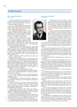
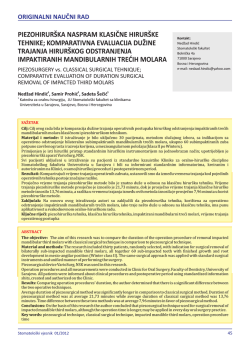
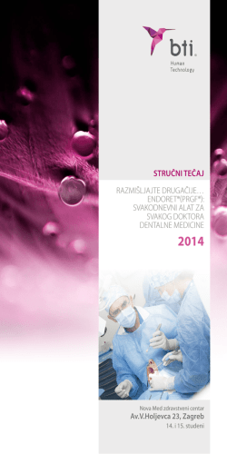
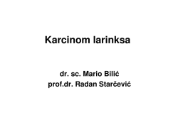
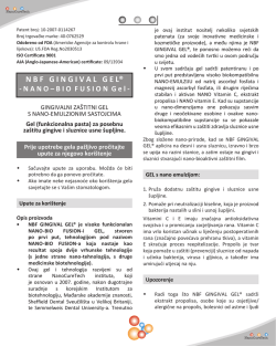
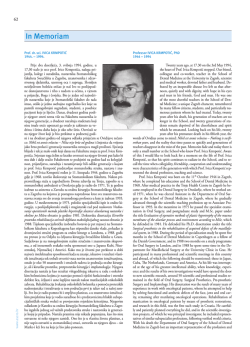
307
ACTA
STOMATOLOGICA
CROATICA
Acta Stomatol Croat. 2012;46(4):307-311.
PRIKAZ SLUČAJA
CASE REPORT
www.ascro.hr
Alaa M. Shuibat1, Emre Bariş1, Begüm Karan2, Tarkan Yetişyiği3
Gingivalni eruptivni hemangiom kao metastaza karcinoma
pluća
Metastasis from Lung Cancer Presenting as Pyogenic
Granuloma in the Lower Gingiva
1
Odsjek za oralnu patologiju Stomatološkog fakulteta Gazijeva Sveučilišta, Ankara, Turska
Gazi University, Faculty of Dentistry Department of Oral Pathology, Ankara-Turkey
2
Odsjek za oralnu maksilofacijalnu kirurgiju Stomatološkog fakulteta Gazijeva sveučilišta, Ankara, Turska
Gazi University, Faculty of Dentistry Department of Oral and Maxillofacial Surgery, Ankara-Turkey
3
Onkološka bolnica Dr. Abdurrahman Yurtaslan, Odjel za medicinsku onkologiju, Ankara, Turska
Dr. Abdurrahman Yurtaslan Oncology Hospital, Department of Medical Oncology, Ankara-Turkey
Sažetak
Metastatski tumori zahvaćaju usnu šupljinu, iako je primarno mjesto metastaza u plućima i prsima. Metastaze karcinoma pluća rijetko se pojavljuju na desnima. Češće su u donjoj čeljusti, odnosno u njezinu posteriornom dijelu. Upravo zato što se rijetko pojavljuju, metastaze u usnoj šupljini teško se dijagnosticiraju i još teže liječe. Vrlo često imaju blage simptome slične zubobolji.
U ovom prikazu opisan je slučaj pacijenta s dijagnozom karcinoma pluća metastaziranim u meka
oralna tkiva. Nakon što su otkrivene gingivalne metastaze, pacijent je upućen na detaljni liječnički pregled i CT-snimanje te su pronađene metastaze u lijevoj ključnoj kosti. Ovaj prikaz pokazuje
koliko je važan stomatološki tim kod dijagnosticiranja metastaza i upućivanja pacijenta na daljnju
obradu. Isto tako treba istaknuti važnost histomorfoloških i imunohistokemijskih tehnika, posebice dijagnosticiranja citokeratina 7, citokeratina 20 i TTF-a 1.
Zaprimljen: 17. kolovoza 2012.
Prihvaćen: 21. studenoga 2012.
Adresa za dopisivanje
Alaa Mohamed Shuibat
Gazi University, Faculty of Dentistry
Department of Oral Pathology
8. Cadde 84. Sokak, 06510
Emek – Ankara – TURKEY
tel: 0090 312 203 4382
faks: 0090 312 223 9226
e-adresa: [email protected]
Uvod
Introduction
Metastaze u usnoj šupljini rijetke su i čine oko jedan posto svih oralnih maligniteta. Glavni izvor svih metastaza čeljusti i okolnih mekih tkiva usne šupljine čine karcinomi.
Njihova najčešća lokacija je posteriorni dio mandibule, meka tkiva usne šupljine, posebice gingiva, sluznica na alveolima i jezik (1). Matastaze na desnima mogu utjecati na funkciju oralnih struktura, govor i prehranu. Adekvatna lokalna
intervencija i liječenje mogu poboljšati prehranu pacijenta i
kvalitetu njegova života (2). U ovom prikazu opisan je pacijent s dijagnosticiranim adenokarcinomom pluća koji je metastazirao na gingivu mandibule u obliku eruptivnog hemangioma.
Metastasis to the oral cavity is uncommon, accounting approximately for 1% of all oral malignancies. Carcinomas are the most common types of metastatic cancer of the
mouth and jaws. However metastases to the jaw bone, especially to the posterior mandible, are more common and oral
soft tissues may be affected, particularly the gingiva, alveolar mucosa and the tongue (1). Gingival metastases can affect oral function, speech and nutrition. Adequate local control and treatment improve nutritional status and quality of
life (2). We report a case of metastatic lung adenocarcinoma
to the mandibular gingiva with clinical appearance similar to
pyogenic granuloma.
Prikaz slučaja
Case report
Pacijent u dobi od 51 godine upućen je u Zavod za oralnu i maksilofacijalnu kirurgiju Gazijeva sveučilišta sa simptomima otečene gingive i krvarenja u donjem lijevom dijelu
mandibule. Na ortopanu se vidjela resorpcija alveolarne kosti
A 51-year-old Turkish male was referred to Gazi University, Faculty of Dentistry, Department of Oral and Maxillofacial Surgery with the main complaint of gingival swelling
and bleeding in the lower left mandibular region. A pan-
www.ascro.hr
Ključne riječi
adenokarcinom; tumori pluća; tumori gingive, sekundarni; metastaze tumora; granulom, piogeni
www.ascro.hr
308
Shuibat i sur.
Metastaze karcinoma pluća na desnima
i uznapredovali parodontitis (slika 1. a). Pacijent se nije žalio
na druge simptome. Submandibularni limfni čvorovi lijeve
strane bili su povećani, bolni i pomični. Oralnim pregledom
ustanovljena je ulcerirana tvorba slična eruptivnom hemangiomu (slika 1. b). Kliničko-patološki nalaz pokazao je da je
bolesniku dijagnosticiran primarni adenokarcinom pluća sedam mjeseci prije promjena na desnima te da je u međuvremenu bio na kemoterapiji (cisplatin + gemcitabin) i terapiji bisfosfonatima.
Tvorba je potpuno uklonjena i poslana na PHD-analizu
u Zavod za oralnu patologiju. Makroskopski se vidjela tvorba veličine 3,5 x 1,5 x 0,9 cm s velikim ulceracijskim površinama. Mikroskopski je uočen infiltrativni alveolarni maligni
tumor ispod ulcerirane oralne sluznice. Histopatološki nalaz
otkrio je stanični pleomorfizam, hiperkromatske jezgre i stanice s više jezgri (slika 2. a). Također su pronađene tumorske
stanice koje su sadržavale citoplazmične eozinofilne globule. Iako nije bilo znakova nekroze, tkivo je sadržavalo mnogo stanica u mitozi (slika 2. b). Tumorske stanice i tubuli iz
uzorka bili su pozitivni na mucin. Imunohistokemijska analiza bila je pozitivna na citokeratin 7 (slika 3. a), citokeratin
20 (slika 3. b), EMA-u (slika 3. c), pankeratin i TTF1 (slika
3. d), ali negativna na vimentin, desmin i s-100.
Ti nalazi pokazuju da je ovaj tumor metastatski adenokarcinom iz pluća. Pacijent je podvrgnut gastroskopiji, kolonoskopiji i CT-u abdomena kako bi se isključio adenokarcinom debelog crijeva. Krajnja dijagnoza za tu gingivalnu
tvorbu bila je metastaza adenokarcinoma pluća. Rendgenska
snimka prsnog koša otkrila je homogenu leziju na lijevom
gornjem plućnom lobusu, uz dijagnozu slabo diferenciranog
bronhioalveolarnog adenokarcinoma koji je prema imunohistokemijskom profilu bio nalik na gingivnu tvorbu.
Nakon postavljene dijagnoze gingivalne tvorbe pacijent
je poslan na daljnje pretrage (MR glave i vrata, CT prsnog
koša i zdjelice, tj. toraksa i pelvisa) te mu je otkrivena još jedna metastatska nakupina u lijevoj ključnoj kosti. Obavljene
su terapije zračenjem i kemoterapija, ali je pacijent nažalost
umro zbog kolapsa respiratornog sustava 70 dana nakon što
mu je bila otkrivena gingivalna metastaza.
oramic radiograph showed severe periodontitis and alveolar
bone resorption of the jaws (Figure 1 A). There were no other
symptoms and no recent history of pain. Left submandibular
lymph nodes were enlarged, tender and mobile, other lymph
nodes were not involved. Oral examination revealed ulcerated gingival mass similar to pyogenic granuloma (Figure 1
B). The clinical-pathological finding indicated that the patient had primary adenocarcinoma of the lung for 7 months
before the appearance of gingival mass. He received chemotherapy (cisplatin + gemcitabine) and bisphosfonate.
The mass was completely excised and sent to the Department of Oral Pathology for histopathological examination. Macroscopically the mass was 3.5 x 1.5 x 0.9 cm, which
showed large areas of ulceration. Microscopically infiltrative
malignant tumor which had an alveolar pattern was observed
under largely ulcerated oral mucosa. Histopathological features showed cellular pleomorphism, nuclear hyperchromatism and clear cells with multiple nucleoli (Figure 2A). It was
also seen that some tumor cells contained intracytoplasmic
eosinophilic globules. Although there was no necrosis of the
tumor, it showed a large number of atypical mitotic figures
(Figure 2B). Mucin positivity was observed in both tumor
cells and duct like spaces. Immunohistochemical evaluation
demonstrated positive findings of cytokeratin-7 (Figure 3A),
cytokeratin-20 (Figure 3B), EMA (Figure 3C), pan-keratin
and TTF1 (Figure 3D), but negative findings of vimentin,
desmin and s-100.
These findings strongly suggest that this tumor was a
metastatic adenocarcinoma from the lung. Despite of that,
the patient underwent gastroscopy, colonoscopy and CT abdomen to exclude large intestine adenocarcinoma. The final
diagnosis of this gingival mass was metastatic adenocarcinoma from the lung. Chest X-ray and pathological reports documented homogeneous lesion on the left upper pulmonary
lobe with diagnosis of low differentiation bronchioloalveolar
adenocarcinoma, which is similar to the pattern and immunohistochemical profile of gingival mass.
After the final diagnosis of the gingival mass, the patient
underwent further investigations (MRI head and neck, CT
chest and pelvis) which detected another metastatic mass in
the left clavicular bone. Radiotherapy and salvage chemotherapy were carried out, but unfortunately the patient died
from respiratory collapse 70 days after the detection of gingival metastasis.
Rasprava
Discussion
Rijetke su udaljene metastaze u predjelu usta (3). Njihovo su najčešće primarno mjesto pluća (4). Hirshberg i suradnici proučili su britansku literaturu o metastazama u usnoj
šupljini i čeljustima od 1916. do 2007. te zaključili da je 112
od 673 metastatske tvorbe posljedica metastaza iz pluća. Na
čeljusnim kostima pronađeno ih je 58, a 54 su nastale na
oralnoj sluznici (1). Od svih metastaza na oralnoj sluznici,
najčešće su one na pričvrsnoj gingivi te na jeziku (1).
Metastaziranje je kompleksni biološki proces u kojem tumorske stanice moraju probiti niz zapreka. Patogeneza oralnih metastaza je nejasna. Prva faza metastaziranja uključuje
Distant metastasis to oral region is a very uncommon
condition (3). The most common primary site was the lung
(4). Hirshberg et al. reviewed the English literature related
to oral cavity and jawbones metastases during the period between 1916 and 2007, and reported that 112 of 673 metastatic tumors were from lung cancer, 58 metastatic cases were
to jaw bones and 54 to oral mucosa (1). In the oral soft tissue, the attached gingiva was the most common affected site
followed by the tongue (1).
Metastasizing is a complex process, the biological basis of
which requires tumor cells to breach a sequence of barriers.
Shuibat et al.
Lung Cancer Metastasis to the Gingiva
309
1
2
3
Slika 1. a: Izrazita resorpcija kosti u maksili u mandibuli vidljiva na ortopantomogramu; b: Klinički izgled ulcerirane gingivne tvorbe na
mandibuli
Figure 1 A: Severe bone resorption in both maxilla and mandible on panoramic radiograph. B: clinical view of ulcerated gingival mass in the
mandible after extraction
Slika 2. a: Infiltrativni maligni tumor ispod oralnog epitela; b: Pleomorfne tumorske stanice s tubularnim strukturama uz mnogobrojne
atipične mitoze (Hematoxylin – Eosin A: x 40, B: x 400)
Figure 2 A: Infiltrative malignant tumor was observed beneath the oral epithelium. B: Pleomorphic tumor cells were constituted of duct like
structures and include number of atypical mitoses. (Hematoxylin-Eosin A: x40, B: x400)
Slika 3. Imunohistokemijski profil tumorskih stanica s citokeratinom 7 (++) (A), citokeratinom 20 (+) (b), EMA-om (+) i TTF-om 1 (+) (d), (ABC,
a: x 400, b: x 200, c: x 200, d: x 200)
Figure 3 Immunohistochemical profile of Tumor cells were cytokeratin 7 (++) (A), cytokeratin 20 (+) (B), EMA (+) (C) and TTF1 (+) (D). (ABC, A:
x400, B: x200, C: x200, D: x200)
www.ascro.hr
4
www.ascro.hr
310
Shuibat i sur.
Metastaze karcinoma pluća na desnima
odvajanje tumorskih stanica od primarnog tumora te njihov
prijenos limfnim ili krvnim žilama. Druga faza je izlazak iz
limfnih/krvnih žila te invazija okolnog tkiva. Jedan od uzroka privlačenja metastatskih stanica na mjesto izlaska iz žile
jest lokalna upala. U ovom slučaju to bi mogla biti upala parodonta. Metastaze se najčešće pojavljuju u mandibuli i to u
premolarnoj regiji (5, 6). Prelazak metastatskih stanica znatno je olakšan prisutnošću adhezivnih molekula. Proteolitički enzimi koji sudjeluju u upali parodonta, kao što su različite kolagenaze i elastaze, razgrađuju ekstracelularni matriks te
pridonose adheziji tumorskih stanica. Ako je dijagnosticiran
maligni tumor, pacijenta treba upozoriti na kvalitetu oralne
higijene te prevenirati upalu parodonta kako bi se smanjila
incidencija oralnih metastaza (7, 8).
Diferencijalna dijagnoza tih invazivnih gingivalnih tvorbi
uključuje odontogene infekcije, eruptivni hemangiom, periferni granulom velikih stanica i periferni osificirajući fibrom.
Biopsija je obvezatna kako bi se postavila konačna dijagnoza
i isključio/potvrdio primarni malignitet (9, 10, 11).
Adenokarcinom je maligni epitelni tumor s diferenciranim žljezdanim tkivom i stanicama koje izlučuju mucin.
Raste na različitim mjestima te postoji acinusni, papilarni, bronhoalveolarni te solidni s izlučivanjem mucina (12).
Bronhoalveolarni rast zabilježen je na plućima i gingivalnoj
tvorbi.
Histološki izgled metastatskih karcinoma znatno varira te
reflektira tip i diferencijaciju tumora. Diferencijalna izraženost citokeratina 7 i 20 te TTF-a 1 može biti korisna u određivanju izvora metastatskog karcinoma. Za plućni adenokarcinom svojstven je nalaz citokeratina (+) i citokeratina 20 (-),
a za adenokarcinom debelog crijeva nalaz citokeratina 7 (-) i
citokeratina (+) (13). Imunohistokemijski profil TTF1 izražen je u dvije trećine adenokarcinoma (14). U ovom slučaju
nalaz citokeratina 7 i citokeratina 20 bio je pozitivan. Zahvaljujući nalazu citokeratina i uz pomoć TTF-a 1, CT-a abdomena i kolonoskopije dijagnosticirana je metastaza plućnog
adenokarcinoma.
Prosječno preživljavanje s dijagnosticiranim karcinomom
pluća i oralnim metastazama jest četiri mjeseca (15). Naš pacijent umro je zbog kolapsa respiratornog sustava 70 dana
nakon što su mu dijagnosticirane oralne metastaze.
Pathogenesis of oral metastasis is unclear. The first stage implies the separation of tumor cells from the primary tumor
and their transport by lymphatic or blood vessels. Secondly,
they cross the vascular wall and invade surrounding tissues.
The role of local inflammation (here periodontal inflammation) in the attraction of metastatic cells has been suggested.
Secondary deposits occur more commonly in the mandible
than the maxilla, the premolar region is the most frequent
site to be affected (5,6).Their passages in gingival tissues
would be facilitated by the greatest permeability of vessels
and the presence of adhesive molecules. Proteolytic enzymes
such as collagenase and elastase which are implied in periodontitis allow degradation of the extracellular matrix by the
tumor cells and support their adhesion. When a malignant
tumor is diagnosed, promotion of oral hygiene and control
of the periodontal inflammation could thus reduce the incidence of oral metastases (7,8).
Differential diagnosis of this rapidly growing gingival
mass includes odontogenic infections, pyogenic granuloma,
peripheral giant cell granuloma and peripheral ossifying fibroma. Biopsy is mandatory for definitive diagnosis and primary malignancy must be considered (9-11).
Adinocarcinoma is a malignant epithelial tumor with
glandular differentiation or mucin production by tumor
cells. Adenocarcinomas grow in various patterns, including
acinar, papillary, bronchoalveolar and solid with mucin formation (12). Bronchoalveolar patterns were observed in both
lung and gingival masses.
The histological appearance of metastatic carcinoma can
be extremely variable, reflecting tumor type and grade of tumor differentiation. Differential expression of cytokeratin 7,
20 and TTF1 may be useful in establishing the origin of a
metastatic carcinoma. While Cytokeratin 7 (+) and Cytokeratin 20 (-) are seen in lung adenocarcinoma, Cytokeratin
7 (-) and Cytokeratin 20 (+) are seen in colon adenocarcinoma (13). TTF1 immunohistochemical profile is expressed by
two thirds of lung adenocarcinomas (14). Both Cytokeratin
7 and 20 positivity were observed in this case; however with
the aid of TTF1, CT abdomen and colonoscopy we confirmed metastasis of lung adenocarcinoma.
The average survival time of lung cancer cases with gingival metastasis was only 4 months (15). Our patient died
from respiratory collapse 70 days after the appearance of gingival mass.
Zaključak
Conclusion
Klinički oralne metastaze karcinoma pluća mogu izgledati kao eruptivni hemangiom. Indicirani su detaljni klinički i histopatološki pregledi. Rano otkrivanje maligne lezije te
pravodobno liječenje znatno utječu na ishod liječenja.
The clinical features of oral metastasis from the lung
mimic reactive gingival lesions such as pyogenic granuloma.
Careful clinical and histopathological examinations are mandatory. Early detection of the type of malignancy and appropriate treatment are of paramount importance to the treatment outcome.
Sukob interesa
Conflict of interest
Autor izjavljuje da nije u sukobu interesa. Za ovaj članak
ni od koga nije dobio novac.
The author declares that he has no conflict of interest. No
funding was taken.
Shuibat et al.
Lung Cancer Metastasis to the Gingiva
Abstract
Metastatic tumors involve the oral cavity, and the most common primary sites are lungs and
breasts. Lung cancer metastasis to the gingiva is quite unusual; metastases to the jaw bone, especially to the posterior mandible are more common. Because of their rarity, metastases to oral
cavity are challenging to diagnose and difficult to treat. They often have vague symptoms that
mimic dental infection. In this paper, a case of a patient with lung cancer, who developed metastasis to the oral soft tissue, is presented. After the detection of gingival metastasis, the patient underwent complete general examination and CT scans which detected another metastasis in the left clavicular bone. The role of dental team in diagnosis and directing the treatment of
these patients is important. On the other hand, the importance of histomorphology and immunohistochemical techniques especially cytokeratin 7, cytokeratin 20 and TTF1 in the final diagnosis
should also be emphasized.
311
Received: August 17, 2012
Accepted: November 21, 2012
Address for correspondence
Alaa Mohamed Shuibat
Gazi University, Faculty of Dentistry
Department of Oral Pathology
8. Cadde 84. Sokak, 06510
Emek-Ankara-Turkey
Tel: 0090 312 203 4382
Fax: 0090 312 223 9226
[email protected]
Key words
Adenocarcinoma; Lung Neoplasms;
Gingival Neoplasms, Secondary;
Neoplasm Metastasis; Granuloma,
Pyogenic
References
9. Perlmutter S, Buchner A, Smukler H. Metastasis to the gingiva.
Report of a case of metastasis from the breast and review of the
literature. Oral Surg Oral Med Oral Pathol. 1974 Nov;38(5):74954.
10. Barr CE, Dym H, Weingarten LA. Metastatic mucous-producing adenocarcinoma of the gingiva. J Am Dent Assoc. 1980
Jul;101(1):53-4.
11. Staalsen NH, Nielsen JS. Bronchogenic metastasis to the gingiva.
Oral Surg Oral Med Oral Pathol. 1992 Nov;74(5):561-2.
12. Kumar V, Abbas AK, Fausto N, Aster JC. Robbins and Cotran pathologic basis of disease. Philadelphia: Saunders Elsevier; 2010. p.
723-4.
13. Campbell F, Herrington CS. Application of cytokeratin 7 and 20
immunohistochemistry to diagnostic pathology. Curr Diagn
Pathol. 2001;7(2):113-22.
14. Kaufmann O, Dietel M. Thyroid transcription factor-1 is the superior immunohistochemical marker for pulmonary adenocarcinomas and large cell carcinomas compared to surfactant proteins A
and B. Histopathology. 2000 Jan;36(1):8-16.
15. Tanaka M, Hiraki A, Ueoka H, Bessho A, Kiura K, Takigawa N et
al. Gingival metastasis in lung cancer. Oncol Rep. 2002 MayJun;9(3):571-4.
www.ascro.hr
1. Hirshberg A, Shnaiderman-Shapiro A, Kaplan I, Berger R. Metastatic tumours to the oral cavity - pathogenesis and analysis of
673 cases. Oral Oncol. 2008 Aug;44(8):743-52.
2. Watanabe E, Touge H, Tokuyasu H, Kawasaki Y. Gingival metastasis of adenocarcinoma from the lung. Respiratory Medicine
CME. 2008;1(2):103-6.
3. Ellis GL, Jensen JL, Reingold IM, Barr RJ. Malignant neoplasms
metastatic to gingivae. Oral Surg Oral Med Oral Pathol. 1977
Aug;44(2):238-45.
4. Pereira CM, de Abreu Alves F, Corrêa ME, Lima CS, de Almeida
OP. Mouth metastasis of peripheral primitive neuroectodermal
tumor. Oral Dis. 2005 Jan;11(1):44-5.
5. Moharil RB, Khandekar S, Dive A. Metastatic lung malignancy to mandibular gingiva. Indian J Dent Res. 2010 JulSep;21(3):449-51.
6. Rajappa S, Loya AC, Rao RD, Rao SI, Surath A, Srihari U. Metastasis to oral cavity: A report of two cases and review of literature.
Indian J Med Pediatr Oncol 2005;26(2):43-6.
7. Lamster IB, Karabin SD. Periodontal disease activity. Curr Opin
Dent. 1992 Mar;2:39-52.
8. Auerbach R, Lu WC, Pardon E, Gumkowski F, Kaminska G, Kaminski M. Specificity of adhesion between murine tumor cells and
capillary endothelium: an in vitro correlate of preferential metastasis in vivo. Cancer Res. 1987 Mar 15;47(6):1492-6.
© Copyright 2026 Paperzz