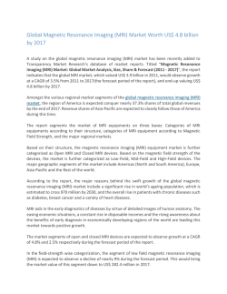
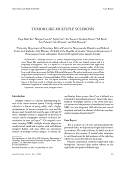
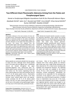
Acta Clin Croat 2010; 49:33-41
Original Scientific Paper
MAGNETIC RESONANCE IMAGING
IN THE DIAGNOSIS OF MALIGNANT TUMORS OF THE
MAXILLOFACIAL REGION
Dijana Podoreški1, Ivan Krolo1, Mirko Ivkić2, Miljenko Marotti1, Vilma Kosović1 and Klaudija Višković 3
1
University Department of Diagnostic and Interventional Radiology, 2University Department of ENT, Head and
Neck Surgery, Sestre milosrdnice University Hospital; 3Department of Radiology and Ultrasonography, Dr. Fran
Mihaljević University Hospital for Infectious Diseases, Zagreb, Croatia
SUMMARY – Magnetic resonance imaging (MRI) is a radiological imaging method that has
not yet found routine application in the detection and assessment of malignant tumors of the maxillofacial region. The aim of this study was to evaluate MRI in the detection, diagnosis and differential diagnosis of malignant tumors of the maxillofacial region. This prospective study included 42
patients with clinically confi rmed malignant tumors of the maxillofacial region. All patients were
examined by MRI. Th is imaging method was evaluated for the ability to detect tumor location
and to analyze dimensions and structure of the tumor, bone involvement, effect on neurovascular
structures and extension to soft tissues. MRI results were compared with histopathologic and intraoperative findings as the ‘gold standard’ methods. MRI identified all of the clinically confi rmed
tumors. The sensitivity for tumor location was 94.4%, tumor necrosis 93%, hemorrhage 93.3% and
bone involvement 91.4%. The specificity for tumor location was 84%, tumor necrosis 92.8%, hemorrhage 92.8% and bone involvement 85.7%. In the evaluation of soft tissue tumor extension to
bone structures, MRI sensitivity and specificity was 94.4% and 88.2%, respectively. The sensitivity
for perineural infi ltration and tumor involvement of vascular structures was 91% and 91.6%, respectively. The specificity for perineural infi ltration was 96.7% and for tumor involvement of vascular
structures 88%. MRI sensitivity and specificity for intracranial extension, intraorbital propagation,
extension to pterygopalatine fossa and other surrounding anatomic spaces was 93.5% and 90.9%,
respectively. MRI proved advantageous for the ability to distinguish neurovascular structures from
adjacent soft tissues without the use of intravenous contrast media. Study results demonstrated MRI
to provide valuable information in the analysis of tumor structure, bone involvement and extension
to soft tissues of malignant tumors of the maxillofacial region.
Key words: Maxillary neoplasms – radiography, diagnosis; Magnetic resonance imaging – utilization;
Neoplasms invasioness – diagnosis
Introduction
Malignant tumors of the maxillofacial region include many different types of tumors. According to
Correspondence to: Dijana Podoreški, MD, University Department of Diagnostic and Interventional Radiology, Sestre milosrdnice University Hospital, Vinogradska c. 29, HR-10000 Zagreb, Croatia
E-mail: [email protected]
Received January 25, 2010, accepted February 26, 2010
Acta Clin Croat, Vol. 48, No. 3, 2009
Book Acta 1-2010.indb 33
the World Health Organization (WHO), head and
neck tumors are anatomically classified into several
groups: nasal cavity and paranasal sinuses, nasopharynx, hypopharynx, larynx and trachea, oral cavity and
oropharynx, salivary glands, odontogenic tumors, and
ear and paraganglionic system1.
Malignant tumors of the mandible and maxilla are
grouped into primary tumors that originate within the
mandible and maxilla and secondary tumors that in33
14.6.2010 9:22:54
Dijana Podoreški et al.
volve the mandible and maxilla secondarily. The most
common malignant tumors of the mandible include
squamous cell carcinoma (SCC) of the oral cavity, i.e.
SCC of the gingival mandible and floor of the mouth
that invade the mandible secondarily2. Metastatic
tumors are not uncommon malignant lesions in the
mandible. Tobacco and alcohol consumption are the
most common predisposing factors2. SCC accounts
for more than 95% of the tumors that arise from oral
mucosa3. In the nasal cavity, 85% of SCC occur in
the lateral wall and floor3. Carcinoma of the septum is
felt to be rare. Eighty percent of paranasal sinus SCC
occur in the maxillary antrum3. Adenoid cystic carcinoma of the sinonasal region is a rare cancer that accounts for 10% of all malignancies at this site4. Sinonasal adenocarcinoma typically arises from the middle
turbinate or ethmoid sinus and less commonly from
the antrum1. Adenocarcinoma occurs predominantly
in men and is associated with wood dust exposure1.
Nearly 20% of malignant melanomas in the human
body occur in the head and neck, and the face represents 3.5% of total body surface5.
About 10% of osteosarcoma cases occur in the head
and neck region, with the majority involving the mandible1. The maxilla is the second most common head
and neck site, however, cases of the maxillary and
ethmoid sinus involvement have also been reported6.
In the mandible, osteosarcoma accounts for about 4%
of all primary malignant tumors7. Bone involvement
shows highest incidence in the second decade of life.
The etiology and precise pathogenesis of the disease
remain unknown7. Chondrosarcoma is a malignant
tumor of cartilage that rarely involves the sinonasal
region8. Esthesioneuroblastoma is a rare, malignant
neoplasm arising from the olfactory neuroepithelium
in the upper nasal cavity1.
Patients and Methods
This prospective study included 42 patients with
clinically confirmed malignant tumors of the maxillofacial region. All patients were examined by magnetic resonance imaging (MRI; Siemens Magnetom
Avanto, 1.5 Tesla, Berlin, Germany) at University
Department of Diagnostic and Interventional Radiology, Sestre milosrdnice University Hospital in Zagreb
between May 2007 and December 2009.
34
Book Acta 1-2010.indb 34
MRI in the diagnosis of malignant tumors
MRI was performed using standard head coil to
obtain T1-weighted images with and without contrast medium and T2-weighted images in axial, coronal and sagittal planes; T1-weighted images with fat
saturation and contrast media (FS-T1) and Short Tau
Inversion Recovery T2-weighted images (STIR-T2)
in coronal planes. For T1-weighted images the field
of view (FOV) was 210-230 mm and for T2-weighted
images and STIR-T2 it was 130-200 mm, with matrix
size of 256x256 pixels. Section thickness varied from
1 to 3 mm and each patient received the paramagnetic
contrast medium, gadolinium-diethylenetriamine
penta-acetic acid (Gd-DTPA) in a dose of 0.1 mmol/
kg. Data were archived in picture archiving and communication systems (PACS).
The images were evaluated for tumor location and
dimensions. We analyzed structural homogeneities
and inhomogeneities, and detected zones of necrosis
and hemorrhage. Bone involvement analysis included
osteosclerosis, osteolysis, cortical expansion, and cortical thinning or destruction. Soft tissue tumors were
analyzed for their extension to cortical bone and bone
marrow. Tumor extension to soft tissues was considered as involvement of the adjacent neurovascular
structures, fat tissue, muscles and anatomic spaces.
MRI findings were compared with histopathologic and intraoperative findings as the ‘gold standard’
methods.
The specificity and sensitivity were analyzed by
statistical processing of the results. Sensitivity was defined as the ability of MRI to detect the presence of
pathology and was calculated as follows19:
Sensitivity = ((True Positive/(True Positive + False
Negative)) x 100.
Specificity was defined as the ability of imaging to
detect the absence of pathology and was calculated as
follows:
Specificity = ((True Negative/(True Negative +
False Negative)) x 100.
Results
Tumor pathology was found in 18 female and 24
male patients, mean age 62.3 and 57.3 years, respectively. The youngest patient was a 20-year-old man
with osteosarcoma of the mandible, and the oldest
was an 80-year-old man with adenocarcinoma of the
nasal cavity. The smallest tumor measured 15 mm
Acta Clin Croat, Vol. 49, No. 1, 2010
14.6.2010 9:22:55
Dijana Podoreški et al.
MRI in the diagnosis of malignant tumors
Table1. Histopathologic findings of the tumors (N=42)
Histopathology
Sublingual SCC
SCC of maxillary sinus
SCC of ethmoid sinus
SCC of gingival maxilla
SCC of gingival mandible
SCC of gingival maxilla and hard palate
Adenocarcinoma of nasal fossa
Osteosarcoma of mandible
Malignant melanoma
Number of patients
6
5
2
7
9
2
5
5
1
%
14.3
11.9
4.8
16.6
21.4
4.8
11.9
11.9
2.4
SCC = squamous cell carcinoma
and the largest tumor measured 65 mm in longest diameter, yielding a mean tumor diameter of 34 mm.
Histopathologic findings of the tumors are shown in
Table 1.
In the evaluation of tumor location, MRI showed
94.4% sensitivity and 84% specificity. Tumor location
was presented in sagittal scan, on a T1-weighted image
(Fig. 1) (sinonasal adenocarcinoma located in the nasal fossa, typically arising from the middle turbinate).
On measuring tumor height and width, the absolute
Fig. 1. Sagittal contrast-enhanced T1-weighted image
shows sinonasal adenocarcinoma located in the nasal fossa,
typically arising from the middle turbinate (white arrows).
Acta Clin Croat, Vol. 49, No. 1, 2010
Book Acta 1-2010.indb 35
range of deviation was 4 mm (minimum – 2 mm and
maximum 2 mm). Study results showed a limitation
of MRI on measuring the height and width of tumor
lesion. The main reasons were smaller artifact disturbances from dental restorative materials. On measuring tumor depth, the absolute range of deviation was
14 mm (minimum – 3 and maximum 11). The heavy
horizontal artifacts were the reason for MRI limitation on evaluating tumor depth. In one patient, the
measurement error was 11 mm.
MRI analysis of tumor structure showed structural homogeneities in seven patients. Tumors with
zones of necrosis were found in 26 patients, whereas
20 patients had tumors with zones of hemorrhage,
which was in accordance with histopathologic and intraoperative findings. The sensitivity and specificity of
MRI on detecting necrotic zones was 93% and 92.8%,
and for hemorrhage 93.3% and 92.8%, respectively.
Thirty seven patients had tumors originating in
soft tissue structures, including SCC of the oral cavity
(n=6), SCC of the gingival maxilla (n=7), SCC of the
gingival mandible (n=9), SCC of the gingival maxilla
and hard palate (n=2), SCC of the maxillary and ethmoid sinuses (n=7), sinonasal adenocarcinoma (n=5)
and malignant melanoma (n=1). We analyzed cortical and bone marrow extension of the tumor. MRI
detected the extension of soft tissue tumors to bone
structures with 94.4% sensitivity and 88.2% specificity. Five patients had osteosarcoma of the mandible.
Bone involvement analysis included osteosclerosis,
osteolysis, cortical expansion, and cortical thinning
or destruction. MRI sensitivity for bone involvement
was 91.4% and specificity 85.7% (Figs. 2-4).
35
14.6.2010 9:22:55
Dijana Podoreški et al.
MRI in the diagnosis of malignant tumors
Fig. 2. Axial T2-weighted image shows destruction of the
mandible, interrupted corticalis and intramedullary tumor extension (white arrows). Histopathology confirmed
ulcerative squamous cell carcinoma of the gingival mandible.
Fig. 4. Axial contrast-enhanced T1-weighted image
shows osteosarcoma of the mandible. Inhomogeneous well
contrast-enhanced tumor mass with osteosclerosis, interrupted cortical bone and decrease in the signal of the bone
marrow, which appears grey because of its involvement by
the tumor (white arrows).
Fig. 3. Sagittal contrast-enhanced T1-weighted image
shows malignant melanoma with a zone of necrosis (black
arrow). Cutaneous and subcutaneous fat tissue, facial and
perioral muscles are infiltrated by the tumor (arrows). The
bone and cartilage structures of the nose are destroyed.
36
Book Acta 1-2010.indb 36
Fig. 5. Coronal contrast-enhanced T1-weighted image
shows squamous cell carcinoma of ethmoid cells. Inhomogeneous tumor mass without intracranial extension is seen
(white arrows). Intraorbital tumor propagation pressing
medial orbicular muscle is shown (empty white arrow).
Acta Clin Croat, Vol. 49, No. 1, 2010
14.6.2010 9:22:55
Dijana Podoreški et al.
MRI in the diagnosis of malignant tumors
a
b
Fig. 6. Squamous cell carcinoma of the maxillary sinus: (a) coronal contrast-enhanced T1-weighted image showing
tumor extension to the left nasal fossa, ethmoid cells and intraorbital propagation with dislocation of inferior orbicular
muscle and optic nerve (white arrows); (b) axial contrast-enhanced T1-weighted image shows suppressed of carotid arteries due to tumor propagation to the carotid space (empty white arrow). The tumor spread to the buccal, parapharyngeal
and pharyngeal spaces and pterygopalatine fossa (white arrows), and to bone structure of the maxilla and hard palate
(black arrow).
The extension of maxillofacial malignant tumor to
neurovascular structures, fat tissue, muscles and anatomical spaces by MRI showed positive findings in 39
patients, which corresponded well with the intraoperative and histopathologic findings. On the analysis of
perineural infi ltration, MRI showed 91% sensitivity
and 96.7% specificity. Tumor involvement of vascular
structures was found in 22 patients, yielding a 91.6%
specificity and 88% sensitivity. MRI showed 93.5%
sensitivity and 90.9% specificity in the evaluation of
intracranial extension, intraorbital propagation, and
extension to pterygopalatine and infratemporal fossa
and other surrounding anatomic spaces (Figs. 5-9).
Discussion
Fig. 7. Axial contrast-enhanced T1-weighted image shows
squamous cell carcinoma of the gingival maxilla and hard
palate. Involvement of branches of the superior alveolar
nerve by the tumor (black arrows) is shown, which caused
facial pain on the left side.
Acta Clin Croat, Vol. 49, No. 1, 2010
Book Acta 1-2010.indb 37
In daily practice at our hospital, we use multisclice
computed tomography (MSCT) in the analysis of
malignant tumors of the maxillofacial region, which
is complementary with MRI9. MSCT acquisition is
much faster with multislice scanners, between 1-3
minutes including contrast administration. MSCT
study is less time consuming than MR examination.
37
14.6.2010 9:22:56
Dijana Podoreški et al.
MRI in the diagnosis of malignant tumors
Fig. 8a
Fig. 8b
Fig. 8. Relapse of maxillary sinus squamous cell carcinoma
20 years after irradiation and surgery for tongue cancer:
(a, b) coronal T1-weighted image with fat saturation
and sagittal contrast-enhanced T1 weighted image show
perineural infiltration through inferior orbital fissure
(white arrows). Pterygomaxillary space and pterygopalatine fossa are involved by the tumor (empty white arrow).
Thickened cranial dura mater is well contrast-enhanced
after irradiation (left-up white arrow); (c) coronal STIR
image shows high signal in mastoid cells (empty white arrow) due to perineural infiltration of mandibular nerve
and dysfunction of motor tensor muscle of tympanic membrane.
Fig. 8c
38
Book Acta 1-2010.indb 38
On our MR machine, the average time is half an hour.
The quality of MR image can be compromised by long
time of examination due to swallowing artifacts.
In a study reported by Belkin et al., persistent
limitation in the head and neck MSCT analysis was
caused by metal artifacts from dental restorative material10. Artifacts caused by metallic objects such as dental crowns, dental implants and metallic orthodontic
appliances are a common problem in head and neck
MRI11. We had five patients with these artifacts. The
patients with dentures that can be taken out, or patients without metal dental fi llings are not affected by
these problems and analysis was much better.
Because of a slightly superior spatial resolution of
MSCT, early cortical involvement by tumor was often more easily seen by MSCT than by MRI12. The
MSCT evaluation of marrow involvement required
recognition of a subtle increase in bone marrow density12. In the present study, the high signal involvement
of cortical bone and inhomogeneous decrease of bone
marrow signal pointed to the diagnosis of bone infi ltration (Figs. 2 and 4). Our MRI results were sensitive
for both medullary and cortical bone involvement.
Several authors compared MRI and MSCT in the
preoperative evaluation of osteosarcoma and primary
bone tumors12. They agree that MRI is superior to
MSCT for determining tumor margins towards normal adjacent muscle, while MSCT is considered superior for the evaluation of subtle cortical changes and
identification and characterization of mineralized tuActa Clin Croat, Vol. 49, No. 1, 2010
14.6.2010 9:22:56
Dijana Podoreški et al.
MRI in the diagnosis of malignant tumors
Fig. 9b
Fig. 9a
mor matrix12. In their ability to determine intramedullary tumor extent, MRI and MSCT are usually
comparable, but generally, MSCT will define better
the margins of tumors that are highly sclerotic, and
MRI can be preferable for detection of osteolytic lesions13. In the evaluation of bone involvement, MSCT
has advantages in tumor osteosclerosis detection,
which is a well known limitation of MRI13.
Fig. 9. Highly aggressive sinonasal undifferentiated carcinoma: (a, b) coronal contrast-enhanced T1 weighted images show tumor extent to the masticator space, masticatory muscles and lymph nodes of the head and neck (black
arrows). Perineural infiltration along the third division
of the trigeminal nerve, through oval hole involving the
left cavernous sinus, (white arrow). Denervation atrophy
of the masticatory muscles due to perineural infiltration
(white arrow); (c) coronal contrast-enhanced T1 weighted
image shows perivascular involvement of the internal
carotid artery in the neck and cavernous part (white arrows). Blood vessel was easily identified as a black area
of the signal void, ‘flow void’ phenomenon (empty white
arrow).
Acta Clin Croat, Vol. 49, No. 1, 2010
Book Acta 1-2010.indb 39
MRI had great effect on the assessment and staging of soft tissue masses14. As the imaging modality
of choice for assessment of oropharyngeal, mouth and
tongue soft tissue masses, MRI has shown superiority
to MSCT due to better soft tissue contrast15. The ex-
Fig. 9c
39
14.6.2010 9:22:57
Dijana Podoreški et al.
tent of tumor invasion into the mandible and maxilla
and further extension into the adjacent face and neck
spaces and structures are causes of special concerns
and may lead to poor prognosis16. Involvement of the
pterygopalatine fossa and masticator space enables invasion along the neurovascular bundles into the cavernous sinus via maxillary and mandibular nerves16.
MRI showed excellent presentation of tumor extension into anatomical spaces (Figs. 5-9).
MRI improved soft tissue visualization without the
use of intravenous contrast medium, and distinguishes
blood vessels and nerves from adjacent soft tissue and
tumor lesions17. Blood vessel was easily identified as
a black area of signal void, ‘flow void’ phenomenon17.
In spite of this MRI advantage, in our study we used
paramagnetic intravenous contrast media to achieve
better contrast resolution.
MRI demonstrated advantage compared to
MSCT in the evaluation of neurovascular structures
from adjacent soft tissues18. MRI is more sensitive and
specific than MSCT in detecting perineural spread19.
MRI excellently presented perineural spread and vascular involvement caused by tumor (Figs. 7-9).
Appropriate assesment of the extent of tumor infi ltration to the surrounding bone, neurovascular and
other soft tissue structures is critical for proper planning of surgical treatment, which is often mutilating
for the patient. The value of our study is to help surgeons chosse an appropriate therapeutic optin
Conclusion
Malignant tumors of the maxillofacial region are
relatively rare, but, for radiologists, their differentiation
and evaluation is not easy because many different types
of tumors are present in this region. In our study that
comprised 42 patients we analyzed the possibilities of
MRI in the detection, diagnosis and differential diagnosis of malignant tumors of the maxillofacial region.
MRI results were compared with intraoperative
and histopathologic findings and were statistically
analyzed. We determined the specificity of MRI in
assessing tumor localization (84%), establishing the
zone of necrosis (92.8%) and hemorrhage (92.8%),
bone involvement (85.7%), perineural infi ltration
(96.7%), involvement of vascular structures (88%),
and tumor spread to the bone (88.2%) and soft tissue
40
Book Acta 1-2010.indb 40
MRI in the diagnosis of malignant tumors
structures and anatomic spaces (90.9%). The sensitivity of MRI was as follows: localization 94.4%, necrotic
zones 93%, hemorrhage 93.3%, perineural infiltration
91%, involvement of vascular structures 91.6%, bone
involvement 91.4%, extension of soft tissue tumors to
bone structures 94.4%, and extension of soft tissue
tumors to the surrounding soft tissue and anatomic
spaces 93.5%.
MRI is a relatively new radiological imaging
method that has not yet found routine application in
the detection and assessment of malignant tumors of
the maxillofacial region. The results of our study confirmed the high value of MR detection, differentiation and assessment of maxillofacial region malignant
tumors. At our hospital, MRI has been established in
preoperative treatment in some patients and our intention is to introduce MRI as a routine preoperative
examination in all patients with tumors of the maxillofacial region.
References
1. BARNES L, EVESON JW, REICHART P, SIDRANSKY
D. World Health Organisation classification of tumors. Pathology & Genetics. Head and neck tumors. Lyon: International Agency for Research on Cancer Press, 2005.
2. WEBER AL, BUI C, KANEDA T. Malignant tumors of the
mandible and maxilla. Neuroimaging Clin North Am 2003;
13:509-24.
3. RAJESH A, KHAN A, KENDALL C, HAYTER J,
CHERRYMAN G. Can magnetic resonance imaging replace
single photon computed tomography and computed tomography in detecting bony invasion in patients with oral squamous
cell carcinoma. Br J Oral Maxillofac Surg 2008;46:11-4.
4. LUPINETTI AD, ROBERTS DB, WILLIAMS MD,
KUPFERMAN ME, ROSENTHAL DI, DeMONTE F, et al. Sinonasal adenoid cystic carcinoma. Cancer
2007;110:2726-31.
5. LARSON DL, LARSON JD. Head and neck melanoma.
Clin Plast Surg 2010;37:73-3.
6. FERNANDES R, NIKITAKIS GN, PAZOKI A, ORD
AR. Osteogenic sarcoma of the jaw: a 10-year experience.
Am Assoc Oral Maxillofac Surg 2007;65:1286-91.
7. CHINDIA LM. Osteosarcoma of the jaw bones. Oral Oncol
2001;37:545-7.
8. YEO JK, SOO AI, GYE-YEON L, HO JC, HYUN JP,
MIN SK, et al. Myxoid chondrosarcoma of the sinonasal cavity in a child: a case report. Korean J Radiol 2007;8:452-5.
9. CHAVDA VS, OLLIFF JFC. The sinuses. In: SUTTON D,
ed. Textbook of radiology and imaging. Vol 2, 7th rev. ed. Fali
grad: Elsevier, Churchill Livingstone, 2003;1519-29.
Acta Clin Croat, Vol. 49, No. 1, 2010
14.6.2010 9:22:58
Dijana Podoreški et al.
MRI in the diagnosis of malignant tumors
10. BELKIN AB, PAPAGEORGE BM, FAKITSAS J,
BANKOFF M. A comparative study of magnetic resonance
imaging versus computed tomography for the evaluation of
maxillary and mandibular tumors. J Oral Maxillofac Surg
1988;46:1039-47.
11. COSTA A, APPENZELLER S, YASUDA C, PEREIRA
F, ZANARDI V, CENDES F. Artifacts in brain magnetic
resonance imaging due to metallic dental objects. Med Oral
Pathol Oral Cir Bucal 2009;20:Pub Med PMID:19300374.
12. ZIMMER WD, BERQUIST TH, Mc LEOD RA, SIM
FH, PRITCHARD DJ, SHIVES TC, et al. Bone tumors:
magnetic resonance imaging versus computed tomography.
Radiology 1985;155:709-18.
13. AISEN AM, MARTEL W, BRAUNSTEIN EM, Mc
MILLIN KI, PHILLIPS WA, KLING TF. MRI and CT
evaluation of primary bone and soft-tissue tumors. Am J Radiol 1986;146:749-56.
14. Mc ENERY KW, MURPHY WA. Magnetic resonance imaging. In: RESNICK D, ed. Bone and joint imaging. 2nd ed.
Philadelphia: Saunders, 1996; 84-92.
16. KIMURA Y, SUMI M, ARIJI Y, ARIJI Y, NAKAMURA
T. Deep extension from carcinoma arising from the gingiva: CT and MR imaging features. AJN Am J Neuroradiol
2002;23:468-472.
17. WILLIAMS LS, MANCUSO AA, MENDENHALL
WA. Perineural spread of cutaneous squamous and basal cell
carcinoma: CT and MR detection and its impact on patient
management and prognosis. Int Radiat Oncol Biol Phys
2001;49:1061-9.
18. GINSBERG LW. MR imaging of perineural tumor spread.
Magn Reson Imaging Clin North Am 2004;14:663-77.
19. HANNA E, VURAL E, PROKOPAKIS E, CARRAU
R, SNYDERMAN C, WEISSMAN J. The sensitivity and
specificity of high-resolution imaging in evaluating perineural spread of adenoid cystic carcinoma to the skull base. Arch
Otolaryngol Head Neck Surg 2007;133:541-5.
20. HARNSBERGER HC, HUDGINS PA, WIGGINS RH,
HUDGINS PA, MICHEL MA, DAVIDSON HC, et al.
Diagnostic imaging: head and neck. 1st ed. Canada grad?:
Amirsys, 2008.
15. KASSEL EE, KELLER MA, KUCHARCZYK W. MRI
of the floor of the mouth, tongue and orohypopharynx. Radiol Clin North Am 1989;27:331-51.
Sažetak
MAGNETSKA REZONANCIJA U DIJAGNOSTICI ZLOĆUDNIH TUMORA MAKSILOFACIJALNOG
PODRUČJA
D. Podoreški, I. Krolo, M. Ivkić, M. Marotti, V. Kosović i K. Višković
Magnetska rezonancija (MRI) je radiologijska slikovna metoda koja još uvijek nije pronašla rutinsku primjenu u otkrivanju i procjeni malignih tumora maksilofacijalnog područja. Svrha ove studije je bila pokazati vrijednost MRI u otkrivanju, dijagnostici i diferencijalnoj dijagnostici malignih tumora maksilofacijalnog područja. Ova prospektivna studija
uključila je 42 bolesnika s klinički potvrđenim zloćudnim tumorom maksilofacijalnog područja. Svi bolesnici su bili obrađeni pomoću MRI. Analizirala se lokalizacija, dimenzije i struktura tumora, zahvaćenost koštanih struktura, utjecaj na
neurovaskularne strukture, proširenost na okolne mekotkivne strukture i prostore. Rezultati su se uspoređivali s patohistologijskim i intraoperacijskim nalazom kao „zlatnim“ standardom. MRI je prikazao sve klinički potvrđene tumore. U procjeni lokalizacije tumora osjetljivost je bila 94,4%; zone nekroze 93%; zone krvarenja 93,3%; te promjene koštane strukture
91,4%. U procjeni lokalizacije tumora specifičnost je bila 84%; zone nekroze 92,8%; zone krvarenja 92,8%; te promjene
koštane strukture 85,7%. U procjeni zahvaćenosti koštanih struktura mekotkivnim tumorskim procesom osjetljivost MRI
je bila 94,4%, a specifičnost 88,2%. Osjetljivost kod procjene perineuralne infi ltracije je bila 91%, a utjecaja tumora na vaskularne strukture 91,6%. Specifičnost kod procjene perineuralne infi ltracije je bila 96,7%, a utjecaja tumora na vaskularne
strukture 88%. MRI je pokazao osjetljivost od 93,5% i specifičnost od 90,9% za intrakranijsku i intraorbitalnu propagaciju,
prodor u pterigopalatinalnu udubinu i druge okolne anatomske prostore. Prednost MRI je u sposobnosti razlikovanja neuralnih i vaskularnih struktura bez primjene intravenskog kontrastnog sredstva. Naši rezultati su potvrdili vrlo važnu ulogu
MRI u procjeni strukture tumora, koštanih promjena i proširenosti tumora u okolne strukture i prostore.
Ključne riječi: Novotvorine maksile – dijagnostika; Novotvorine maksile – radiografija; Magnetska rezonancija – upotreba;
Invazivnost novotvorine – dijagnostika
Acta Clin Croat, Vol. 49, No. 1, 2010
Book Acta 1-2010.indb 41
41
14.6.2010 9:22:58
© Copyright 2026 Paperzz