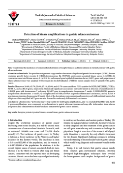
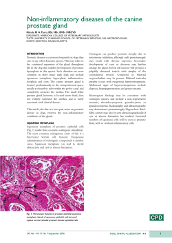
Kocatepe Tıp Dergisi
Kocatepe Medical Journal
2014;15(3):345-8
CASE PRESENTATION / OLGU SUNUMU
Two Different Giant Pleomorphic Adenoma Arising from the Palate and
Parapharyngeal Space
Damak ve Parafarengeal Bölgeden Kaynaklanan Farklı İki Dev Pleomorfik Adenom Olgusu
Abdulkadir BUCAK1, Şahin ULU1, Mustafa Said TEKİN1, Emre KAÇAR2, Orhan Kemal KAHVECİ1,
Nurten HAKTANIR3, Abdullah AYÇİÇEK1
1
Afyon Kocatepe University Faculty of Medicine, Department of Otorhinolaryngology, Afyonkarahisar
2
Afyon Kocatepe University Faculty of Medicine, Department of Radiology, Afyonkarahisar
3
Afyon Kocatepe University Faculty of Medicine, Department of Plastic and Reconstructive Surgery, Afyonkarahisar
Geliş Tarihi / Received: 11.09.2013
ABSTRACT
Pleomorphic adenoma is the most common of minor salivary gland tumors. Pleomorphic adenoma of minor salivary
glands are commonly located on the palate nevertheless
rarely may originate from the parapharyngeal region. Pleomorphic adenomas on the palate show slow growth over
the years. The tumors that originate from the
parapharyngeal region could reach great dimensions. In this
case report, an orphan and neglected geriatric patient who
had giant pleomorphic adenoma on the palate and a young
patient with a giant pleomorphic adenoma that was excised
by transcervical approach without additional surgical procedures at the parapharyngeal region, were presented.
Keywords: Pleomorphic adenoma; minor salivary gland;
palate; parapharyngeal space.
Kabul Tarihi / Accepted: 06.11.2013
ÖZET
Minör tükürük bezi kaynaklı tümörler arasında en sık görülen pleomorfik adenomdur. Minör tükürük bezi kaynaklı
pleomorfik adenom en sık damakta görülüp, parafarengeal
bölgede nadiren karşımıza çıkar. Damak pleomorfik adenomları yıllar içinde yavaş büyüme gösterirler.
Parafarengeal bölgeden kaynaklananlar büyük boyutlara
ulaşabilirler. Bu makalede kimsesiz ve bakımsız kalmış
geriatrik bir hastada damak bölgesinde yerleşik oldukça
büyük boyutlara gelmiş pleomorfik adenom olgusu ile genç
bir hastada yine oldukça büyük boyutlara ulaşmış ve ek
cerrahi prosedüre ihtiyaç kalmadan transservikal yaklaşımla
çıkartılabilmiş plemorfik adenom olgusu sunuldu.
Anahtar Kelimeler: Pleomorfik adenom; minör tükrük bezi;
sert damak; parafarengeal bölge.
INTRODUCTION
Salivary glands are commonly divided into two groups
as major and minor. Although minor salivary glands
exist at the plate with the highest rate, a substantial
amount are located throughout the upper
respiratory-digestive tract submucosal layer (1).
Salivary gland tumors constitute 3 % of all head and
neck tumors (2). The tumors originating from the
minor salivary gland rare cases and constitute only 10
to 25 % of all salivary gland tumors. Pleomorphic
adenoma (PA) (or benign mixt tumor) is the most
common minor salivary gland tumor and approximately constitute the 65 % of all salivary gland
tumors, the 40 % of minor salivary gland tumors. It
can be seen at the palate and parapharyngeal region,
as it may occur in the nasal cavity, paranasal sinuses
Yazışma Adresi / Correspondence: Abdulkadir BUCAK, M.D.
Department of Otorhinolaryngology, Afyon Kocatepe University
Faculty of Medicine Afyonkarahisar [email protected]
and larynx. Most of the patients with PA that
originate from minor salivary glands are between
ages 40-60 years and a slight female dominance has
been detected (1). PA is mixed tumor including both
epithelial and mesodermal elements. The epithelial
cell islets and cords are available in the myxoid
stroma. PA has a thin and incomplete capsule and
extensions termed pseudopods. Pseudopods are
considered to be responsible for the postoperative
recurrence. If the capsule is torn or an insufficient
resection size is done during the operation, a residual
tissue or implantation may occur (3). In this study,
two cases were presented. One of them, a homeless
and neglected geriatric patient was brought to our
clinic by the home care service stuff, who had giant
pleomorphic adenoma on his palate and other one, a
young patient with a giant pleomorphic adenoma at
the parapharyngeal region.
346
Bucak et al.
CASE PRESENTATION- 1
Seventy-eight-year-old male patient was brought to
the clinic by home care service stuff. The detailed
anamnesis from patient was not possible due to
massive dementia and no accessible collateral kin.
The main complaints of nursing home practitioners
were spoilt of speech fluency, feeding difficulty and
snoring and a giant mass in-mouth. On the physical
examination, immobile, non-pulsatile, painless mass,
covered with broken mucosa, medium-hard in consistency, with an approximately 6x4x3 cm in size, was
detected on right paramedian location in hard-plate
(Figure I). There was no lymphadenopathy in the
neck. At the computerized tomography (CT) scanning,
solid mass which was depleting to palatine bone but
not invading was detected. Following, the mass was
removed en bloc with the hard palate periosteum by
intraoral approach. The patient's definitive
pathological result was reported as PA. At the early
postoperative period, the complaints related to mass
disappeared. The wound healing was completed at
the end of a month. There was no recurrence for
three months. But, the patient died a cardiovascular
disorder in the postoperative forth month.
Figure I: Giant pleomorphic adenoma in the hardplate.
Figure III: Heterogeneous hypodense solid mass at
the T1 weighted magnetic resonance imaging.
CASE PRESENTATION- 2
Figure II: Giant pleomorphic adenoma in the left
parapharyngeal region.
Twenty-nine-year-old male patient was referred to
our clinic with gradual increasing complaints in the
throat for a year, such as painless swelling, dysphagia,
snoring, sleep apnea, and respiratory distress. The
patient had no history of systemic disease. On the
physical examination, a mass which was pushing the
soft palate and the tonsilla palatina medially was
detected at the left parapharyngeal region. It had a
smooth surface, hard consistency and was not giving
pulsation on the bimanual palpation. The size of the
mass was approximately 6x4 cm in size (Figure II).
There was no lymphadenopathy in the neck. Cranial
nerve functions were normal. At the magnetic
resonance imaging (MRI) of the neck; heterogeneous
hypodense solid mass, approximately 7x6x4 cm in
size, starting from the neighborhood of inferior level
Kocatepe Tıp Dergisi 2014;15(3):345-8
Kocatepe Tıp Dergisi, Cilt 12 No:3, Eylül 2011
347
Two Different Giant Pleomorphic Adenoma
İki Farklı Dev Pleomorfik Adenom Olgusu
of pterygoid plates superiorly, extending to the left
submandibular gland medial neighborhood at the
caudal and causing narrowing in the airway column
were determined at the left parapharyngeal region
(Figure III). The mass were removed en bloc with
transcervical approach without additional surgical
procedures, under general anesthesia. There were no
complications of vascular and neural tissues. The
patient's definitive pathological result was reported
as PA. There is no recurrence for one year, at the
patient.
DISCUSSION
PA is a benign tumor in terms of both appearance and
behaviour but in proportion to the residence time in
the body can show malignant transformation to 1,69,4 % , if it exists for a long time without treatment
(4).
PA is the most common minor salivary gland tumor and most commonly originates from palate,
uncommonly seems on the parapharyngeal region
(5). Parapharyngeal tumors rarely emerge and constitute to 0,5 % of all head and neck tumors (6). The 80
% of them are benign and PA is most common of all
parafarengeal tumors (7). Parapharyngeal space is in
the form of an inverted pyramid, is divided into two
by the styloid process of temporal bone, termed
prestyloid and poststyloid space (8, 9). Adipose tissue, lymph nodes and the deep lobe of parotid gland
are located in the prestyloid region and the carotid
artery, internal jugular vein, IX., X., XI. and XII. cranial
nerves, sympathetic chain and lymph nodes located
in the poststyloid region (9, 10). Masses in the
poststyloid region including neurogenic tumors,
paragangliomas, vascular tumors and aneurysms have
usually benign character (11). Many different types of
mass can be seen in the prestyloid region. The most
common tumor seen in this region is PA, originated
from the deep lobe of parotid gland (12).
Parapharyngeal region tumors were classified according to the localization of the carotid artery in some
publications. Tumors pushing the carotid artery posteriorly called prestyloid and tumors pushing anteriorly called poststyloid (8). Our case had pleomorphic
adenoma located in the prestyloid region and pushing
the carotid artery posteriorly. Parapharyngeal region
tumors don’t give symptom until reaching the sufficient size, so early diagnosis cannot usually be possi-
ble (6). Symptoms usually begin to appear when the
tumor size is on 2,5-3,0 cm (13). Foreign body sensation in the throat, difficulty in swallowing, otological
symptoms, volume change and trismus are the main
symptoms (8). Conversely, the tumors of the palate
including PA early create awareness and rarely reach
a size larger than 1-2 cm. To our opinion, the tumor
had reached to 6 cm size in patient 1 due to
neglection as he had dementia. He had been cured
following the attention home care service. In such a
tumor in this patient can affect the survival due to
obstruction of feeding.
Salivary gland tumors are usually diagnosed by
history, physical examination, histopathological and
radiological assessment. CT and MRI provide useful
information about the extension to superficial and
deep tissues, size and placement of the tumor. While,
CT is superior in detecting the bony structure about
the hard plate, MRI has a higher resolution for soft
tissue, so is superior in evaluation of parapharyngeal
space tumors. Another important issue is the
evaluation of the fat plan between the deep lobe of
parotid gland and the tumor at parapharyngeal space.
The lesions of the deep portion of the parotid gland
extending to the parapharyngeal region and the lesions originating directly from the parapharyngeal
region can be discriminated with this feature (14).
Following the preoperative diagnostic process,
the fine needle aspiration biopsy can be prefered to
the transoral open biopsy which can cause vascular
damage on the parapharyngeal region tumors (7).
The various surgical approaches to the treatment of
tumors of the parapharyngeal region have been identified.
Transoral,
transcervical,
transcervicaltransmandibuler,
transcervical-transparotid
approaches are most frequently performed surgical
approaches. The surgical approach which provides
the maximum field of view of the tumor for complete
removal, causing minimum functional and aesthetic
disadvantage should be preferred (15). In our case, it
was predicted that the complete excision of the tumor by transoral approach, might be difficult due to
the dominance of anatomical structures and additional surgical procedures such as mandibulectomy
might be required to reach the mass. However, we
have reached completely to the mass by transcervical
approach and excised the submandibular gland and
no additional procedures were required.
Kocatepe Tıp Dergisi 2014;15(3):345-8
Kocatepe Tıp Dergisi, Cilt 12 No:3, Eylül 2011
348
Bucak et al.
REFERENCES
1. Kuo YL, Tu TY, Chang CF, et al. Extra-major salivary
gland pleomorphic adenoma of the head and neck: a
10-year experience and review of the literature. Eur
Arch Otorhinolaryngol 2011;268(7):1035-40.
9. Shirakura S, Tsunoda A, Akita K, et al.
Parapharyngeal space tumors: anatomical and image
analysis findings. Auris Nasus Larynx 2010;37(5):6215.
2. Van der Wal JE, Snow GB, Van der Wall I.
Histological reclassification of 101 intraoral salivary
gland tumors. J Clin Pathol 1992;45(9):834-5.
10. Work WP, Hybels RL. A study of tumors of the
parapharyngeal
space.
Laryngoscope
1974;84(10):1748-55.
3. Shaaban H, Bruce J, Davenport PJ. Recurrent
pleomorphic adenoma of the palate in a child. Br J
Plast Surg 2001;54(3):245-7.
11. Papadogeorgakis N, Petsinis V, Goutzanis L,
Kostakis G, Alexandridis C. Parapharyngeal space
tumors: surgical approaches in a series of 13 cases.
Int J Oral Maxillofac Surg 2010;39(3):243-50.
4. Keller AZ. Residence, age, race and related factors
in the survival and associations with salivary tumors.
Am J Epidemiol 1969;90(4):269-77.
5. Hakeem AH, Hazarika B, Pradhan SA, Kannan R.
Primary pleomorphic adenoma of minor salivary
gland in the parapharyngeal space. World J Surg
Oncol 2009;7(11):85.
6. Batsakis JG, Sneige N. Parapharyngeal and
retropharyngeal space diseases. Ann Otol Rhinol
Laryngol 1989;98(4):320-1.
7. Kesse KW, Howlett DC, Manjaly G. An unusual case
of dysphagia: ectopic salivary gland pleomorphic
adenoma of the parapharyngeal space. J Otolaryngol
2002;31(3):193-5.
12. Avitia S, Hamilton JS, Osborne RF. Deep- lobe
parotid tumor presenting in the parapharyngeal
space. Ear Nose Throat J 2007;86(12):730-1.
13. Khafif A, Segev Y, Kaplan DM, Gil Z, Fliss DM.
Surgical management of parapharyngeal space
tumors: a 10- year review. Otolaryngol Head Neck
Surg 2005;132(3):401-6.
14. Polat Ş, Serin GM, Öztürk O, Üneri C. Pleomorphic
adenomas recurrences within the parapharyngeal
space. J Craniofac Surg 2011;22(3):1124-8.
15. Dimitrijevic MV, Jesic SD, Mikic AA, Arsovic NA,
Tomanovic NR. Parapharyngeal space tumors: 61 case
reviews. Int J Oral Maxillofac Surg 2010;39(10):983-9.
8. Giddings CE, Bray D, Rimmer J, Williamson P.
Pleomorphic adenoma and severe obstructive sleep
apnoea. J Laryngol Otol 2005;119(3):226-9.
Kocatepe Tıp Dergisi 2014;15(3):345-8
Kocatepe Tıp Dergisi, Cilt 12 No:3, Eylül 2011
© Copyright 2026 Paperzz