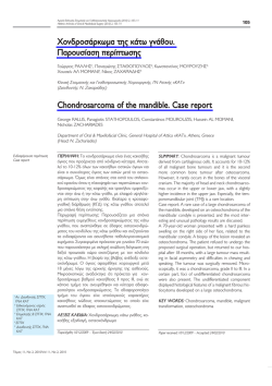
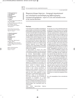
Αρχεία Ελληνικής Στοματικής και Γναθοπροσωπικής Χειρουργικής (2011) 2, 113-120
Hellenic Archives of Oral & Maxillofacial Surgery (2011) 2, 113-120
113
Νευρειλήμμωμα προωτιαίας χώρας σε παιδιατρικό
ασθενή. Παρουσίαση περίπτωσης
Ιωάννης ΙΑΤΡΟΥ1, Διονύσιος ΦΩΤΟΠΟΥΛΟΣ2, Νάντια ΘΕΟΛΟΓΗ-ΛΥΓΙΔΑΚΗ3, Αικατερίνη ΜΙΧΑΗΛ4
Κλινική Στοματικής και Γναθοπροσωπικής Χειρουργικής, Οδοντιατρική Σχολή Πανεπιστημίου Αθηνών
(Διευθυντής: Καθηγητής Κ. Αλεξανδρίδης)
στο Νοσοκομείο Παίδων «Π & Α Κυριακού» (Υπεύθυνος: Αναπλ. Καθηγητής Ι. Ιατρού)
Eργαστήριο Παθολογοανατομίας Νοσοκομείου Παίδων «Π & Α Κυριακού» (Διευθύντρια: Α. Μιχαήλ)
Preauricular facial nerve neurilemmoma in a paediatric
patient. A Case report
Iοannis IATROU, Dionisios FOTOPOULOS, Nadia THEOLOGIE-LYGIDAKIS, Ekaterini MICHAIL
Oral & Maxillofacial Surgery Clinic. Dental School, University of Athens (Head: Professor C. Αlexandridis)
“P and A Kyriakou” Children’s Hospital (Director: Associate Professor I. Iatrou)
Histopathology Department, Children’s Hospital (Head: A. Michail)
Eνδιαφέρουσα περίπτωση
Case report
ΠΕΡΙΛΗΨΗ: Το νευρειλήμμωμα ή σβάννωμα είναι καλοήθης όγκος του νευρικού ιστού. Συνήθως είναι ασυμπτωματικός και περιβάλλεται από κάψα. Προέρχεται
από τα κύτταρα του ελύτρου του Schwann των νεύρων
που πολλαπλασιαζόμενα σχηματίζουν τον όγκο και
απωθούν τους νευράξονες στην περιφέρεια.
Σκοπός της εργασίας είναι η παρουσίαση μιας περίπτωσης νευρειλημμώματος της προωτιαίας χώρας σε
ασθενή 13 ετών.
Περιγραφή περίπτωσης: Πρόκειται για κορίτσι 13 ετών
με σφαιροειδή όγκο προωτιαίας χώρας διαμέτρου
4cm, σχετικά ευκίνητο, με σύσταση ελαστική και ανώδυνο. Μετά την βιοψία που κατέδειξε καλοήθεια και
πιθανή νευρογενή προέλευση ή οζώδη περιτονιίτιδα,
έγινε χειρουργική αφαίρεση του όγκου με προωτιαία
προσπέλαση, σε υγιή όρια και χωρίς διεγχειρητικές ή
μετεγχειρητικές επιπλοκές. Ιστολογικά η βλάβη ήταν
συμβατή με νευρειλήμμωμα.
Το αξιοσημείωτο στην προκειμένη περίπτωση είναι η
σπανιότητα σε σχέση με τη φύση και τη θέση του
όγκου αλλά και η ηλικία της ασθενούς.
ΛΕΞΕΙΣ ΚΛΕΙΔΙΑ: Νευρειλήμμωμα, σβάννωμα, προσωπικό νεύρο, παιδική ηλικία.
1
Αναπλ. Καθηγητής ΣΓΠΧ,
ΕΚΠΑ
2
ΣΓΠΧ, Υπ. Διδάκτορας
3
Λέκτορας ΣΓΠΧ, ΕΚΠΑ
4
Διευθύντρια ΕΣΥ, Παθολογοανατόμος
Παρελήφθη: 11/04/2011 - Έγινε δεκτή: 15/05/2011
Τόμος 12, Νο 2, 2011/Vol 12, No 2, 2011
SUMMARY: Neurilemmomas, also known as schwannomas, are benign tumours of neurogenic origin. They
are usually asymptomatic encapsulated tumours, composed of Schwann nerve sheath cells, which multiply
and form the tumour, pushing the axons aside.
The aim of this paper is to present a case of neurilemmoma of the preauricular region in a 13-year-old
patient, and review the relevant literature.
Case report: A 13-year-old female patient presented
with a round, elastic and relatively mobile painless
swelling in the left preauricular region, which was 4cm
in diameter. Biopsy results indicated a benign tumour of
neurogenic origin or a nodular fasciitis, which was then
removed surgically through a preauricular incision, in
healthy margins and without any intra- or postoperative
complications. Histological examination of the lesion
revealed a neurilemmoma (of ancient type).
The tumour’s rare origin and location, as well as the
patient’s young age make this case noteworthy.
KEY WORDS: Neurilemmoma, Schwannoma, facial
nerve, childhood
Paper received: 11/04/2011 - Accepted: 15/05/2011
114
Ιατρού Ι. και συν./Iatrou Ι. et al.
ΕΙΣΑΓΩΓΗ
INTRODUCTION
Οι καλοήθεις όγκοι νευρογενούς προέλευσης είναι
σπάνιοι και διακρίνονται σε νευρειλημμώματα, νευροϊνώματα και εκδηλώσεις της νευροϊνωμάτωσης (Kaban
και Troulis, 2004). Το νευρειλήμμωμα ή σβάννωμα είναι καλοήθης όγκος ενώ αναφέρεται ότι μπορεί να
εμφανίσει και κακοήθη εξαλλαγή (McMenamin και
Fletcher, 2001).
Το νευρειλήμμωμα αρχικά περιγράφηκε από τον
Verocay το 1910 ο οποίος και διατύπωσε την άποψη
ότι είναι όγκος που προέρχεται από τα κύτταρα του
ελύτρου του Schwann που πολλαπλασιαζόμενα σχηματίζουν τον όγκο και απωθούν τους νευράξονες στην
περιφέρεια (Samet και συν. 1985).
Κλινικά εμφανίζεται ως ασυμπτωματικός, ευκίνητος και
συνήθως μικρού μεγέθους όγκος ο οποίος καλύπτεται
από φυσιολογικό βλεννογόνο ή δέρμα. Αναπτύσσεται
σε άτομα κάθε ηλικίας ενώ η περιοχή κεφαλής και τραχήλου αποτελεί συνήθη θέση εντόπισης (Bochlogyros
και συν. 1992, Αγγελόπουλος και συν. 2000, Leu και
Chang, 2002). Ιστολογικά τα νευρειλημμώματα περιβάλλονται από κάψα ινώδους συνδετικού ιστού. Αποτελούνται από κύτταρα του Schwann τα οποία οργανώνονται σε δύο κυτταρικά πρότυπα, τους τύπους
Antoni–A και Antoni–B. Στον πρώτο παρατηρούνται
πυκνές δέσμες επιμήκων κυττάρων με ατρακτοειδείς
πυρήνες που διατάσσονται πασαλοειδώς σε παράλληλες σειρές. Οι στοίχοι κυττάρων αφορίζουν μία ακύτταρη περιοχή με λεπτές ίνες κολλαγόνου και κυτταροπλασματικές προσεκβολές με αναδιπλώσεις της κυτταρικής μεμβράνης. Οι σχηματισμοί αυτοί αποτελούν τα
σωμάτια Verocay. Στον τύπο Antoni– B παρατηρείται
μικρότερος αριθμός κυττάρων τα οποία διατάσσονται
ακανόνιστα σε υπόστρωμα μυξωματώδους σύστασης
που περιέχει κυστικούς σχηματισμούς και αυξημένο
αριθμό αγγείων. Η μορφολογία των κυττάρων αλλά και
του στρώματος ενίοτε αποκλίνει από αυτή που παρουσιάζουν τα τυπικά σβαννώματα και έτσι αναδεικνύονται
διάφοροι ιστολογικοί τύποι όπως κυτταροβριθές,
αρχέγονο, αδενικό, δικτυωτό, πεπαλαιωμένο (:ancient)
κ.α. (Samet και συν. 1985, Kumar και συν. 1996, Αγγελόπουλος και συν. 2000).
Σκοπός της εργασίας είναι να περιγραφεί μια περίπτωση νευρειλημμώματος σε ασθενή 13 ετών, με εντόπιση
στην προωτιαία χώρα και να γίνει μία σύντομη βιβλιογραφική ανασκόπηση.
Benign tumours of neurogenic origin are rare and can
be divided into neurilemmomas, neurofibromas and
manifestations of neurofibromatosis (Kaban and
Troulis, 2004). A neurilemmoma, also known as
schwannoma, is a benign tumour, which can reportedly transform into a malignant tumour (McMenamin and
Fletcher, 2001).
Neurilemmomas were first described in 1910 by Verocay, who expressed the opinion that they are tumours
composed of Schwann sheath cells, which multiply and
form the tumour, pushing the axons aside (Samet et al.
1985).
Clinically, they present as asymptomatic, elastic and
usually small tumours, covered with normal mucosa or
skin. They can occur in individuals of any age, while the
head and neck region is the most frequent site
(Bochlogyros et al. 1992, Aggelopoulos et al. 2000, Leu
and Chang, 2002). Histologically, neurilemmomas are
surrounded by a fibrous connective tissue sheath. They
consist of Schwann cells, organised in two cell patterns:
the Antoni–A and Antoni–B types. In the first, densely
packed elongated cells can be observed, with spindleshaped nuclei, which are aligned in a parallel manner,
producing a palisading appearance. These cell rows
outline an acellular area occupied by thin collagen fibers
and cytoplasmic projections, composed of reduplicated
basement membrane. These formations are the socalled Verocay bodies. In the Antoni–B type, there is a
smaller number of cells, scattered within a myxomatous
stroma, containing cystic formations and an increased
number of vessels. In some cases, the cell and stroma
morphology diverges from typical schwannomas,
resulting in different histological variants such as hypercellular, primitive, glandular, plexiform, ancient etc.
schwannomas (Samet et al. 1985, Kumar et al. 1996,
Aggelopoulos et al. 2000).
The aim of this paper is to present a case of neurilemmoma of the preauricular region in a 13-year-old
patient, and to briefly review the relevant literature.
ΠΕΡΙΓΡΑΦΗ ΠΕΡΙΠΤΩΣΗΣ
Η ασθενής Θ.Κ., 13 ετών, παραπέμφθηκε στην κλινική
Στοματικής και Γναθοπροσωπικής Χειρουργικής από
συναδέλφους άλλης ειδικότητας (ΩΡΛ) για περαιτέρω
διερεύνηση διόγκωσης στην αριστερή προωτιαία χώρα
και τελική χειρουργική αντιμετώπιση. Είχε προηγηθεί
CASE REPORT
A 13-year-old patient (initials: Th.K.) was referred to the
Clinic of Oral and Maxillofacial Surgery by an ENT specialist to have a swelling in the left preauricular region
further examined and surgically treated. This was preceded by a biopsy, during which the removal of a small
tissue sample caused significant bleeding that led to the
discontinuation of the procedure. Histological examination suggested the possibility of nodular fasciitis, without
excluding the possibility of a tumour of neurogenic origin, while, due to the bleeding that had been noticed
before, a vascular tumour was also considered possible.
Αρχεία Ελληνικής Στοματικής & Γναθοπροσωπικής Χειρουργικής/
Hellenic Archives of Oral and Maxillofacial Surgery
Νευρειλήμμωμα προωτιαίας χώρας/Preauricular facial nerve neurilemmoma
βιοψία κατά την οποία, αφού λήφθηκε μικρό ιστοτεμάχιο, παρατηρήθηκε έντονη αιμορραγία με αποτέλεσμα
τη διακοπή της διαδικασίας. Η ιστολογική εξέταση ανέφερε πιθανότητα οζώδους περιτονιίδος χωρίς όμως να
αποκλείει νευρογενή προέλευση του όγκου ενώ λόγω
της προηγηθείσας αιμορραγίας δεν αποκλειόταν και η
πιθανότητα αγγειακού όγκου.
Κλινικά η διόγκωση ήταν σφαιροειδής, ελαστικής σύστασης, ευκίνητη και ανώδυνη διαμέτρου περίπου 4cm
(Εικ. 1-2). Η λειτουργικότητα του προσωπικού νεύρου
ήταν φυσιολογική.
Ο προεγχειρητικός έλεγχος περιελάμβανε αιματολογικό και απεικονιστικό έλεγχο με μαγνητική τομογραφία
για επακριβή καθορισμό των ορίων και του μεγέθους
της βλάβης (Εικ. 3).
Η ασθενής υπό γενική αναισθησία υποβλήθηκε σε ολική εξαίρεση του όγκου. Η προσπέλαση έγινε με προωτιαία τομή παρωτιδεκτομής, με υπογνάθια επέκταση.
Ακολούθησε προσεκτική παρασκευή και αποκόλληση
του όγκου από τους υποκείμενους ιστούς χωρίς σημαντική δυσκολία καθώς απαιτήθηκε μόνο η παρασκευή
και προστασία του βυκανητικού κλάδου του προσωπι-
Εικ. 1,2: Κλινική εικόνα διόγκωσης στην προωτιαία χώρα.
Fig. 1,2: Clinical pictures of the preauricular swelling.
Εικ. 4: Διεγχειρητική εικόνα.
Fig. 4: Surgical approach.
Τόμος 12, Νο 2, 2011/Vol 12, No 2, 2011
115
Clinically, the swelling was spherical, elastic, mobile and
painless, measuring approximately 4cm in diameter (Fig.
1-2). The functionality of the facial nerve was normal.
Preoperative examination included blood tests and CTscans, in order to determine the lesion’s exact limits
and size (Fig. 3).
Under general anesthesia, the patient underwent total
tumour resection, through a preauricular parotidectomy incision with a submandibular extension. The tumour was then carefully dissected and removed from
the adjacent tissues, without particular difficulty, since
only the buccal branch of the facial nerve, at the root
exit, had to be dissected and protected from the splitting of the central branch. Intraoperatively, the great
auricular nerve was exposed and dissected. After a thin
strip of skin was removed at the margins of the incision,
corresponding to the tumour, the wound was sutured
in layers, and a vacuum drain was fixed in place (Fig. 45). The specimen was sent for histological examination.
Right after the operation, the function of the facial
nerve was normal, and the patient’s postoperative
course was free of complication. Five years postopera-
Εικ. 3: Προεγχειρητική μαγνητική τομογραφία.
Fig. 3: Preoperative MRI.
Εικ. 5: Η συρραφή.
Fig. 5: Suturing of tissues
116
Ιατρού Ι. και συν./Iatrou Ι. et al.
Εικ. 6-8: Η ασθενής άμεσα μετεγχειρητικά χωρίς συμπτώματα πάρεσης του προσωπικού νεύρου.
Fig. 6-8: Immediately postoperative images of the girl without any symptoms of facial nerve paresis.
κού νεύρου στην έκφυσή του από το διχασμό του
κεντρικού στελέχους. Διεγχειρητικά έγινε αποκάλυψη
και παρασκευή του μείζονος ωτιαίου νεύρου. Αφού
αφαιρέθηκε στενή λωρίδα δέρματος στα όρια της
τομής αντίστοιχα με τον όγκο, ακολούθησε η συρραφή του τραύματος κατά στρώματα και η τοποθέτηση
παροχέτευσης κενού (Εικ. 4-5). Το παρασκεύασμα
στάλθηκε για ιστολογική εξέταση.
Άμεσα μετεγχειρητικά η λειτουργία του προσωπικού
νεύρου ήταν φυσιολογική ενώ η μετεγχειρητική πορεία
υπήρξε ομαλή. Πέντε χρόνια μετά την επέμβαση η
ασθενής δεν παρουσιάζει κανένα πρόβλημα στην
περιοχή της επέμβασης ενώ παρακολουθείται τακτικά
(Εικ. 6-8).
Ιστολογικά ο όγκος είχε εξαιρεθεί πλήρως και είχε διαστάσεις 5x4x3 cm. Περιοριζόταν από σαφή ινώδη
κάψα με παρουσία μικρού αριθμού λεμφοκυττάρων και
αναπτυσσόταν εν τω βάθει του χορίου και του υποδόριου λιπώδους ιστού. Ο όγκος παρουσίαζε περιφερειακά ατρακτοειδή κύτταρα με συχνά κεκαμμένο πυρήνα
και μετρίως ηωσινόφιλο κυτταρόπλασμα. Τα κύτταρα
διατάσσονταν σε διαπλεκόμενες ή συνεστραμμένες
δέσμες χωρίς συγκεκριμένο πρότυπο ανάπτυξης, ενώ
κεντρικότερα οι κυτταρικές αθροίσεις έχαναν τη συνοχή τους με αύξηση του μυξωματώδους υποστρώματος
χωρίς να παρουσιάζεται εικόνα ατυπίας, πολυμορφία ή
παρουσία μιτώσεων (Εικ. 9). Η ανοσοϊστοχημική μελέτη έδειξε έντονη παρουσία αντιγόνων έναντι αντισωμάτων VIM, SMA & S-100pr, ενώ αρνητική ήταν η απάντηση στους δείκτες HHF–35, Desmin GFAP, CD 57,
CD 34 ,CD 31, Internexin A1, Ker AE1 & EMA. Η ανωτέρω μορφολογία σε συνδυασμό με τον ανοσοϊστοχημικό έλεγχο συνηγορούσε με τη νευρογενή προέλευση του όγκου και ήταν συμβατή με νευρειλήμμωμα,
πεπαλαιωμένο ως προς τον ιστολογικό τύπο.
Εικ. 9: Παρατηρούνται ατρακτοειδή κύτταρα χωρίς εικόνα ατυπίας,
πολυμορφίας ή μιτώσεων.
Fig. 9: Bands of spindle shaped cells without any mitotic figures,
cellular pleomorphism or atypia.
tively, the site of the operation is healthy, and the
patient receives regular follow-ups (Fig. 6-8).
Histologically, the tumour was completely removed
and measured 5x4x3 cm. It was surrounded by a distinctive fibrous capsule, where a small number of lymphocytes were present, and had been developing deep
within the dermis and subcutaneous fat tissue. The
tumour had spindle-shaped cells at its periphery, which
in many cases had curved nuclei, and a mildly
eosinophilic cytoplasm. The cells were arranged in
interlacing or twisted bundles, without a specific pattern of development, while, more centrally, the cell
clusters were less cohesive, with increased myxomatous stroma and without features of atypia, diversity or
presence of mitoses (Fig. 9). The immunohistochemical
study revealed a significant presence of antigens against
VIM, SMA & S-100pr antibodies, while there was a negative response to the following indices: HHF–35,
Desmin GFAP, CD 57, CD 34, CD 31, Internexin A1,
Αρχεία Ελληνικής Στοματικής & Γναθοπροσωπικής Χειρουργικής/
Hellenic Archives of Oral and Maxillofacial Surgery
Νευρειλήμμωμα προωτιαίας χώρας/Preauricular facial nerve neurilemmoma
ΣΥΖΗΤΗΣΗ
Τα νευρειλημμώματα είναι ασυμπτωματικοί, ευκίνητοι,
βραδέως αυξανόμενοι, συνήθως καλοήθεις και σπανίως κακοήθεις όγκοι νευρογενούς προέλευσης. Στη
στοματογναθοπροσωπική περιοχή αναπτύσσονται σε
οποιοδήποτε από τα κρανιακά ή νωτιαία νεύρα (Leu
και Chang, 2002, Cunningham και Warner, 2003, Kaban
και Troulis, 2004).
Η περιοχή κεφαλής και τραχήλου αποτελεί τη συνήθη
περιοχή εντόπισής τους, με περίπου το ένα τρίτο των
αναφερόμενων περιπτώσεων να παρουσιάζεται σε
αυτή (Leu και Chang, 2002). Ειδικότερα μπορεί να
παρατηρηθούν στην περιοχή του προσώπου, του εγκεφαλικού κρανίου, του οφθαλμικού κόγχου, της ρινικής
και στοματικής κοιλότητας και του λάρυγγα
(Bochlogyros και συν. 1992, Shah, 2003). Όσον αφορά
στην στοματική κοιλότητα, συχνότερα ανευρίσκονται
στη γλώσσα, την υπερώα, τις παρειές κ.α. (Αγγελόπουλος και συν. 2000). Τα νευρειλημμώματα σπανίως μπορεί να εμφανιστούν και ενδοοστικά, συνοδευόμενα
από πόνο και παραισθησία της αντίστοιχης περιοχής
(Marzola και συν. 1988, Bochlogyros και συν. 1992).
Λίγες είναι οι περιπτώσεις στη διεθνή βιβλιογραφία
όπου γίνεται αναφορά σε νευρειλημμώματα που
παρουσιάζονται ως διογκώσεις στην παρωτιδική περιοχή, κυρίως από κλάδους του προσωπικού νεύρου.
Ακόμη πιο σπάνιες είναι οι αναφορές σε παιδιατρικούς
ασθενείς, όπως και στη δική μας περίπτωση, όπου και
αναφέρονται συνήθως ως μεμονωμένα περιστατικά
(Elahi και συν. 1995, Kumar και συν. 1996, Malpuech και
συν. 1997, Gupta και συν. 1997, Jayaraj και συν. 1997,
Maly και συν. 2003, Wu και συν. 2008).
Ο Kun και συν. (1993) σε σειρά 49 ασθενών με νευρειλήμμωμα στην περιοχή του τραχήλου και της στοματογναθοπροσωπικής περιοχής, αναφέρουν μόνο
δύο περιστατικά σε παιδιατρικούς ασθενείς, με εντόπιση ενδοκογχική και κροταφική. Ο Kumar και συν.
(1996) δημοσιεύουν για πρώτη φορά στην βρετανική
βιβλιογραφία περίπτωση νευρειλημμώματος προσωπικού νεύρου στην παρωτίδα σε παιδιατρικό ασθενή. Ο
Malpuech και συν. (1996) αναφέρουν επίσης μία περίπτωση εξάχρονου ασθενούς με νευρειλήμμωμα στην
παρωτιδική περιοχή. Ο Gupta και συν. (1997) αναφέρουν περίπτωση εννιάχρονου ασθενούς με νευρειλήμμωμα στον υποκροτάφιο βόθρο και επέκταση στο
ηθμοειδές οστό με κλινική εικόνα όγκου στο πρόσωπο.
Ο De Campora και συν. (1999) αναφέρουν την εμπειρία τους από την αντιμετώπιση 35 νευρογενών όγκων
σε παιδιά ηλικίας 8 έως 16 ετών. Εξ αυτών οι 26 ήταν
νευρειλημμώματα στην περιοχή κεφαλής και τραχήλου.
Τέλος ο Wu και συν. (2008) αναφέρουν επίσης περίπτωση δεκάχρονου ασθενούς με νευρειλήμμωμα παρειάς.
Μία τέτοια μεμονωμένη περίπτωση νευρειλημμώματος
Τόμος 12, Νο 2, 2011/Vol 12, No 2, 2011
117
Ker AE1 & EMA. The morphology described above,
together with the immunohistochemical results, indicated a tumour of neurogenic origin and was compatible
with neurilemmoma of ancient type.
DISCUSSION
Neurilemmomas are asymptomatic, mobile, slowlygrowing tumours of neurogenic origin, which are usually benign and less frequently malignant. In the maxillofacial region, neurilemmomas can develop in any of the
cranial or spinal nerves (Leu and Chang, 2002,
Cunningham and Warner, 2003, Kaban and Troulis,
2004).
The head and neck region is the most common site for
neurilemmomas, accounting for almost one-third of all
reported cases (Leu and Chang, 2002). More precisely,
they might be located in the face, cranial cavity, orbit,
nasal and oral cavities, and larynx (Bochlogyros et al.
1992, Shah, 2003). In the oral cavity, they are usually
located in the tongue, palate, cheeks etc. (Aggelopoulos et al. 2000). Rarely, neurilemmomas can also
occur intraosseously, accompanied by pain and paraesthesia of the adjacent area (Marzola et all. 1988,
Bochlogyros et all. 1992).
In the international bibliography, only a small number of
cases are reported, in which neurilemmomas present as
swellings in the parotid region, originating mainly from
branches of the facial nerve. Paediatric patients are
even fewer, and are usually reported as individual cases
(Elahi et al. 1995, Kumar et al. 1996, Malpuech et al.
1997, Gupta et al. 1997, Jayaraj et al. 1997, Maly et al.
2003, Wu et al. 2008).
In a series of 49 patients with neurilemmomas of the
neck and the oral-maxillofacial regions, Kun et al.
(1993) reported only two paediatric patients, with
intraorbital and temporal locations. In the British literature, Kumar et al. (1996) were the first to report a case
of intraparotid facial nerve neurilemmoma in a paediatric patient. Malpuech et al. (1996) also reported a
case of a 6-year-old patient with an intraparotid
neurilemmoma. Gupta et al. (1997) reported a case of
a 9-year-old patient with a neurilemmoma in the subtemporal fossa, extending to the ethmoid bone, with
the clinical appearance of a facial tumour. De Campora
et al. (1999) also reported their experience from treating 35 neurogenic tumours in children between 8 and
16 years of age. Twenty-six of them were neurilemmomas in the head and neck region. Last, Wu et al.
(2008) reported a case of a 10-year-old patient with a
neurilemmoma of the cheek.
The case presented in this paper is also an individual
case of facial nerve neurilemmoma in a young female
patient. The location of the swelling in the preauricular
area did not provide clear indications about the
118
του προσωπικού νεύρου σε νεαρή ασθενή, είναι και
αυτή που περιγράφεται στην παρούσα εργασία. Η
εντόπιση της διόγκωσης στην προωτιαία χώρα δεν
καθόριζε σαφώς και την προέλευση του όγκου. Διεγχειρητικά αποδείχθηκε ότι ο όγκος ήταν σε επαφή με
τον βυκανητικό κλάδο του προσωπικού νεύρου, ο
οποίος και παρασκευάστηκε. Εν τούτοις δεν μπορεί να
αποκλεισθεί περίπτωση να εκπορευόταν ο όγκος από
άλλον αισθητικό κλάδο της περιοχής.
Σύμφωνα με την βιβλιογραφία, τα νευρειλημμώματα
αναπτύσσονται σε άτομα κάθε ηλικίας σχεδόν, με μέσο
όρο ηλικίας τα 44,2 έτη και με το 55,1 % των ασθενών
να βρίσκονται στην τρίτη με τέταρτη δεκαετία της ζωής
(Bochlogyros και συν. 1992, Kun και συν. 1993). Η
περίπτωση που παρουσιάστηκε αφορά ασθενή 13 ετών, ηλικία που αναφέρεται και στη διεθνή βιβλιογραφία.
Γενικά δεν παρατηρείται προτίμηση ως προς το φύλο,
παρά το ότι αναφέρεται κάποια διαφορά ως προς την
εντόπιση των όγκων. Στην περιοχή του τραχήλου
εμφανίζεται συχνότερα στις γυναίκες (2/1), ενώ στην
στοματογναθοπροσωπική περιοχή παρατηρείται προτίμηση στο ανδρικό φύλο (2,6/1) (Kun και συν. 1993).
Παρά την παραπάνω σχέση φύλου / εντόπισης που
αναφέρεται στη βιβλιογραφία η νεαρή ασθενής της
εργασίας μας εμφάνιζε τη διόγκωση στην προωτιαία
χώρα.
Η σπανιότητα εμφάνισης νευρολειμμωμάτων σε παιδιατρικούς ασθενείς μπορεί να οδηγήσει σε διαγνωστικά προβλήματα. Η διαγνωστική προσέγγιση εκτός από
ένα λεπτομερές ιστορικό και κλινική εξέταση, περιλαμβάνει εργαστηριακές και απεικονιστικές εξετάσεις
κυρίως μαγνητική τομογραφία για απεικόνιση των
μαλακών μορίων (Shimizu και συν. 2005). Η βιοψία δια
αναρροφήσεως με λεπτή βελόνη (FNA) είναι μια διαγνωστική μέθοδος που χρησιμοποιείται ευρύτατα τα
τελευταία χρόνια, ιδιαίτερα για το διαχωρισμό μεταξύ
φλεγμονώδους και νεοπλασματικής βλάβης (Papadogeorgakis και συν. 2000, Maly και συν. 2003, Ιατρού και
συν. 2004). Στην προκειμένη περίπτωση δεν χρησιμοποιήθηκε, παρά το γεγονός ότι ο όγκος ήταν εύκολα
προσπελάσιμος για FNA, διότι με την αρχική, έστω
ανεπαρκή βιοψία είχε ήδη αποκλειστεί κακοήθεια αλλά
και η μικρή ασθενής είχε ταλαιπωρηθεί αρκετά. Για την
τελική διαγνωστική διερεύνηση εξ άλλου, τα τελευταία
χρόνια σημαντική βοήθεια παρέχει και ο ανοσοϊστοχημικός έλεγχος.
Η διαφορική διάγνωση των νευρειλημμωμάτων στην
στοματογναθοπροσωπική περιοχή είναι αρκετά δύσκολη περιλαμβάνοντας βλάβες φλεγμονώδους αιτιολογίας, όπως σιαλαδενίτιδα αλλά και νεοπλάσματα καλοήθη ή και κακοήθη αδενικής και μεσεγχυματικής
προέλευσης, όπως πλειόμορφα αδενώματα, αιμαγγειώματα, τα οποία αποτελούν και τους συχνότερα εμφανιζόμενους καλοήθεις όγκους της παιδικής ηλικίας, νευ-
Ιατρού Ι. και συν./Iatrou Ι. et al.
tumour’s origin. Intraoperatively, it was proved that the
tumour contacted the buccal branch of the facial nerve,
which had to be carefully dissected. However, the possibility that the tumour could have arisen from another
aesthetic branch of the area cannot be excluded.
According to the relevant literature, neurilemmomas
can occur in individuals of any age, with an average age
of 44.2 years and 55.1% of the patients being in the
third to fourth decade of life (Bochlogyros et al. 1992,
Kun et al. 1993). The case presented here involves a
13-year-old patient, an age which is also reported in the
international literature.
Generally, no preference regarding gender has been
observed, although some differences in location are
reported. In the neck region, neurilemmomas occur
more often in women (2/1), while in the oral-maxillofacial region they are more common in men (2.6/1)
(Kun et al. 1993). Despite this relation between gender
and location that is reported in the literature, our
young patient had a swelling in the preauricular area.
The rare incidence of neurilemmomas in paediatric
patients might lead to diagnostic problems. Besides
detailed medical history-taking and clinical examination,
the diagnostic approach also includes laboratory and
imaging tests, mainly CT-scans, to obtain images of the
soft tissues (Shimizu et al. 2005). Fine needle aspiration
(FNA) biopsy has been used widely in the recent years,
especially to distinguish between inflammatory and
neoplastic lesions (Papadogeorgakis et al. 2000, Maly et
al. 2003, Iatrou et al. 2004). In the case here presented,
it was not used despite the fact that the tumour was
easily accessible by FNA, because the initial, rather
inadequate biopsy had excluded the possibility of a
malignancy, and the young patient had already suffered
enough. Besides, in the recent years, immunohistochemical studies have proved to be a significant tool to
establish the final diagnosis.
Differential diagnosis of neurilemmomas in the oralmaxillofacial region is quite difficult, including lesions of
inflammatory origin, such as glandular infections, as well
as benign neoplasms or even malignant neoplasms of
lymphoid and mesenchymal origin, for example pleomorphic adenomas, hemangiomas, which are the most
frequently occurring benign tumours in childhood, neurofibromas, adenoid cystic carcinomas, lymphomas,
metastatic tumours etc. (Iatrou et al. 2004, Kaban and
Troulis, 2004).
Surgical resection of the tumour is the treatment of
choice. In most cases, the tumour does not damage the
nerve itself because it is derived from nerve sheath
cells, where it is also attached. The surgical removal of
the tumour should be carried out in healthy margins,
and if the nerve has been injured, it should be sutured
(Katz and McAlpin, 1993, Lee et al. 2007).
In case the nerve is involved with the tumour, and a
Αρχεία Ελληνικής Στοματικής & Γναθοπροσωπικής Χειρουργικής/
Hellenic Archives of Oral and Maxillofacial Surgery
Νευρειλήμμωμα προωτιαίας χώρας/Preauricular facial nerve neurilemmoma
ροϊνώματα, αδενοκυστικά καρκινώματα, λεμφώματα,
μεταστατικούς όγκους (Ιατρού και συν. 2004, Kaban και
Troulis, 2004).
Θεραπεία εκλογής θεωρείται η χειρουργική εξαίρεση
του όγκου. Ό όγκος δεν προκαλεί συνήθως βλάβη στο
ίδιο το νεύρο διότι προέρχεται από τα κύτταρα του
νευρικού ελύτρου όπου και προσφύεται. Προτείνεται η
χειρουργική αφαίρεση των όγκων σε έδαφος υγιών
ιστών και εφόσον το νεύρο έχει τραυματιστεί να γίνεται η συρραφή του (Katz και McAlpin, 1993, Lee και
συν. 2007). Σε περιπτώσεις εμπλοκής του νεύρου στον
όγκο και αφαίρεσης τμήματός του, προτείνεται η αποκατάσταση με αυτόλογο νευρικό μόσχευμα όπως το
φρενικό ή το μείζον ωτιαίο νεύρο (Caughey και συν.
2004, Marchioni και συν. 2007, Salemis και συν. 2008).
Έχοντας υπόψη αυτή την πιθανότητα, στην περίπτωση
που παρουσιάζεται έγινε παρασκευή του μείζονος ωτιαίου, το οποίο τελικά δεν χρειάστηκε να χρησιμοποιηθεί. Αναφέρεται επίσης ότι σε περιπτώσεις όπου η
ολική αφαίρεση του όγκου δεν είναι εφικτή ή απαιτείται
θυσία μεγάλου μέρους υγιών ιστών και απαραιτήτων
δομών, τότε μπορεί να μείνει τμήμα όγκου χωρίς τον
κίνδυνο υποτροπών (Caughey και συν. 2004).
ΣΥΜΠΕΡΑΣΜΑΤΑ
Η εμφάνιση των νευρειλημμωμάτων σε παιδιατρικούς
ασθενείς είναι εξαιρετικά σπάνια. Η προεγχειρητική
διάγνωση είναι δύσκολη, ενώ η θεραπευτική αντιμετώπιση είναι η χειρουργική εξαίρεση του όγκου σε υγιή
όρια. Σε περίπτωση τραυματισμού του εμπλεκομένου
νεύρου απαιτείται η συρραφή του ή η χρήση νευρικού
αυτομοσχεύματος.
Το νευρειλήμμωμα που παρουσιάστηκε στην προωτιαία χώρα σε νεαρή ασθενή εκτιμήθηκε ότι προήλθε
από το βυκανητικό κλάδο του προσωπικού νεύρου και
η ολική εξαίρεσή του πραγματοποιήθηκε μετά την
παρασκευή αυτού. Η άμεση και απώτερη μετεγχειρητική παρακολούθηση σε διάστημα πέντε ετών δεν κατέδειξε κάποια επιπλοκή.
ΒΙΒΛΙΟΓΡΑΦΙΑ/REFERENCES
Aggelopoulos A, Papanikolaou S, Aggelopoulou E.: Modern Oral and
Maxillofacial Pathology. Pub Litsas, 3rd Ed Athens, 2000, 410-412
Bochlogyros PN, Kanakis P, Tsikou-Papafrangou N, Chase D: A large,
painless mass in the submandibular space. J Oral Maxillofac Surg
50 (11): 1213-6, 1992
Caughey RJ, May M, Schaitkin BM: Intraparotid facial nerve schwannoma: diagnosis and management. Otolaryngol Head Neck Surg
130 (5): 586-92, 2004
Cunningham L, Warner M: Schwannoma of the vagus nerve first diagnosed as a parotid tumor. J Oral Maxillofac Surg 6 (1): 141-44,
2003
Τόμος 12, Νο 2, 2011/Vol 12, No 2, 2011
119
part of it has to be removed, it should be reconstructed with the use of an autologous nerve graft, such as
the phrenic or the great auricular nerve (Caughey et al.
2004, Marchioni et al. 2007, Salemis et al. 2008).
Considering this possibility, in the case presented here,
the great auricular nerve was dissected, but finally was
not used. It is also reported that in cases where the
complete removal of the tumour is not possible or a
large part of the soft tissues or essential structures have
to be sacrificed, then a part of the tumour can be left
behind, without the risk of complications (Caughey et
al. 2004).
CONCLUSIONS
Neurilemmomas are very rare in paediatric patients.
Preoperative diagnosis is difficult, while their treatment
involves the surgical resection of the tumour with
healthy margins. In case the nerve involved gets injured,
it should be sutured or reconstructed using a nerve
autograft.
The neurilemmoma that occurred in the preauricular
region of our young patient was identified as originating
from the buccal branch of the facial nerve and was
completely removed after the dissection of the latter.
In the immediate postoperative follow-up period, as
well as the 5-year follow-up period that followed, no
complications were observed.
De Campora E, Radici M, de Campora L: Neurogenic tumors of the
head and neck in children. Int J Pediatr Otorhinolaryngol 5 (49)
Suppl 1: 231-35, 1999
Elahi MM, Audet N, Rochon L, Black MJ: Intraparotid facial nerve
schwannoma. J Otolaryngol 24 (6): 364-7, 1995
Gupta AK, Singh I, Mann SB, Banerjee AK: Schwannoma of intratemporal fossa in a young child. Indian J Pediatr 64(1): 108-11, 1997
Iatrou I. Aggelopoulos A, Alexandridis C: Oral and maxillofacial surgery in children. Modern oral and maxillofacial surgery. Pub
Aggelopoulos A. Athens 2004, 980-3
Jayaraj SM, Levine T, Frosh AC, Almeyda JS: Ancient schwannoma
masquerading as parotid pleomorphic adenoma. J Laryngol Otol
111(11): 1088-90, 1997
120
Kaban LB, Troulis MJ: Pediatric Oral and Maxillofacial Surgery.
Elsevier, Philadelphia USA 2004, 207
Katz AD, McAlpin C: Face and neck neurogenic neoplasms. Am J
Surg 166 (4): 421- 3, 1993
Kumar BN, Wals R.M, Walter NM, Path MRC: Intraparotid facial
nerve schwannoma in a child. J Laryngology Otology. 110: 11691170, 1996
Kun Z, Qi DY, Zhang KH: A comparison between the clinical behavior of neurilemmomas in the neck and oral and maxillofacial
region. J Oral Maxillofac Surg 51(7): 769-71, 1993
Lee JD, Kim SH, Song MH, Lee HK, Lee WS: Management of facial
nerve schwannoma in patients with favorable facial function.
Laryngoscope 117(6):1063-8, 2007
Leu YS, Chang KC: Extracranial head and neck schwannomas: a
review of 8 years experience. Acta Otolaryngol 122(4): 435-7,
2002
Malpuech F, Gueroult JM, Collin JF, Bony S, Rerolle S, Mondie JM, Peri
G: Schwannoma of the parotid region. A propos of a case
report. Rev Stomatol Chir Maxillofac 97(1): 22-5, 1996
Maly B, Maly A, Doviner V, Reinhartz T, Sherman Y: Fine needle aspiration biopsy of intraparotid schwannoma. A case report. Acta
Cytol 47(6): 1131-4, 2003
Marchoni D, Alicandri C, Presutti L: Intraparotid facial nerve schwannoma: Literature review and classification proposal. J Laryngol
Otol 121. 707-712, 2007
Διεύθυνση επικοινωνίας:
Ιατρού Ιωάννης
Ασκληπιού 40
114 71 Αθήνα
Τηλ.: 210-3635034, 6932 204700
e-mail: [email protected]
Ιατρού Ι. και συν./Iatrou Ι. et al.
Marzola C, Borguetti MJ, Consolaro A: Neurilemmoma of the
mandible. J Oral Maxillofac Surg. 46. 330, 1988
McMenamin ME, Fletcher CD: Expanding the spectrum of malignant
change in schwannomas: epithelioid malignant change, epithelioid malignant peripheral nerve sheath tumor, and epithelioid
angiosarcoma: a study of 17 cases. Am J Surg Pathol. 25(1): 1325, 2001
Papadogeorgakis N, Goutzanis L, Marti K, Koutselini E, Gorgis G,
Aggelopoulos A: The diagnostic value of FNA in parotid tumors.
Hellenic Arch Oral Maxillofac Surg 1(4): 313-18, 2000
Salemis NS, Karameris A, Gourgiotis S, Stavrinou P, Nazos K, Vlastarakos P, Tsiambas E, Tsohataridis E: Large intraparotid facial
nerve schwannoma: case report and review of the literature. Int
J Oral Maxillofac Surg 37(7):679-81, 2008
Samet A, Podoshin L, Fradis M, Simon J, Lazarov N, Boss H: Unusual
sites of schwannoma in the head and neck. J Laryngol Otol
99(5): 523-8, 1985
Shah J: Head and Neck Surgery and Oncology. Mosby 3th Edition.
2003, 475
Shimizu K, Iwai H, Ikeda K, Sakaida N, Sawada S: Intraparotid facial
nerve schwannoma: a report of five cases and an analysis of MR
imaging results. Am J Neuroradiol 26(6): 1328-30, 2005
Wu AJ, Jarzembowski J, Morgan Y, Lucas DR: Wanger-Meissner
neurilemmoma of the right cheek. Ann Diagn Pathol 12(3): 2047, 2008
Address:
Iatrou Ioannis
40 Asklipiou str.
114 71 Athens - Greece
Τel.: 210-3635034, 6932 204700
e-mail: [email protected]
Αρχεία Ελληνικής Στοματικής & Γναθοπροσωπικής Χειρουργικής/
Hellenic Archives of Oral and Maxillofacial Surgery
© Copyright 2026 Paperzz