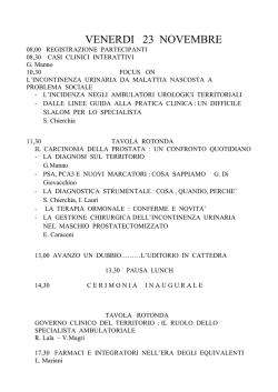
Cop 2014_Layout 1 04/04/14 10:12 Pagina 1
w ess
N o cc a. i t
A u
en w.ai
p
O w
w
ISSN 1124-3562
Poste Italiane S.p.A. - Spedizione in abbonamento postale - D.L. 353/2003 (conv. in L. 27/02/2004 n. 46) Art. 1, comma 1 DCB Milano
Archivio Italiano di Urologia e Andrologia / Archives of Italian Urology and Andrology - Vol. 86; n. 1, March 2014
Vol. 86; n. 1, March 2014
ANDROLOGICAL SCIENCES
Antioxidant cosupplementation therapy with vitamin C, vitamin E, and coenzyme Q10
in patients with oligoasthenozoospermia
Yoshitomo Kobori, Shigeyuki Ota, Ryo Sato, et al.
Is there any effect of insulin resistance on male reproductive system?
Ayhan Verit, Fatma Ferda Verit, Halil Oncel, Halil Ciftci
Evaluation of penile cavernosal artery intima-media thickness in patients
with erectile dysfunction. A new parameter in the diagnosis of vascular erectile dysfunction.
Our experience on 59 cases
Domenico Prezioso, Fabrizio Iacono, Umberto Russo, et al.
Urinary and sexual functions after surgical treatment of penile fracture
concomitant with complete urethral disruption
Ali Abdel Raheem, Hassan El-Tatawy, Ahmed Eissa, et al.
Testicular microlithiasis and dyspermia: Is there any correlation?
Francesco Catanzariti, Ubaldo Cantoro, Vito Lacetera, et al.
Treatment of urethral strictures in balanitis xerotica obliterans (BXO)
using circular buccal mucosal meatoplasy: Experience of 15 cases
Abdulmuttalip Simsek, Sinasi Yavuz Onol, Omer Kurt
Present and future association between obesity and hypogonadism in Italian male
Valentina Boddi, Valeria Barbaro, Paul Mc Nieven, et al.
In vitro effects of PDE5 inhibitor and statin treatment on the contractile responses off
experimental MetS rabbit's cavernous smooth muscle
Yasin Erden, Esat Korgalı, Gokce Dundar, et al.
Penile fracture: Penoscrotal approach with degloving of penis
after Magnetic Resonance Imaging (MRI)
Gabriele Antonini, Patrizio Vicini, Salvatore Sansalone, et al.
First case of bilateral, synchronous anaplastic variant of spermatocytic seminoma treated with
radical orchifunicolectomy as single approach: Case report and review of the literature
Giorgio Gentile, Francesca Giunchi, Riccardo Schiavina, et al.
Penile strangulation: An unusual sexual practice that often presents an urological emergency
Lucio Dell’Atti
Rare case of intra-testicular adenomatoid tumour
Filippo Migliorini, Roberto Baldassarre, Walter Artibani, et al.
Use of inflatable penile prostheses AMS CX with momentary squeeze in a patient
with Peyronie’s disease after removal of two previously implanted penile prostheses
Patrizio Vicini, Ferdinando De Marco, Gabriele Antonini, et al.
Surgical repair of the iatrogenic falsepassage in the treatment
of trauma-induced posterior urethral injuries
Faruk Dog˘an, Ali Feyzullah S¸ahin, Tevfik Sarıkaya, Alper Dırık
The impact of non-urologic drugs on sexual function in men
Ferdinando Fusco, Marco Franco, Nicola Longo, et al.
Practical recommendations for performing ultrasound scanning
in the urological and andrological fields
Pasquale Martino, Andrea Benedetto Galosi, Marco Bitelli, et al.
Sahin CR_Stesura Seveso 26/03/14 10:47 Pagina 48
DOI: 10.4081/aiua.2014.1.48
CASE REPORT
Surgical repair of the iatrogenic falsepassage
in the treatment of trauma-induced posterior urethral injuries
Faruk Dog˘an 1, Ali Feyzullah S¸ahin 1, Tevfik Sarıkaya 2, Alper Dırık 1
1 Department
2 Department
of Urology, S¸ifa University Medicine School, I˙zmir, Turkey
of Urology, Sivas Public Hospital, Sivas, Turkey
Pelvic fracture associated urethral
injury (PFAUI) is a rare and challenging sequel of blunt pelvic trauma. Treatment of iatrogenic false urethral passage (FUP) remains as a challenge for urologists. In this case report we reviewed the
iatrogenic FUP caused by wrong procedures performed
in the treatment of a patient with PFAUI and the treatment of posterior urethral stricture with transperineal
bulbo-prostatic anatomic urethroplasty in the management of FUP. A 37-year-old male patient with PFAUI
had undergone a laparotomy procedure for pelvic bone
fracture, complete urethral rupture, and bladder perforation 8 years ago. After stricture formation, patient
had undergone procedures that caused FUP. Following
operations, he had a low urinary flow rate, and incontinence and urgency even with small amounts of urine.
FUP was diagnosed by voiding cystourethrography and
retrograde urethrography. FUP was fixed with open
urethroplasty with the guidance of flexible antegrade
urethtoscopy. False passage should always be taken
into account in the differential diagnosis of patients
with persistent symptoms that underwent PFAUI therapy. In addition, we believe that in the evaluation of
patients with PFAUI suspected for having a false
passage, bladder neck and urethra should be assessed
by combining routine voiding cystourethrography and
retrograde urethrography with preoperative flexible
cystoscopy via suprapubic route.
trauma may cause a life-long morbidity unless properly
treated. While treatment approach in partial urethral
rupture includes watchful waiting after performing a
simple cystostomy, complete urethral rupture can be
repaired with 3 methods: 1) Realignment of the separated urethral ends over a catheter (urethral realignment),
2) Primary anastomosis of separated urethral ends, and
3) Immediate suprapubic cystostomy and delayed repair
of the resulting stricture on an elective basis (3).
Emergency treatment of PFAUI has not been standardized
in developing countries. On majority of cases wrong procedures cause iatrogenic injuries in addition to trauma
itself (4). Sometimes, careless and/or repeated urethral
dilatations also lead to false urethral passage (FUP) formation, which results in infections and incontinence.
Treatment of iatrogenic FUP remains as a challenge for
urologists (5). Furthermore, there is a paucity of data
about the urethroplasty procedure performed for urethral
stricture formed by FUP. In this case report we reviewed
the iatrogenic FUP caused by wrong procedures performed in the treatment of a patient with PFAUI and the
treatment of posterior urethral stricture with transperinealbulbo-prostatic anatomic urethroplasty in the management of FUP.
Case Report and Figures are posted in Suppementary
materials on www.aiua.it.
KEY WORDS: False urethral passage; flexible cystoscoby; pelvic
fracture; urethral injury.
False passage is an abnormal passage between urinary
bladder and urethra, which is observed in association
with posterior urethral stricture and is caused by a iatrogenic injury resulting from careless and wrong treatment
procedures after trauma. If unnoticed, this false passage
between urethra and bladder causes very frequent complaints; furthermore, repeated endoscopic procedures
(internal urethrotomy), catheterization, or urethral
dilatation become necessary. Following dilatation, hesitancy, incontinence, and urgency persist. Furthermore,
false passage scar tissue around traumatized tissue causes prolonged and chronic infections and hence leads to
stricture formation (6).
Summary
Submitted 30 July 2013; Accepted 31 December 2013
INTRODUCTION
Pelvic fracture associated urethral injury (PFAUI) is a rare
and challenging sequel of blunt pelvic trauma. In 425% of male patients with pelvic bone fractures, simultaneous posterior urethral injury is observed (1). Injury
to posterior urethra may be a simple contusion, or it
may appear as partial or complete rupture (2). Urethral
No conflict of interest declared
48
Archivio Italiano di Urologia e Andrologia 2014; 86, 1
DISCUSSION
Sahin CR_Stesura Seveso 26/03/14 10:47 Pagina 49
Surgical repair of the iatrogenic falsepassage in the treatment of trauma-induced posterior urethral injuries
Methods used for diagnosis of posterior urethral strictures before reconstruction should clearly delineate stricture length and site, determine the anatomy of posterior
urethra and bladder neck, and show false passages and
fistulas if any. Conventional methods include simultaneous CCUG and dynamic retrograde urethrography. It is
necessary to make a pre-treatment assessment via combined radiological and endoscopic methods especially in
obliterated cases where prostatic and proximal urethra
cannot be visualized.
False passage is diagnosed with urethrography. Combined
voiding cystourethrography and retrograde urethrography can clearly show the site and path of the passage (7).
Cystourethroscopy plays a limited role in diagnosis since
normal anatomical structures cannot be localized. This
was also the case in our patient because his false passage
could not be identified despite multiple endoscopic procedures at other medical facilities. A false passage should
be taken into consideration for diagnosis in case normal
anatomical markers like veru montanum, bladder
trigone, and external sphincter could not be seen during
cystourethroscopy. A flexible cystoscope advanced
through a suprapubic route is highly useful for the diagnosis of false passage. While normal bladder neck is a
funnel-shaped, soft, elastic, and of smooth structure, a
false passage is a pale mucosal formation associated with
a vertical circular dense scar and a coarse granulation that
is located close to bladder neck (6).
In conclusion, recurrent urethral strictures develop due
to inappropriate and insufficient initial therapies in
patients with PFAUI with complete urethral separation;
Figure.
Preoperative VCUG and RUG shows the location of a false passage
and urethral stricture. The black arrow shows normal bladder neck
and prostatic urethra, the blue arrow shows the urethral stricture
and the beginning of the intraprostatic false passage, while the white
arrow shows the false passage located close to bladder neck.
as a result, various endoscopic procedures and urethral
dilatations become necessary. Even after these therapies,
hesitancy, incontinence, and urgency are observed. False
passage should always be taken into account in the differential diagnosis of patients with persistent symptoms
that underwent PFAUI therapy. In addition, we believe
that in the evaluation of patients with PFAUI suspected
for having a false passage, bladder neck and urethra
should be assessed by combining routine voiding cystourethrography and retrograde urethrography with preoperative flexible cystoscopy via suprapubic route.
The guidance of a flexible cystoscope via suprapubic
route during the operation is quite helpful. It also guides
procedures of curettage and dissection to determine the
true anatomical structures and to identify the normal
urethral tract, particularly in cases where prostatic urethra is blocked.
REFERENCES
1. Koraitim MM, Marzouk ME, Atta MA, et al. Risk of urethral
injury in pelvic fractures. Br J Urol. 1996; 77: 876-80.
2. Lupu AN, Forrer JH, Smith RB, Kaufman J. Urethral gap in complete disruption of membraneus urethra. Urology. 1987; 29:378-82.
3. Webstre GD, Mathes GL, Selli C. Prostatomembranous urethral
injuries: A review of the literature and a rational approach to their
management. I. Urol. 1983; 130:898.
4. Barbagli G. History and evolution of transpubic urethroplasty: a
lesson for young urologists in training. Eur Urol. 2007; 52:1290-2.
5. Barbagli G, Palminteri E, Lazzeri M, Guazzoni G. One-stage circumferential buccal mucosa graft urethroplasty for bulbous stricture
repair. Urology. 2003; 61:452-5.
6. Qiang Fu, Jiong Zhang, Ying-long Sa, San-bao Jin, Yue-minXu.
Transperineal bulbo-prostatic anastomosis for posterior urethral
stricture associated with false passage: a single-centre experience.
BJU Int. 2011; 108:1352-4.
7. Secrest CL. Staged urethroplasty: indications and techniques. Urol
Clin North Am. 2002; 29:467-75.
Correspondence
Faruk Dog˘an, MD (Corresponding Author)
[email protected]
Alper Dırık, MD
[email protected]
Specialist in Urology
Department of Urology, S¸ifa University Medicine School, Izmir, Turkey
Ali Feyzullah S¸ahin, MD, FEBU
[email protected]
Asistant Professor in Urology
Department of Urology, S¸ifa University Medicine School
Sanayi cad. No:7 Bornova, Izmir, Turkey
Tevfik Sarıkaya, MD
[email protected]
Specialist in Urology
Department of Urology, Sivas Public Hospital, Sivas, Turkey
Archivio Italiano di Urologia e Andrologia 2014; 86, 1
49
Cop 2014_Layout 1 04/04/14 10:12 Pagina 2
9° Congresso Nazionale 2014
22-24 Maggio 2014
Auditorium Pietro d'Abano - Abano Terme (PD)
Segreteria Organizzativa
MEC Congress Srl
Via Gorizia 51 - 95129 Catania
Dr.ssa Antonella Barbagallo
tel. 334 6527204 - email: [email protected]
Società Italiana
Chi intende iscriversi alla SIUrO trova le istruzioni ed i moduli necessari sul sito internet
www.siuro.it
È possibile pagare la quota associativa annuale on-line:
con carta di credito collegandosi direttamente al sito www.siuro.it o tramite bonifico bancario
intestato alla Società Italiana di Urologia Oncologica, presso Banca Popolare di Milano
IBAN: IT08W0558402402000000000539, avendo cura di specificare il nome del socio pagante
Per ottenere ulteriori informazioni è possibile contattare la segreteria
Via Dante 17 - 40126 Bologna
Tel +39 051 349224 Fax +39 051 349224
e-mail: [email protected] www.siuro.it
CHI PUÒ FARNE PARTE
Possono far parte dell'Associazione
con la qualifica di Socio Ordinario gli
Urologi
Specialisti in Urologia e gli specializOspedalità
Gestione Privata
zandi in Urologia operanti in strutture
assistenziali urologiche dell’Ospedalità a gestione Privata; con la qualifica di Socio Corrispondente gli
studiosi italiani o stranieri che abbiano dimostrato un particolare interesse per l'Urologia.
ISCRIZIONE
Iscriversi è semplice, basta scaricare la scheda di adesione presente
sul sito www.urop.it, compilarla in tutte le sue parti prendendo visione dell'informativa sulla privacy ed inviarla via fax al numero 089
771330, allegando la fotocopia di un valido documento di riconoscimento.
QUOTA SOCIALE
La quota sociale per l'anno 2014 è stabilita in € 100,00 e dà diritto
alla ricezione della rivista "Archivio Italiano di Urologia e di Andrologia",
organo ufficiale della Associazione.
Il pagamento della quota sociale 2014 potrà essere effettuato mediante bonifico bancario come segue:
Urop - Banco di Napoli SpA - Ag. 3 - Salerno
IBAN IT72 U 01010 15203 100000004420
Inserire nella causale del bonifico bancario il proprio Nome e Cognome seguito dalla dizione QS2014.
INFORMAZIONI
Per richiedere informazioni contattare il Dr. Stefano Pecoraro
all'indirizzo e-mail [email protected]
oppure al seguente recapito telefonico 333 7451321
Ed _Cop+Ed+fisse 2006 03/04/14 10:36 Pagina II
ll ruolo della SIEUN
La SIEUN (Società Italiana di Ecografia Urologica, Andrologica, Nefrologica) riunisce diversi medici specialisti e non che si occupano
di tutte quelle metodiche in cui gli ultrasuoni vengono utilizzati a scopo diagnostico ed interventistico in ambito uro-nefro-andrologico.
La SIEUN organizza un Congresso Nazionale con cadenza biennale e diverse altre iniziative in genere con cadenza annuale (corsi
monotematici, sessioni scientifiche in occasione dei congressi nazionali delle più importanti società scientifiche in ambito UroNefro-Andrologico).
Dal 2001 la SIEUN è affiliata all’ESUI (European Society of Urological Imaging); pertanto tutti i soci possono partecipare alla iniziative della
Società Europea.
L’Archivio Italiano di Urologia e Andrologia è l’organo ufficiale della SIEUN.
Questa pagina permette una informazione puntuale sulla attività della nostra Società e consente al Consiglio Direttivo della SIEUN
di comunicare non solo ai soci, ma ad una platea più ampia, ogni nuova iniziativa.
Notizie dalla SIEUN
Linee guida sulla Ecografia Urologica ed Andrologica
La versione finale delle linee giuda è online sul sito della Società Italiana di Urologia (www.siu.it).
Corso di Perfezionamento in Ecografia Urologica, Andrologica e Nefrologica
Università di Bari AA. 2013-2014
Anche quest’anno il corso si tiene presso la Urologia 1° Universitaria del Policlinico di Bari, coordinato dal Prof. a.c. P. Martino.
Tra i docenti figurano diversi soci della SIEUN.
Sito web: www.uniba.it area formazione post laurea.
Informazioni: Prof. Pasquale Martino - e mail: [email protected] - tel. 0805594101
Sig. Giacoma Loverro - e mail: [email protected] - tel. 080.5578719
Società
Italiana
di Ecografia
Urologica
Andrologica
Nefrologica
19° Congresso SIEUN
19° CONGRESSO S.I.E.U.N. 2014
Società Italiana di Ecografia Urologica, Andrologica, Nefrologica
Fermo, 15-17 Maggio 2014
Presidente del Congresso
Andrea B. Galosi
Sede Congressuale
Hotel Royal - Località Casa Bianca - FERMO
Presidente Onorario del Congresso
Giovanni Muzzonigro
Ospedale A. Murri - ASUR Marche AV4 - FERMO
Presidente
Pasquale Martino
Vice Presidenti
Paolo Consonni, Vincenzo Scattoni
Consiglieri
Marco Bitelli, Michele Del Zingaro,
Giuseppe D’Eramo, Andrea Galosi, Roberta Gunelli,
Amelia Liccardo, Giovanni Liguori
Tesoriere
Michele Barbera
Si terrà a Fermo
dal 15 al 17 Maggio 2014.
Maggiori informazioni verranno inserite
sul sito SIEUN (www.sieun.it).
Presidente del Congresso
è il dott. Andrea Benedetto Galosi.
ECM
Evento in procedura di accreditamento ECM
Modalità di partecipazione
Iscrizioni on line: www.congressare.it
sez. Meeting ECM 2014
Segreteria Organizzativa e Provider ECM N.2295
PROMISE GROUP srl
Via Ghino Valenti, 2 - 60131 Ancona
Tel. 071.202123 - Fax 071.202447
e-mail: [email protected]
web: www.congressare.it
Segretario
Silvano Palazzo
www.congressare.it/sieun 2014
I PUNTI SIEUN (una possibilità di incontro tra Soci SIEUN e di contatto con altri specialisti)
Presso i punti SIEUN i nostri soci potranno essere continuamente informati su tutte le attività e le iniziative della Società
e rinnovare il pagamento della quota associativa.
I PROSSIMI APPUNTAMENTI SIEUN
La SIEUN nel 2014 sarà presente con relazioni, moderazioni e letture nei congressi
delle più prestigiose Società scientifiche di Urologia, Andrologia ed Ecografia:
Corso Regionale SIEUN Teorico-pratico di Ecografia Andrologica – Roma, 15 Febbraio 2014
25° Congresso Interregionale della Società Apulo-Lucana di Urologia – Bari, 7-8 Marzo 2014
19° Congresso Nazionale Società Italiana di Ecografia Urologica, Andrologica, Nefrologica (SIEUN) – Fermo (AN), 15-17 Maggio 2014
IX° Congresso Nazionale U.R.O.P. – Abano Terme (PD), 22-24 Maggio 2014
XXX Congresso Nazionale SIA – Maratea (PZ), 28-31 Maggio 2014
XXI Congresso Nazionale AURO.it – Roma, 4-6 Giugno 2014
38° Congresso SIUD – Milano, 19-21 Giugno 2014
XXIV Congresso Nazionale SIUrO – Bologna, 22-24 Giugno 2014
87° Congresso Nazionale SIU – Firenze, 27-30 Settembre 2014
RINNOVO PAGAMENTO QUOTA 2014
La segreteria della Società
ELLERRE
CENTRE
QUOTE ASSOCIATIVE 2014
A partire dal 1 Gennaio 2014 le quote sono:
Socio ordinario - Euro 70,00
Socio Junior - Euro 35,00
Via S. Matarrese, 47/G - 70124 BARI
Coordinate: Conto Corrente n. 2053691 c/o Monte dei Paschi di Siena Intestato a:
Tel. 080.5045353
Fax
080.5045362
"S.I.E.U.N.
Soc. It. di Ecografia Urologica Nefrologica"
4
II Archivio Italiano di Urologia e Andrologia 2012, 84,IBAN:
www.ellerrecentre.it E-mail: [email protected]
IT 72 M 01030 04001 000002053691 - BIC: PASCITM1BA1
è a disposizione per ulteriori informazioni.
Inviare copia dell’avvenuto bonifico per e-mail: [email protected] o fax 080.5045362
Cop 2014_Layout 1 04/04/14 10:12 Pagina 2
9° Congresso Nazionale 2014
22-24 Maggio 2014
Auditorium Pietro d'Abano - Abano Terme (PD)
Segreteria Organizzativa
MEC Congress Srl
Via Gorizia 51 - 95129 Catania
Dr.ssa Antonella Barbagallo
tel. 334 6527204 - email: [email protected]
Società Italiana
Chi intende iscriversi alla SIUrO trova le istruzioni ed i moduli necessari sul sito internet
www.siuro.it
È possibile pagare la quota associativa annuale on-line:
con carta di credito collegandosi direttamente al sito www.siuro.it o tramite bonifico bancario
intestato alla Società Italiana di Urologia Oncologica, presso Banca Popolare di Milano
IBAN: IT08W0558402402000000000539, avendo cura di specificare il nome del socio pagante
Per ottenere ulteriori informazioni è possibile contattare la segreteria
Via Dante 17 - 40126 Bologna
Tel +39 051 349224 Fax +39 051 349224
e-mail: [email protected] www.siuro.it
CHI PUÒ FARNE PARTE
Possono far parte dell'Associazione
con la qualifica di Socio Ordinario gli
Urologi
Specialisti in Urologia e gli specializOspedalità
Gestione Privata
zandi in Urologia operanti in strutture
assistenziali urologiche dell’Ospedalità a gestione Privata; con la qualifica di Socio Corrispondente gli
studiosi italiani o stranieri che abbiano dimostrato un particolare interesse per l'Urologia.
ISCRIZIONE
Iscriversi è semplice, basta scaricare la scheda di adesione presente
sul sito www.urop.it, compilarla in tutte le sue parti prendendo visione dell'informativa sulla privacy ed inviarla via fax al numero 089
771330, allegando la fotocopia di un valido documento di riconoscimento.
QUOTA SOCIALE
La quota sociale per l'anno 2014 è stabilita in € 100,00 e dà diritto
alla ricezione della rivista "Archivio Italiano di Urologia e di Andrologia",
organo ufficiale della Associazione.
Il pagamento della quota sociale 2014 potrà essere effettuato mediante bonifico bancario come segue:
Urop - Banco di Napoli SpA - Ag. 3 - Salerno
IBAN IT72 U 01010 15203 100000004420
Inserire nella causale del bonifico bancario il proprio Nome e Cognome seguito dalla dizione QS2014.
INFORMAZIONI
Per richiedere informazioni contattare il Dr. Stefano Pecoraro
all'indirizzo e-mail [email protected]
oppure al seguente recapito telefonico 333 7451321
Cop 2014_Layout 1 04/04/14 10:12 Pagina 1
w ess
N o cc a. i t
A u
en w.ai
p
O w
w
ISSN 1124-3562
Poste Italiane S.p.A. - Spedizione in abbonamento postale - D.L. 353/2003 (conv. in L. 27/02/2004 n. 46) Art. 1, comma 1 DCB Milano
Archivio Italiano di Urologia e Andrologia / Archives of Italian Urology and Andrology - Vol. 86; n. 1, March 2014
Vol. 86; n. 1, March 2014
ANDROLOGICAL SCIENCES
Antioxidant cosupplementation therapy with vitamin C, vitamin E, and coenzyme Q10
in patients with oligoasthenozoospermia
Yoshitomo Kobori, Shigeyuki Ota, Ryo Sato, et al.
Is there any effect of insulin resistance on male reproductive system?
Ayhan Verit, Fatma Ferda Verit, Halil Oncel, Halil Ciftci
Evaluation of penile cavernosal artery intima-media thickness in patients
with erectile dysfunction. A new parameter in the diagnosis of vascular erectile dysfunction.
Our experience on 59 cases
Domenico Prezioso, Fabrizio Iacono, Umberto Russo, et al.
Urinary and sexual functions after surgical treatment of penile fracture
concomitant with complete urethral disruption
Ali Abdel Raheem, Hassan El-Tatawy, Ahmed Eissa, et al.
Testicular microlithiasis and dyspermia: Is there any correlation?
Francesco Catanzariti, Ubaldo Cantoro, Vito Lacetera, et al.
Treatment of urethral strictures in balanitis xerotica obliterans (BXO)
using circular buccal mucosal meatoplasy: Experience of 15 cases
Abdulmuttalip Simsek, Sinasi Yavuz Onol, Omer Kurt
Present and future association between obesity and hypogonadism in Italian male
Valentina Boddi, Valeria Barbaro, Paul Mc Nieven, et al.
In vitro effects of PDE5 inhibitor and statin treatment on the contractile responses off
experimental MetS rabbit's cavernous smooth muscle
Yasin Erden, Esat Korgalı, Gokce Dundar, et al.
Penile fracture: Penoscrotal approach with degloving of penis
after Magnetic Resonance Imaging (MRI)
Gabriele Antonini, Patrizio Vicini, Salvatore Sansalone, et al.
First case of bilateral, synchronous anaplastic variant of spermatocytic seminoma treated with
radical orchifunicolectomy as single approach: Case report and review of the literature
Giorgio Gentile, Francesca Giunchi, Riccardo Schiavina, et al.
Penile strangulation: An unusual sexual practice that often presents an urological emergency
Lucio Dell’Atti
Rare case of intra-testicular adenomatoid tumour
Filippo Migliorini, Roberto Baldassarre, Walter Artibani, et al.
Use of inflatable penile prostheses AMS CX with momentary squeeze in a patient
with Peyronie’s disease after removal of two previously implanted penile prostheses
Patrizio Vicini, Ferdinando De Marco, Gabriele Antonini, et al.
Surgical repair of the iatrogenic falsepassage in the treatment
of trauma-induced posterior urethral injuries
Faruk Dog˘an, Ali Feyzullah S¸ahin, Tevfik Sarıkaya, Alper Dırık
The impact of non-urologic drugs on sexual function in men
Ferdinando Fusco, Marco Franco, Nicola Longo, et al.
Practical recommendations for performing ultrasound scanning
in the urological and andrological fields
Pasquale Martino, Andrea Benedetto Galosi, Marco Bitelli, et al.
Istr_Stesura Seveso 03/04/14 10:32 Pagina 79
INSTRUCTIONS
AUTHORS’ RESPONSIBILITIES
Manuscripts are accepted with the understanding that they have not been published or submitted for publication in any other journal.
Authors must submit the results of clinical and experimental studies conducted according to the Helsinki Declaration on clinical research and to the Ethical
Code on animal research set forth by WHO (WHO Chronicle 1985; 39:51).
The Authors must obtain permission to reproduce figures, tables and text from
previously published material. Written permission must be obtained from the
original copyright holder (generally the Publisher).
MANUSCRIPT PRESENTATION
Authors must submit the text (MAC and WINDOWS Microsoft Word are accepted) and illustrations by e-mail ([email protected]).
Manuscripts must be written in English language in accordance with the
“Uniform Requirements for Manuscripts submitted to biomedical journals” defined by
The International Committee of Medical Journal Editors (http://www.ICMJE.org).
Manuscripts in Italian language can be published after translation (expenses will be charged to the Authors). Manuscripts should be typed double spaced
with wide margins.
They must be subdivided into the following sections:
TITLE PAGE
It must contain:
a) title;
b) a short (no more than 40 characters) running head title;
c) first, middle and last name of each Author without abbreviations;
d) University or Hospital, and Department of each Author;
e) last name, address and e-mail of all the Authors
f) corresponding Author
g) phone and/or fax number to facilitate communication;
h) acknowledgement of financial support;
i) list of abbreviations.
SUMMARY
The Authors must submit a long English summary (300 words, 2000 characters).
Subheadings are needed as follows: Objective(s), Material and method(s),
Result(s), Conclusion(s).
After the summary, three to ten key words must appear, taken from the standard
Index Medicus terminology.
TEXT
For original articles concerning experimental or clinical studies and case
reviews, the following standard scheme must be followed:
Summary - Key Words - Introduction - Material and Methods - Results Discussion - Conclusions - References - Tables - Legends - Figures.
SIZE OF MANUSCRIPTS
Literature reviews, Editorials and Original articles concerning experimental or
clinical studies should not exceed 20 typewritten pages including figures, tables,
and reference list (references are to be limited to 20, additional references may be
submitted as supplementary materials for posting on www.aiua.it.).
Case reports, Notes on surgical technique, Short Communications and
Letters to the editors should not exceed 1000 words (summary included) with
only one table or figure, and no more than three references. No more than five
authors are permitted.
As an accompaniment to Case reports, Notes on surgical technique and Short
Communications manuscripts for the print version of Archivio Italiano di
Urologia e Andrologia (AIUA), authors may submit supplementary materials
for posting on www.aiua.it.
The material is subject to the same editorial standards and peer-review procedures as the print publication.
FAST-TRACK PEER REVIEW
We offer fast-track peer review and publication of controlled trials that we judge
of importance to practice or research. If you wish to discuss your proposed submission, please write ([email protected]) or call our editorial office in Milan (+39
02 70608091).
REFERENCES
References must be sorted in order of quotation and numbered with arabic digits
between parentheses. Only the references quoted in the text can be listed. Journal
titles must be abbreviated as in the Index Medicus. Only studies published on
easily retrieved sources can be quoted. Unpublished studies cannot be quoted,
however articles “in press” can be listed with the proper indication of the journal
title, year and possibly volume.
References must be listed as follows:
TO
AUTHORS
BOOK
CHAPTERS
Authors of the chapters - Complete chapter title. In: Book Editor, complete
Book Title, Edition number. City of publication: Publisher, Publication year:
first page of chapter in the book.
Example: Sagawa K. The use of central theory and system analysis. In: Bergel DH
(Ed), Cardiovascular dynamics. 2nd ed. London: Academic Press Inc., 1964; 115.
TABLES
Tables must be aimed to make comprehension of the written text easier. They
must be numbered in Arabic digits and referred to in the text by progressive
numbers. Every table must be accompanied by a brief title. The meaning of
any abbreviations must be explained at the bottom of the table itself.
(If sent by surface mail tables must be clearly printed with every table typed
on a separate sheet).
FIGURES
(Graphics, algorithms, photographs, drawings).
Figures must be numbered and quoted in the text by number.
The meaning of all symbols, abbreviations or letters must be indicated. Histology
photograph legends must include the enlargement ratio and the staining method.
Legends must be collected in one or more separate pages.
Please follow these instructions when preparing files:
• Do not include any illustrations as part of your text file.
• Do not prepare any figures in Word as they are not workable.
• Line illustrations must be submitted at 600 DPI.
• Halftones and color photos should be submitted at a minimum of 300 DPI.
• Power Point files cannot be uploaded.
• If at all possible please avoid transmitting electronic files in JPEG format. If this
is unavoidable please be sure to save the JPEG at the highest quality available
and at the correct resolution for the type of artwork it is
• PDF files for individual figures may be uploaded.
MANUSCRIPT
REVIEW
Only manuscript written according to the above mentioned rules will be considered. All submitted manuscripts are evaluated by the Editorial Board and/or
by two referees designated by the Editors. The Authors are informed in a time
as short as possible on whether the paper has been accepted, rejected or if a
revision is deemed necessary.
The Editors reserve the right to make editorial and literary corrections with
the goal of making the article clearer or more concise, without altering its
contents. Submission of a manuscript implies acceptation of all above rules.
PROOFS
Authors are responsible for ensuring that all manuscripts are accurately typed
before final submission. Galley proofs will be sent to the first Author. Proofs
should be returned within seven days from receipt.
REPRINTS
A copy of the issue in which the article appears will be provided free of charge.
Reprints are not provided. The PDF file is only for personal use.
OPEN ACCESS
All papers published in Archivio Italiano di Urologia e Andrologia (AIUA) are
peer reviewed and upon acceptance will be immediately and permanently free
for everyone to read and download (Open Access). Open Access means making
your papers available to anyone, at any time. In order to cover the costs associated with Open Access publication, a fee has to be pay by the author or research
funder to cover the costs associated with publication.
As of January 1, 2014 the Authors of Case Reports, Notes on Surgical
Technique, Short communications and Letters to the Editors published in
Archivio Italiano di Urologia e Andrologia (AIUA) will be charged a publication fee of 200 Euros + VAT.
Reviews and Original papers will continue to be published free of charge.
Authors who will pay a fee for Open Access publication will use an exclusive
licensing agreement, where they will retain copyright alongside scholarly usage
rights and Edizioni Scripta Manent will be granted publishing and distribution
rights.
PAYMENT
OF ARTICLE PROCESSING CHARGE
JOURNAL ARTICLES
The submitting authors must confirm, at the time of submission, that they will
organize payment should the article be accepted for publication. Following peer
review, once a manuscript has received editorial acceptance in principle, the submitting author needs to arrange payment of the article-processing charge.
Once formatting checks are completed, and payment of the article-processing
charge has been received, the article will be published.
BOOKS
Payment can be made by any of the following methods:
1) Invoice payment to the bank account Edizioni Scripta Manent s.n.c.
Via E. Bassini, 41 - 20133 Milano
IBAN: IT 23 K 02008 01749 000100472830
2) PayPal international e-commerce payment connecting to
https://www.paypal.com/cgi-bin/webscr?cmd=_s-xclick&hosted_button_id=ZUPFHQWA7WRY2
Name, surname and tax codes (CF and P.IVA for Italy) are to be attached.
A receipt will be sent once payment has been processed.
All Authors if there are six or fewer, otherwise the first three, followed by “et al.”.
Complete names for Work Groups or Committees. Complete title in the original
language.
Title of the journal following Index Medicus rules. Year of publication; Volume
number: First page.
Example: Starzl T, Iwatsuki S, Shaw BW, et al. Left hepatic trisegmentectomy Surg
Gynecol Obstet. 1982; 155:21.
Authors - Complete title in the original language. Edition number (if later than
the first). City of publication: Publisher, Year of publication.
Example: Bergel DIA. Cardiovascular dynamics. 2nd ed. London: Academic Press
Inc., 1974.
Istruz_Cop+Ed+fisse 2006 24/06/13 11:14 Pagina 57
NEW WEBSITE:
www.aiua.it
W!
E
N
Pages 352 - The cost of a printed copy is € 30 plus shipping.
Pdf price € 30,00
To purchase go to: http://www.urologyplanet.it/
The stone disease is extremely diverse – from single stone formers
with spontaneous stone passage to staghorn stones and stone diseases associated with severe metabolic abnormalities such as
renal tubular acidosis and primary hyperoxaluria. It is obvious that
these different conditions should be dealt with selectively. In this
context sharing knowledge on clinical practices – tips and tricks –
between practising stone clinicians becomes especially important,
and the most effective way that tips and tricks may be lifted up
into an academic sphere is through international knowledge
exchange. It is with this respect that this handbook on stone management plays a particular important role by creating a platform for
exchange of clinical expertise. The contributors are international
renowned stone experts, sharing with us their views of different approaches to the great variety of
urinary stone diseases.
The handbook offers a unique synthesis of theory and tips-and tricks-technology, bridging basic
science and daily clinical practice.
Palle Osther, MD, PhD
Professor of Urology
Chairman, EAU Section of Urolithiasis (eULIS)
© Copyright 2026 Paperzz