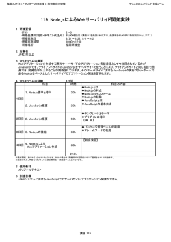
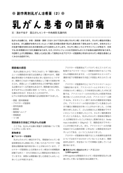
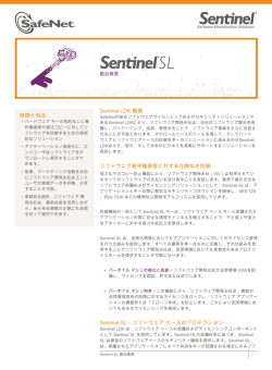
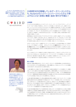
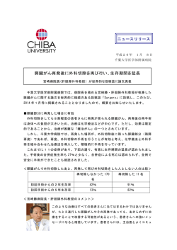
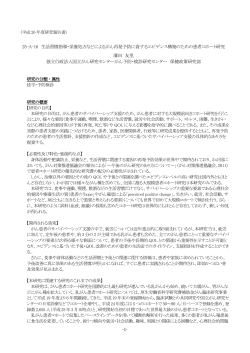
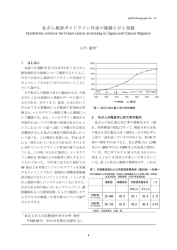
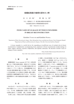
日医大医会誌 2010; 6(3)
111
―綜 説―
乳腺内視鏡手術と 3D-CT 乳腺リンパ管造影<その 2>
3D-CT 乳腺リンパ管造影によるセンチネルリンパ節生検
山下 浩二
日本医科大学大学院医学研究科機能制御再生外科学
Video-Assisted Breast Surgery and 3-Dimensional Computed Tomographic Mammary Lymphography〈2〉
Sentinel Node Biopsy with 3D-CT Mammary Lymphography
Koji Yamashita
Department of Biological Regulation and Regenerative Surgery, Graduate School of Medicine, Nippon Medical School
Abstract
Background: I have studied endoscopic surgery for breast diseases and 3-dimensional (3D)
computed tomographic (CT) lymphography for sentinel node (SN) biopsy. In this second in a
series of reports, I explain the techniques of 3D-CT lymphography. 3D-CT lymphography can
show the detailed lymphatic flow from the breast tumor toward the SNs and the exact local
relation between axillary lymph nodes. I have developed this 3D image-processing system to
more precisely depict the anatomical structures of the mammary lymphovascular system. This
system allows us to systematically collect axillary lymph nodes, including SNs.
Methods: 3D-CT lymphography was performed to mark SNs on the skin on the day
before surgery. Above the tumor and near the areola, 2 ml of Iopamiron 300 was injected
subcutaneously. Sixteen-channel multidetector-row helical CT scan images were obtained 1
minute after injection to detect SNs, and after 3 and 5 minutes to observe lymph flow into the
venous angle. The scan images were reconstructed to produce 3D images. SN biopsy was
performed with the dye-staining method and endoscopy.
Results: 3D-CT lymphography accurately showed lymphatic flow from the tumor to SNs.
We classified the relationship between the lymph ducts and the drained SNs into 4 patterns.
Following up 3 and 5 minutes after injection of the contrast agent, we can follow the lymph
ducts beyond the SN into the second and third nodes toward the venous angle with the
complex plexus. The figure of the axillary nodes shows 5 beads-like grouped nodes. 3D-CT
lymphography can also recognize the metastatic patterns of the enhanced lymph node. These
patterns will predict the metastasis before SN biopsy. 3D-CT lymphography can also be used
to detect lymph flow from the arm to avoid harming the arm lymph channel during axillary
node dissection and SN biopsy.
Conclusions: With 3D-CT lymphography, we can more accurately and precisely recognize
lymph flow and the positional relations of SN and axillary nodes to surrounding anatomical
structures.
(日本医科大学医学会雑誌 2010; 6: 111―117)
Key words: breast cancer, endoscopic surgery, sentinel node biopsy, 3-dimensional computed
tomographic lymphography, aesthetic results
Correspondence to Koji Yamashita, Department of Biological Regulation and Regenerative Surgery, Graduate School
of Medicine, Nippon Medical School, 1―1―5 Sendagi, Bunkyo-ku, Tokyo 113―8603, Japan
E-mail: [email protected]
Journal Website(http:!
!
www.nms.ac.jp!
jmanms!
)
112
日医大医会誌 2010; 6(3)
3D-CT LG の撮影方法
はじめに
乳癌の縮小手術に貢献するため,われわれが開発・
MDCT は,16 列マルチディテクタ 3D-CT スキャ
実施してきた二つの研究のうち,前回は乳腺内視鏡手
ナ(Toshiba Aquilion 16;Toshiba Medical Systems
術について紹介したが,2 回目の今回は,センチネル
Corporation, Tochigi, Japan)
,造影剤はイオパミロン
リンパ節生検の精度向上を目的として開発した 3D-
300(Iopamidol 300 mg!
mL;Nihon Shering, Osaka,
CT リンパ管造影について紹介する.3D-CT リンパ管
Japan)を使用した.体位は,手術時の体位に近づけ
造影は,乳腺腫瘍からセンチネルリンパ節までのリン
て,仰臥位,上肢を 90˚ 外転位に保持する.腫瘍直上
パ管を詳細に描出でき,色素やアイソトープよりも正
と乳輪縁の 2 カ所に,1% キシロカイン各 0.5 mL を
確なセンチネルリンパ節の同定を実現した.
注射して局所麻酔を行い,造影剤各 2 mL を皮下に注
早期乳癌において,腋窩リンパ節転移の有無は,予
射する.内胸リンパ節を観察する場合には腫瘍の背側
後を規定する因子であり,今後の治療を決定するのに
にも注射する.造影剤注入後,1 分,3 分(腋窩リン
重要である.しかし,腋窩リンパ節郭清は,肩関節拘
パ節を追跡する際には 5 分)に,3 mm スライス厚で
縮・上肢浮腫・麻痺などの合併症を起こし QOL を悪
CT 像を撮影する.SN は横断面 CT 像で容易に観察
化させる.センチネルリンパ節(SN)は,腫瘍から
することができ,その位置を CT のレーザーポイン
1,2
のリンパ流を受ける最初のリンパ節と定義され ,SN
生検によって,腋窩リンパ節の転移を判断し,腋窩郭
清を省略するための情報を得ることができる.SN を
検出する一般的方法は色素法3,4 とアイソトープ法5,6 で
ある.マルチディテクタ 3 次元 CT 乳腺リンパ管造影
(3D-CT LG)では,より正確にセンチネルリンパ節
を同定することができ7,8,術前に体表にマーキングす
ることにより確実な SNB が可能となる9.
3D-CT LG は,リンパ流とリンパ節との詳細な関係
を描出することができ10,後期相では,SN から静脈
角までの腋窩リンパ路構造を描出できる11,12.3D-CT
LG をもとに系統的にリンパ節を採取することによ
図 1 CTレーザーポインターによるセンチネルリンパ
節位置の体表マーキング
り,
たとえ SN に転移があっても,不要なリンパ節郭清
を減らすことができ,合併症を減らすことができる.
a
b
図2 3
DCT乳腺リンパ管造影 (文献 1
0より)
造影剤を乳輪縁と腫瘍直上の皮内に注射し,センチネルリンパ節(SLN)への正確なリンパ流を
描出できる.
日医大医会誌 2010; 6(3)
113
ターを使用して体表に術前マーキングをする(図 1)
.
乳房内リンパ経路(乳房〜腋窩)
さらに,横断像から Volume Rendering 法により 3DCT 像を再構成することにより,より精細なリンパ管
乳房内のリンパ流は,乳房のほぼ全体から乳輪周囲
とリンパ節との関係を観察できる(図 2)
.
手術は,SNB を色素法で行う.1% インドシアニン
のリンパ叢へ求心的に流れ,乳頭乳輪周囲を環状に周
グリーン各 2 mL を造影剤と同部位に注入し,20 分後
回し,その後腋窩方向へ 3〜4 本の主幹リンパ管によ
に採取を行う.10 mm 硬性内視鏡にオプティカルト
り流出し,最初に腋窩 SN へと辿り着く(図 6)
.こ
ロカーのビジポート(Tyco Healthcare Japan, Tokyo,
のリンパ経路は,各個人によって偏倚が著しく,一様
Japan,図 3)
を使用し,腋窩の体表マーキングに 1 cm
に腫瘍から腋窩へのリンパ流を規定することは危険で
の皮膚切開を施し,ビジポートを挿入して内視鏡下に
ある.3D-CT LGでは,このリンパ流の様子を各個人
緑色に染色されたリンパ管とリンパ節を観察する(図
で明快に描出することが可能であり,より正確な SN
4)
.SN および第 2 群,第 3 群の腋窩リンパ節を内視
を同定することができる.主幹リンパ管の数と SN の
鏡下に採取することができる.術中迅速病理診断を行
数との関係を 4 分類したものが,図 7 である.一管対
い,SN 転移の有無を判断し,陰性は腋窩温存し,陽
一 節 が 約 60%,一 管 対 多 節 が 2%,多 管 対 一 節 が
性は皮切 2.5 cm に広げ内視鏡下に腋窩郭清(level I+
II)を行う3.乳房温存手術も乳腺内視鏡手術により実
施している13.内視鏡的センチネルリンパ節生検後の
整容性は良好で,腋窩のわずか 1 cm の傷はほとんど
目立たない(図 5)
.
図 4 センチネルリンパ節(SN)と流入するリンパ管
(LD)
のビジポートを通した内視鏡像
(文献 1
2より)
矢印は,緑染した SNと LDを指している.立体的位置関
係は 3
DCT LGで把握しているので,リンパ管内の緑色
素を追跡していけば,容易に SNを発見できる.
図 3 オプティカルトロカーのビジポート
(Tyc
oHe
a
l
t
h
c
a
r
eJ
a
pa
n,To
kyo
,J
a
pa
n)
a
b
図 5 乳腺内視鏡手術(Vi
de
o
a
s
s
i
s
t
e
dbr
e
a
s
ts
ur
ge
r
y:VABS)によるセンチネルリンパ節生検
(SNB)の整容性.矢印は,SNBの腋窩創瘢痕を指す.(文献 1
0より)
a:3
5歳女性,1
8
0
° 左扇状乳腺部分切除術後 1年+ SNBbyVABS.
b:3
7歳女性,左円柱状乳腺部分切除術後 1年+ SNBbyVABS.
114
日医大医会誌 2010; 6(3)
a
b
図6 3
DCT LGによるセンチネルリンパ節(SN)とリンパ管(LD)の描出(文献 1
2より)
造影剤を乳輪縁と腫瘍直上の皮内に注射することにより,3
DCT LGは,腫瘍から SNへの正確な
リンパ流を描出できる.腫瘍からのリンパ流は,乳輪縁方向と腋窩方向へと分かれる.乳輪縁の
リンパ流は腫瘍から注ぎ込み,乳頭周囲を周回し,後に腋窩へと流れていく.この腋窩へのリン
パ管は,腫瘍から直接腋窩に流れるリンパ管とは別のものであった.
図 7 リンパ管(LD)とセンチネルリンパ節(SN)との 4型の関係(文献 1
0より)
リンパ流のパターンは,これらの 4型に分類できる.矢印は SNを指す.各型に含まれる患者数は,a.
4
0
名,b.
1名,c
.
1
3名,d.
1
2名であった.
20%,多管対多節が 18% であった10.SN が複数ある
造影剤は腋窩リンパ節群を流れ,静脈角まで到達する
場合に,偽陰性の発生する可能性が高くなると考えら
ことを描出することが可能である11,12(図 8)
.また,
れ,3D-CT LG はより正確な SN の同定を可能にする
この腋窩リンパ節群を 5 群に分類することができ,3
ことができる.
群まで 80%,5 群までは 30% で描出できた.この 5
群における転移状況を検討すると,SN 転移陽性 40
腋窩内リンパ経路
例のうち,SN のみ転移は 21 例であり,第 2・3 群転
移陰性例にほかの腋窩リンパ節に転移は観察されな
3D-CT LG において,造影剤注射後わずか 1 分で SN
かった.SN に転移があっても,第 2 群・第 3 群に転
に到達するが,さらに 3 分,5 分と観察を続けると,
移がなければ,ほかの腋窩リンパ節に転移がないと判
日医大医会誌 2010; 6(3)
115
図8 3
DCTLGのイオパミドール注射後 1
,
3
,
5分後の経
時的観察(文献 1
1より)
造影剤はセンチネルリンパ節(SN)を越えて,次のリン
パ節に流れていく.3
Dレンダリング画面で大小胸筋を
部分的に除去していくと,3
D腋窩には,5つの数珠状に
連なったリンパ節の集簇を観察することができる.これ
らは,リンパ節転移の順序を表していると考えられる.
矢印は,SNを含めて腋窩リンパ節群を 1~ 5まで番号を
付けて示している.
図9 3
DCTLGによるリンパ節転移の評価
(文献 1
4より)
3
DCT LGは,センチネルリンパ節(SN)が転移してい
るか否かを予測できるかもしれない.癌細胞で占拠され
ていると,3
DCT LGで造影剤はリンパ節の流入部分を
杯状に流れるのが分かるが,リンパ節自身は認識でき
る.ある症例では,リンパ管は転移したリンパ節の周り
を迂回する.しかし,リンパ節への部分的な転移
(微小転
移)の場合には,3
DCT LGで正常とは区別が付かない.
転移を予想するには,リンパ管ルートやリンパ節の造影
パターンをより注意深く検証することが必要である.
するために,上肢浮腫などの術後 QOL を悪化させる
断でき,腋窩温存の可能性が示唆された.
われわれは,
重大な合併症を引き起こす可能性が起こる.上肢に造
内視鏡的に SN 生検および第 2・3 群生検を行うこと
影剤を注入することにより,3D-CT LG で上肢からの
ができるので,より低侵襲に腋窩温存の指標とするこ
リンパ路を描出することができる15(図 10)
.上肢由
とができると考える.
来リンパ路は腋窩静脈周囲を走行し,乳房由来リンパ
路と数カ所で結合する.その合流する部分は腋窩静脈
リンパ節転移パターン
上へ架橋のように伸びるリンパ管として観察されるの
で,その部分を術前に判断することにより,上肢由来
腋窩リンパ節転移が微小である場合には,造影剤は
リンパ流を傷害することを回避できると考える.ま
リンパ節を容易に造影し通過してしまうため,微小転
た,SN に上肢由来リンパ流が直接流入する症例 2 例
移の有無を 3D-CT LGで判断することは難しいが,リ
があり,いずれも SN 生検のみで腋窩温存したにも関
ンパ節内のリンパ流を障害するほどの転移があれば,
わらず,上肢浮腫を引き起こしており,腋窩郭清症例
リンパ節の斑状,蟹爪状,杯状染色をもたらし,リン
と同様の術後リハビリを実施する必要があることが判
パ流が途絶すると,リンパ管の肥大,棍棒状,迂回リ
断できる.
14
ンパ管を観察することができる (図 9)
.非造影非染
色リンパ節であっても 3D-CT LGでは判別可能であ
内胸リンパ節
り,より転移が疑われる.これにより,リンパ節の大
きさだけではなく,転移の有無を判断する目安とする
造影剤を腫瘍背側に注入することにより,内側方向
へのリンパ流を観察しやすくなり,約 20% の症例で
ことができる.
内胸リンパ節(胸骨傍リンパ節)を描出することがで
上肢由来リンパ流
きた16(図 11)
.アイソトープ法による報告では 10%〜
15% の検出率であり,少ない症例ながら,より高率
SN 生検で転移陽性の患者には,従来どおり腋窩リ
に描出することが可能となり,見逃されていた内胸セ
ンパ節郭清を実施することになっているが,その際に
ンチネルリンパ節生検を見直すきっかけになることを
は乳房からだけではなく,上肢からのリンパ流も傷害
期待する.将来,内視鏡的に腋窩アプローチでの内胸
116
日医大医会誌 2010; 6(3)
b
a
c
d
e
図1
0 上肢からのリンパ流は,上腕に造影剤を注射することにより 3
DCT LGで観察できる.この上肢由来
リンパ流は,腋窩静脈に沿って走行して腋窩に入り,種々の位置で乳房由来リンパ流と合流する.こ
れは,腋窩静脈上を交差していくリンパ管と合致する.(文献 1
5より)
リンパ節生検を計画している.
おわりに
まとめ
以上,2 回に分けて,乳腺内視鏡手術と 3D-CT リ
3D-CT LG は,正確で詳細なリンパ流を認識でき,
ンパ管造影について概説した.乳癌の縮小手術にはと
リンパ節マッピングは転移ルートを識別でき,選択的
もに非常に重要な手段であると考えられ,今後さらに
腋窩郭清の実現に有用である.
研究を進めていく必要がある.より多くの乳癌患者に
日医大医会誌 2010; 6(3)
117
図1
1 造影剤を腫瘍の上方と背側に注入すると,リンパ管に流入する.大半は外側方向に流れ,センチネ
ルリンパ節を含む腋窩リンパ節へと流入する.しかし,一部分は内側方向に流れ,胸骨に隣接する
内胸リンパ節へと流入する.これが内胸センチネルリンパ節と考えられる.左図では,乳輪縁か
らの内側リンパ流が一つのリンパ管から分岐し,第 3
・第 4肋間の二つのリンパ節へと流れる.右
図では,乳房の頭内側(A)領域の腫瘍から内側リンパ流が二つのリンパ管から第 2肋間の一つの
リンパ節へと流れる.(文献 1
6より)
利益をもたらすよう励んでいく予定です.
文 献
1.Schwartz GF, Giuliano AE, Veronesi U: Proceedings
of the consensus conference on the role of sentinel
lymph node biopsy in carcinoma of the breast April
19 to 22, 2001, Philadelphia, Pennsylvania. Cancer
2002; 94: 2542―2551.
2.Kuehn T, Bembenek A, Decker T: A concept for the
clinical implementation of sentinel lymph node
biopsy in patients with breast carcinoma with
special regard to quality assurance. Cancer 2005; 103:
451―461.
3.Giuliano AE, Kirgan DM, Guether V: Lymphatic
mapping and sentinel lymphadenectomy for breast
cancer. Ann Surg 1994; 220: 391―398.
4.Borgstein PJ, Meijer S, Pijpers R: Intradermal blue
dye to identify sentinel lymph-node in breast cancer.
Lancet 1997; 384: 149―157.
5.Krag DN, Weaver DL, Alex JC: Surgical resection
and radiolocalization of the sentinel lymph node in
breast cancer using a gamma probe. Surg Oncol
1993; 2: 335―339.
6.Giuliano AE, Kirgan DM, Guenther JM: Lymphatic
mapping and sentinel lymphadenectomy for breast
cancer. Ann Surg 1994; 220: 391―398.
7.Suga K, Ogasawara N, Okada M: Interstitial CT
lymphography-guided localization of breast sentinel
lymph node: preliminary results. Surgery 2003; 133:
170―179.
8.Minato M, Hirose C, Sasa M: 3-Dimensional
computed
tomography
lymphography-guided
identification of sentinel lymph node in breast cancer
patients using subcutaneous injection of nonionic
contrast medium: a clinical trial. J Comput Assist
Tomogr 2004; 28: 46―51.
9.Tangoku A, Yamamoto S, Suga K: Sentinel lymph
node
biopsy
using
computed
tomography-
lymphography in patients with breast cancer.
Surgery 2004; 135: 258―265.
10.Yamashita K, Shimizu K: Video-assisted breast
surgery and sentinel lymph node biopsy guided by
3D-CT lymphography. Surg Endosc 2008; 22: 392―
397.
11.Yamashita K, Shimizu K: Evaluation of sentinel
lymph node metastasis alone guided by threedimensional computed tomographic lymphography
in video-assisted breast surgery. Surg Endosc 2009;
23: 633―640.
12.Yamashita K, Shimizu K: Video-assisted breast
surgery can sample the second and third sentinel
nodes to omit axillary node dissection for sentinelnode positive patients. Surg Endosc 2009; 23: 1574―
1580.
13. Yamashita
K,
Shimizu
K:
Transaxillary
retromammary route approach of video-assisted
breast surgery enables the innerside breast cancer
to be resected for breast-conserving surgery. Am J
Surg 2008; 196: 578―581.
14.Yamashita K, Shimizu K: Five axillary node groups
beyond sentinel node observed by 3 D-CT
lymphography, evaluated for avoiding axillary
dissection with sentinel node metastasis. Annual
Meeting 2008 of American Society of Clinical
Oncology, 2008; Abstract #595.
15.Yamashita K, Shimizu K, Haga S: 3D-CT Mammary
Lymphography Can Help Selective Axillary
Dissection of Breast Lymph Flow Deffered From the
Arm. World J Surg 2010, in print.
16.Yamashita K, Shimizu K, Haga S: Video-assisted
breast surgery and internal mammary sentinel node
biopsy guided by 3D-CT lymphography. Surg
Endosc 2010, in print.
(受付:2009 年 11 月 30 日)
(受理:2010 年 4 月 23 日)
© Copyright 2026 Paperzz