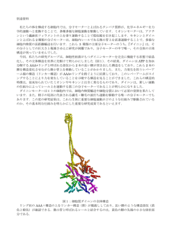
Jpn. J. Protozool. Vol. 42, No. 1. (2009) した一連の試料調製法を用いてトランスクリプトー ム解析を行う(スパイクコントロール実験)。この スパイクコントロール実験により,テトラヒメナ用 試料調製法の各ステップの最適化を行い,手法確立 後にテトラヒメナおよび大腸菌からなる人工共生系 55 のトランスクリプトーム解析を行う。 [文献] 1) Pomposiello, P.J., Bennik, M.H. and Demple, B. (2001) J. Bacteriol., 183(13): 3890-3902. テトラヒメナのADF/cofilin様蛋白質Adf73pの生化学的研究 汐崎 七海,中野 賢太郎,沼田 治(筑波大・構造生物) Biochemical studies of ADF/cofilin-like protein Adf73p in Tetrahymena thermophila Nanami SHIOZAKI, Kentaro NAKANO and Osamu NUMATA (Structural Biosciences, Graduate School of Life and Environmental Sciences, University of Tsukuba) SUMMARY ADF/cofilin-family proteins induce reorganization of the actin cytoskeleton through severing and depolymerizing filamentous actin (F-actin). Recently, the unique mechanism of actin reorganization in protists has become a focus of study because actin rapidly polymerizes and easily disassembles in Apicomplexa species, such as Toxoplasma gondii and Plasmodium falciparum. It has been reported that actin in the ciliate Tetrahymena thermophila also has unusual biochemical properties. Therefore, we studied the role of ADF/cofilin-like protein Adf73p in actin reorganization in T. thermophila. First, we investigated the biochemical action of Adf73p on muscle actin. It was found that Adf73p severed and depolymerized F-actin, although depolymerized G-actin repolymerized in the absence of the actin-capping protein, gelsolin. It is possible that Adf73p might be unable to suppress repolymerization of actin, because it accelerated nucleotide exchange of muscle actin to ATP form. Immunofluorescence microscopy on T. thermophila revealed that Adf73p is localized to the deep fiber, nascent phagosomes, and phagosomes near the cytoproct. This suggests that Adf73p might be required for controlling the actin cytoskeleton involved in membrane dynamics of phagosomes in T. thermophila. [目的] 真核生物の重要な生命活動である細胞質分裂 や細胞運動,そしてファゴサイトーシスにはアクチ ン細胞骨格の再編成が伴う。その制御には多様なア クチン調節タンパク質が働くことが知られている。 と り わ け ア ク チ ン 繊 維 を 切 断・脱 重 合 す る ADF/ cofilinはアクチン細胞骨格の再編成に欠かせない。 ところが最近,原生生物の一群であるマラリア原 虫やトキソプラズマ原虫などのアピコンプレクサの ADF/cofilinがアクチンの重合を促進することで細胞 内のアクチン細胞骨格のダイナミクスを促進すると いうユニーク活性を持つことが報告された。そのた め,アピコンプレクサと近縁な生物群である繊毛虫 のADF/cofilinの性質を調べることは興味深い。そこ で私たちは全ゲノム配列が決定されている代表的な 繊 毛 虫 で あ る テ ト ラ ヒ メ ナ に お い て,そ の ADF/ cofilin (Adf73p)の生化学的活性と細胞内機能について 解析を行った。 [方法] アクチン繊維の切断活性の検討 4 μMの骨格筋アクチンに0.8 μMのAdf73pを加え て,25℃で反応させた。2分後,グリッドのせて,さ らに2分間放置した。その後,2%酢酸ウランで30秒 間染色して,透過型電子顕微鏡で観察した。 アクチン重合速度の測定 ピレンでラベルしたアクチンは重合すると蛍光輝 度が上がる。この性質を利用して,Adf73pのアクチ ン重合への影響について蛍光光度計を用いて測定し た。 F-アクチンの脱重合実験 骨格筋アクチン繊維とAdf73pを混合し,25℃で2時 間放置した。その後,100,000 g,25℃で30分遠心し た。SDS-PAGEで上清と沈澱を電気泳動し,CBB染 色により各分画のアクチン量を比較した。 蛍光抗体法 テトラヒメナを100%冷メタノールを用いて一晩 20℃で固定した。細胞をPBS+0.1% Triton X-100で処 理して,PBS+1%スキムミルクでブロッキングした 後,テトラヒメナACT1抗体とAdf73p抗体と反応させ た。蛍光標識2次抗体と反応後,蛍光顕微鏡を用いて 細胞を観察した。 56 原生動物学雑誌 第 42 巻 第 1 号 2009 年 [結果と考察] テトラヒメナT. thermophilaの大核ゲノ ムにはADF/cofilin様遺伝子が一つだけ存在する。第 37回原生動物学会大会にて,私達はこの遺伝子をク ローニングして大腸菌にテトラヒメナのAdf73pを発 現させたことを報告した。今回,その精製タンパク 質についてウサギ骨格筋のアクチン繊維に対する切 断活性を,電子顕微鏡観察によって調べた。その結 果,Adf73pは脊椎動物や菌類のADF/cofilinと同様に アクチン繊維を切断する活性を示した。さらに,ピ レンラベルしたアクチンを用いた重合測定を行った 結果,Adf73pはアクチンの初期の重合を促進するこ とが認められた。一般的にアクチンが重合する過程 は2つの層に分けることが出来る。すなわち,アクチ ンモノマーが重合核を形成する初期と,その重合核 にアクチンモノマーが次々に重合して伸長する過程 である。重合核は不安定な構造のため,この形成が ア ク チ ン 重 合 反 応 の 律 速 段 階 と な る。お そ ら く Adf73pは重合し始めたアクチン繊維を切断すること で,繊維端の数を増やしてアクチン重合を促進した と考えられる。 次にアクチン繊維を脱重合する活性を調べた。高 速遠心により,アクチンモノマーは上清に,アクチ ン繊維は沈殿に分けられる。一般的なADF/cofilinは アクチン繊維を脱重合するため,その存在下では上 清のアクチン量が増加する。ところが,Adf73pの存 在下では,上清のアクチンの量はほとんど変化しな かった。このとき,Adf73pはアクチン繊維と共沈殿 した。Adf73pは少なくともアクチン繊維に結合する と考えられた。そこで,Adf73pにはアクチン脱重合 活性がないのか調べるために,アクチン繊維端を キャップするゲルゾリンを加えて実験を行った。そ の結果,Adf73pの存在下では上清側のアクチン量が 増えた。この実験結果から,Adf73pは骨格筋アクチ ン繊維と結合して脱重合するのだが,脱重合された アクチンモノマーが再重合してしまうことが考えら れた。このような生化学的活性は,テトラヒメナの Adf73pに特徴的である。以上の実験より,テトラヒ メナのAdf73pはユニークな生化学的性質を持つこと が示唆された。 Adf73pの細胞内機能を調べるために,蛍光抗体法 を用いてAdf73pの細胞内の局在性を解析した。その 結果,Adf73pは口部装置から伸びているディープ ファイバーや,形成直後と排出間近の食胞上に,ア クチンと共局在する様子が観察された。食胞形成に は,Adf73pによるアクチン細胞骨格の制御が重要な 働きを担う可能性が考えられた。 ゾウリムシの大核特異的抗原の性質 田中 健也,藤島 政博(山口大・院理工・環境共生系) Characteristics of macronucleus-specific antigens of Paramecium caudatum Kenya TANAKA and Masahiro FUJISHIMA (Dep. of Env. Sci. and Eng, Grad. Sch. of Sci and Eng. Yamaguchi Univ.) SUMMARY Eight monoclonal antibodies specific for macronuclear proteins of the ciliate Paramecium caudatum were developed, and molecular weight of the antigens, cross-reactivity of the antibodies and timing of appearance of the antigens during nuclear differentiation were elucidated. The properties of these antigens were compared with characteristics of a presumed receptor substance on the macronuclear envelope for lipopolysaccharides of the outer membrane of the infectious form of Holospora obtusa. One of the eight antigens shows characteristics similar to those of the receptor substance. Indirect immunofluorescence microscopy shows that the antigen locates near the macronuclear envelope and appears in the macronuclear anlagen after the appearance of heterochromatic aggregates during the nuclear differentiation process. The molecular weight of the antigen is 30 kDa. Cross-reactivity of the antibody shows that the epitopes are present not only in strains of P. caudatum, but also in P. jenningsi, P. multimicronucleatum, P. tetraurelia P. putrinum P. calkinsi, P. polycaryum and P. nephridiatum. [目的] グラム陰性細菌Holospora obtusaは,繊毛虫 Paramecium caudatumの大核に特異的に共生する核内 共生細菌である。H. obtusaは,宿主の生理状態に適 応してその形態を増殖型と感染型に分化させる。感 染型は,長さが約13 µmで宿主の飢餓状態を引き金に して増殖型から分化し,細胞質,ペリプラズム,侵 入先端の3つの領域から構成される。感染型は,増殖 能力はないが,宿主食胞を経由して細胞質に侵入 し,宿主の2種の核膜を識別して大核に侵入する能力 を持つ。
© Copyright 2026 Paperzz